

doi: 10.1074/jbc.X109.001552.
Epub 2009 May 5.
The ribosome as a conveying thermal ratchet machine
Affiliations
- PMID: 19416977
- PMCID: PMC2755833
- DOI: 10.1074/jbc.X109.001552
Item in Clipboard
The ribosome as a conveying thermal ratchet machine
J Biol Chem.
.
No abstract available
Figures
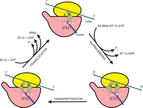
General schematic representation of the elongation cycle of a translating ribosome. Each cycle results in (i) elongation of a growing peptide chain by one amino acid residue (formation of one peptide bond), (ii) hydrolysis of two GTP molecules, (iii) entry of one molecule of aminoacyl (Aa)-tRNA into intersubunit channel, (iv) exit of one molecule of deacylated tRNA from the intersubunit channel, and (v) movement of mRNA chain by three nucleotides toward the 3′-end. Here, as well as in Figs. 3–5, ribosomal particles are shown in the orientation in which the small subunit (yellow) is on the top and the large subunit (red) is on the bottom. The head of the small subunit and the central protuberance of the large subunit are facing the viewer, with the L7/L12 stalk of the large subunit directed to the left. In this orientation, the bound L-like tRNAs must face the viewer by their external angles (“elbows”), as in reality they are facing the head of the small subunit; however, here, as well as in Figs. 3–5, for the sake of better discerning between the A and P site tRNAs, the external angles of their symbolical depictions (green) are shown to be drawn apart. The intersubunit channel accommodating the mRNA and tRNAs is traced by dotted lines. A and P are the tRNA-binding sites on the small subunit, and a, p, and e are the binding sites for their 3′ termini, either acylated or deacylated, on the large subunit. (For the sake of better clarity, the subsites on the small subunit are designated by capital letters, A and P, as originally proposed and usually accepted (59, 94), whereas the large subunit subsites, which are localized within a small area of the PTC and nearby, are designated by lowercase italicized letters, a and p, as proposed elsewhere (92).)
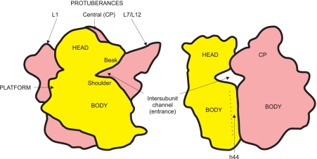
Contours of two ribosomal subunits, the small 30 S (yellow) and the large 50 S (red), associated in the full 70 S ribosome, with designations of some morphological features. Left, the so-called overlap projection when the 30 S subunit is facing the viewer and covers part of the 50 S subunit; right, the lateral projection viewed from the side of the L7/L12 stalk. CP, central protuberance.
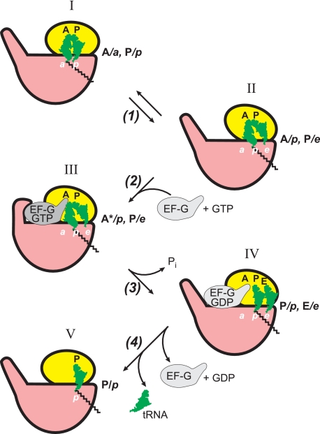
Proposed sequence of events during EF-G/GTP-promoted translocation in terms of tRNA ligand positions and the ribosomal locking-unlocking (closing-opening) concept. The ribosomes in the unlocked form (where in reality the small subunit is rotated relative to the large subunit around the axis perpendicular to the subunit interface) are conditionally depicted with the small subunits somewhat rotated relative to the large subunits in the plane of the figure (positions II–IV).
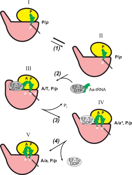
Proposed sequence of events during EF-Tu/GTP-promoted binding of aminoacyl-tRNA in terms of tRNA ligand positions and the ribosome locking-unlocking (closing-opening) concept. Aa, aminoacyl.
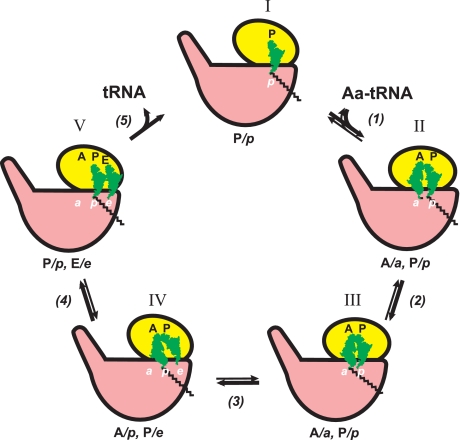
Factor-free elongation cycle and main functional states of the translating ribosome. The ribosomes in the unlocked forms (positions I, IV, and V) are shown with the small subunits somewhat rotated relative to the large subunits in the plane of the figure. In the case of the factor-promoted working cycle, step 1 is catalyzed by EF-Tu with GTP (see Fig. 4), and step 4 involves EF-G with GTP (see Fig. 3). Aa, aminoacyl.
References
-
- Kiesel A., Beloserskii A. (1934) Hoppe-Seyler's Z. Physiol. Chem. 229, 160–166
-
- Belozersky A. N., Dubrovskaya I. I. (1936) Biokhimiya 1, 665–675
-
- Belozersky A. N. (1940) Mikrobiologiya 9, 107–113
-
- Belozersky A. N. (1947) Cold Spring Harbor Symp. Quant. Biol. 12, 1–6
-
- Vischer E., Chargaff E. (1948) J. Biol. Chem. 176, 703–714, 715–734 - PubMed
Publication types
MeSH terms
LinkOut - more resources
Full Text Sources
