. 2009 May;8(5):1086-94.
doi: 10.1158/1535-7163.MCT-08-1166. Epub 2009 May 5.

Transcriptional induction of GRP78/BiP by histone deacetylase inhibitors and resistance to histone deacetylase inhibitor-induced apoptosis

Affiliations

Transcriptional induction of GRP78/BiP by histone deacetylase inhibitors and resistance to histone deacetylase inhibitor-induced apoptosis

Peter Baumeister et al. Mol Cancer Ther. 2009 May.

Abstract

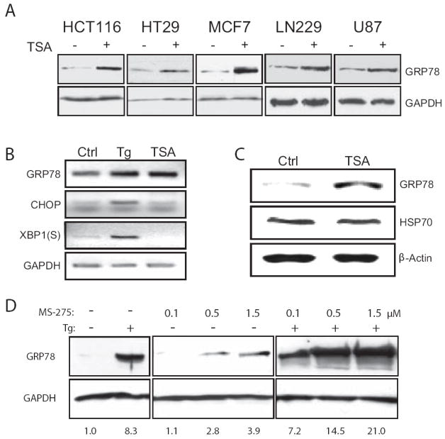
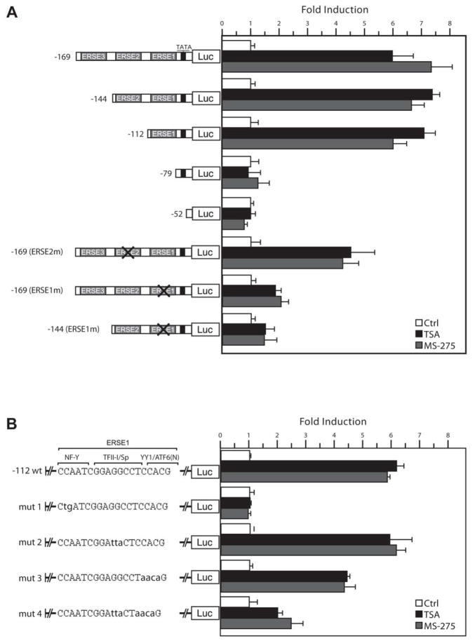
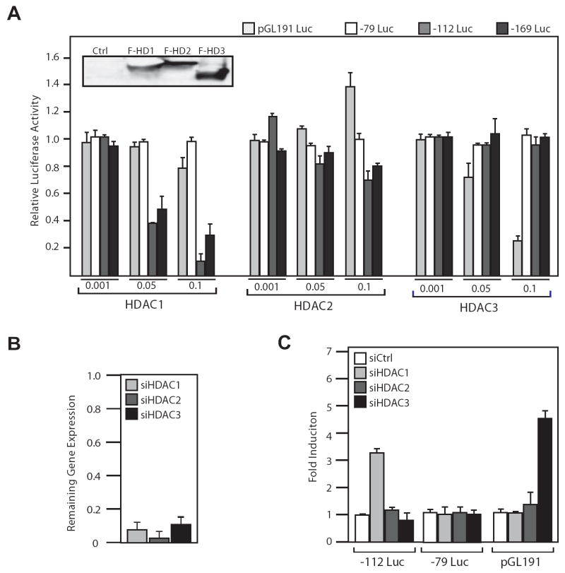
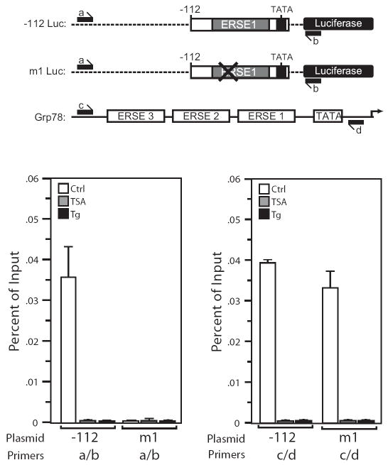
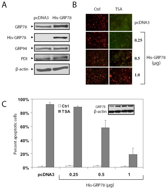
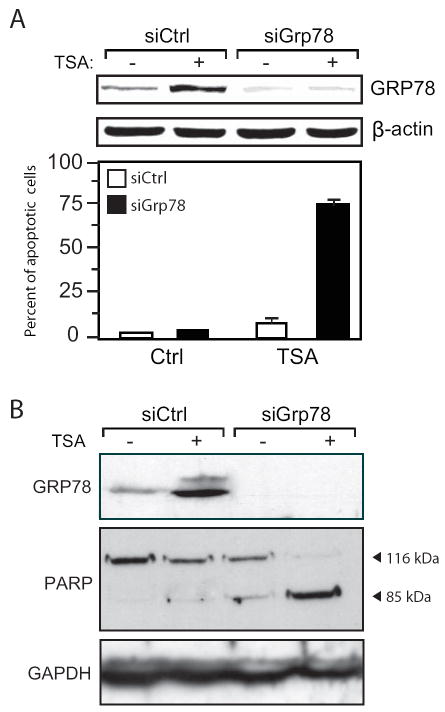
Histone deacetylase (HDAC) inhibitors are emerging as effective therapies in the treatment of cancer, and the role of HDACs in the regulation of promoters is rapidly expanding. GRP78/BiP is a stress inducible endoplasmic reticulum (ER) chaperone with antiapoptotic properties. We present here the mechanism for repression of the Grp78 promoter by HDAC1. Our studies reveal that HDAC inhibitors specifically induce GRP78, and the induction level is amplified by ER stress. Through mutational analysis, we have identified the minimal Grp78 promoter and specific elements responsible for HDAC-mediated repression. We show the involvement of HDAC1 in the negative regulation of the Grp78 promoter not only by its induction in the presence of the HDAC inhibitors trichostatin A and MS-275 but also by exogenous overexpression and small interfering RNA knockdown of specific HDACs. We present the results of chromatin immunoprecipitation analysis that reveals the binding of HDAC1 to the Grp78 promoter before, but not after, ER stress. Furthermore, overexpression of GRP78 confers resistance to HDAC inhibitor-induced apoptosis in cancer cells, and conversely, suppression of GRP78 sensitizes them to HDAC inhibitors. These results define HDAC inhibitors as new agents that up-regulate GRP78 without concomitantly inducing the ER or heat shock stress response, and suppression of GRP78 in tumors may provide a novel, adjunctive option to enhance anticancer therapies that use these compounds.


Figures

Figure 1
Figure 1. Induction of GRP78 by HDAC inhibitors in cancer cell lines and in xenograft models
A, the cancer cell lines were either untreated (-) or treated (+) with TSA for 24 h and GRP78 protein levels were assessed by Western blot, with GAPDH levels serving as a loading control. B, HCT116 cells were treated with either TSA for 12 h or Tg for 4 h and the RNA was isolated and used in the RT-PCR reaction to determine the mRNA level of the UPR indicators Grp78, CHOP and the spliced (S) form of XBP-1. C, xenograft tumors were generated through subcutaneously injection of the human breast cancer cell line MDA-MB-435 in nude mice. Following DMSO (Ctrl) or TSA injection for 4 days, the tumors were harvested and Western blots performed to determine levels of GRP78 and HSP70, with β-actin as a loading control. D, HCT116 cells were either non-treated (-), treated with increasing concentrations of MS-275 for 24 h as indicated, or in combination with Tg for an additional 16 h. GRP78 levels were detected by Western blots with GAPDH as control. The fold increase of GRP78 under each condition after normalization against GAPDH is indicated below each lane.
Figure 2
Figure 2. Mutational analysis to determine HDAC inhibitor response element in the Grp78 promoter
A, luciferase reporter plasmids containing 5’ deletion or specific ERSE mutations of the rat Grp78 promoter as indicated were transiently transfected into HeLa cells, and luciferase activities were monitored 18 h after TSA or MS-275 treatment. For inactivation of ERSE, the CCAAT sequence was mutated. The luciferase activity for each construct in non-treated cells was set as 1, and the fold induction by TSA or MS-275 was plotted with standard deviations. B, the minimal HDAC inhibitor-inducible Grp78 promoter -112Luc was used as a template for successive mutation of putative transcription factor binding sites within the ERSE. The putative transcription factor binding sites are indicated. Following transfection and treatment with TSA or MS-275, the luciferase activities were determined. The fold induction was plotted with standard deviations.
Figure 3
Figure 3. Identification of HDAC1 as a repressor of Grp78 promoter activity
A, flag-tagged HDAC1, 2 and 3 expression plasmids at increasing amounts (μg) as indicated were transfected into HeLa cells along with either -79Luc, -112Luc, -169Luc or the HDAC3-repressed human GDF11 promoter reporter plasmid pGL191Luc. The transfected HDAC levels were determined by Western blot using anti-flag antibody (inset). The cells were harvested 72 h after transfection. The relative luciferase activity was plotted with standard deviations. B, HeLa cells were transfected with siRNA against HDACs 1, 2 or 3 and incubated for 48 h, harvested, and the RNA was isolated and subjected to quantitative RT-PCR to determine mRNA levels of each HDAC in comparison to control siRNA-treated cells. C, HeLa cells were transfected with siRNA against HDACs 1, 2 or 3, along with reporter plasmids -112Luc, -79Luc or pGL191Luc. The cells were harvested after 72 h and luciferase activity was determined. Fold induction in comparison to control siRNA is shown with standard deviations.
Figure 4
Figure 4. The NF-Y binding site in ERSE1 is required for HDAC1 binding to the Grp78 promoter
A, schematic drawing of the luciferase reporter constructs (-112 and mut 1 Luc) and the endogenous Grp78 promoter. The positions of the primer sets used for the ChIP assays are indicated. B, HeLa cells stably transfected with either -112Luc or mut1Luc were either non-treated (Ctrl) or treated with TSA for 12 h or Tg for 4 h and subjected to ChIP assay. Chromatin was immunoprecipitated with anti-HDAC1 antibody or IgG, and the purified DNA fraction was assayed by quantitative PCR with primers specific for detection of HDAC1 binding to plasmid DNA (a/b). Results are shown as percentage of input corrected by IgG, with standard error of the mean. C same as (B) except the primers (c/d) were used to detect HDAC1 binding to the endogenous Grp78 promoter in cells stably transfected with either -112Luc or mut 1 Luc.
Figure 5
Figure 5. Overexpression of GRP78 protects 293T cells from TSA-induced apoptosis
A, cell lysates prepared from 293T cells transfected with either 1.0 μg of a plasmid expressing His-tagged GRP78 or same amount of empty vector pcDNA3 were subjected to Western blot with anti-KDEL, anti-His, and anti-β-actin antibodies. The anti-KDEL antibody recognized the C-terminal KDEL motif of GRP78, GRP94 and protein disulphide isomerase (PDI). B, empty vector pcDNA3 or increasing amounts of the plasmid expressing His-GRP78 (0.25, 0.5 and 1.0 μg) as indicated were transfected to 293T cells. Empty vector was added to adjust the total amount of plasmids to be the same. Twenty-four h after transient transfection, the cells were treated with 500 nM TSA for 48 h and then subjected to mitochondrial membrane potential staining using the JC-1 assay, which detects cells at early stage of apoptosis. Red fluorescence indicates viable cells and green apoptotic cells. C, the percent of apoptotic cells under each condition in (B) was quantitated and plotted against the transfected amount of GRP78. The open and solid bars represent no treatment and TSA treatment respectively. The standard deviations are shown.
Figure 6
Figure 6. Knockdown of GRP78 sensitizes cancer cells to HDAC inhibitors-mediated apoptosis
A, MDA-MB-435 cells were transfected with siRNA against GRP78 (siGrp78) or a random control (siCtrl), and 48 h later, were either non-treated (-) or treated (+) with TSA for 24 h. The upper panel shows GRP78 levels in the transfected cells with β-actin as loading control. Lower panel shows the percent of apoptotic cells under each condition as determined by mitochondrial membrane potential staining using the JC-1 assay. B, HCT116 cells were subjected to the same conditions as (A) and the cell lysates were assayed for PARP cleavage by Western blot. The normal length (116 kDa) and apoptotic, cleaved forms (85 kDa) of PARP are indicated. GAPDH was used as a control.

References

    1. Glozak M, Seto E. Histone deacetylases and cancer. Oncogene. 2007;26:5420–32. - PubMed
    1. Yang X, Seto E. HATs and HDACs: from structure, function and regulation to novel strategies for therapy and prevention. Oncogene. 2007;26:5310–8. - PubMed
    1. Wang J, Bown C, Young L. Differential display PCR reveals novel targets for the mood-stabilizing drug valproate including the molecular chaperone GRP78. Mol Pharmacol. 1999;55:521–7. - PubMed
    1. Lee AS. The glucose-regulated proteins: stress induction and clinical applications. Trends Biochem Sci. 2001;26:504–10. - PubMed
    1. Hendershot LM. The ER function BiP is a master regulator of ER function. Mt Sinai J Med. 2004;71:289–97. - PubMed

Publication types

MeSH terms

Substances