. 2009 Jul 2;114(1):109-18.
doi: 10.1182/blood-2008-12-193375. Epub 2009 May 5.

EBV Zta protein induces the expression of interleukin-13, promoting the proliferation of EBV-infected B cells and lymphoblastoid cell lines

Affiliations

EBV Zta protein induces the expression of interleukin-13, promoting the proliferation of EBV-infected B cells and lymphoblastoid cell lines

Shu-Chun Tsai et al. Blood. .

Abstract

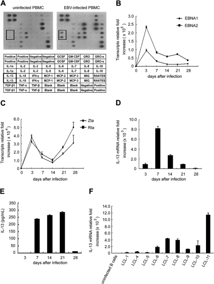
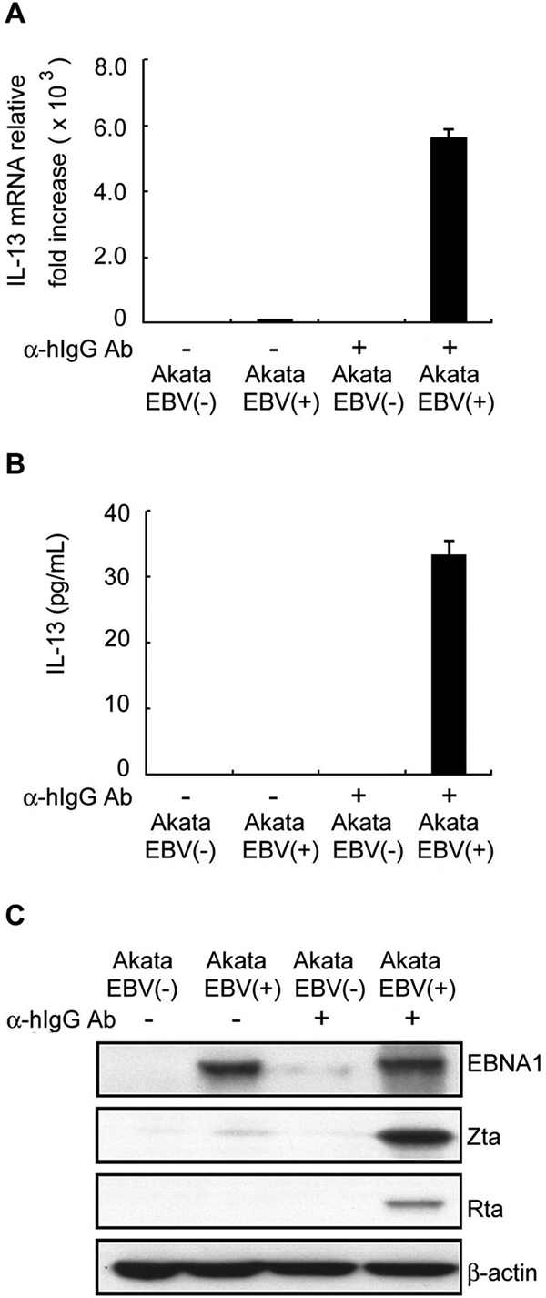
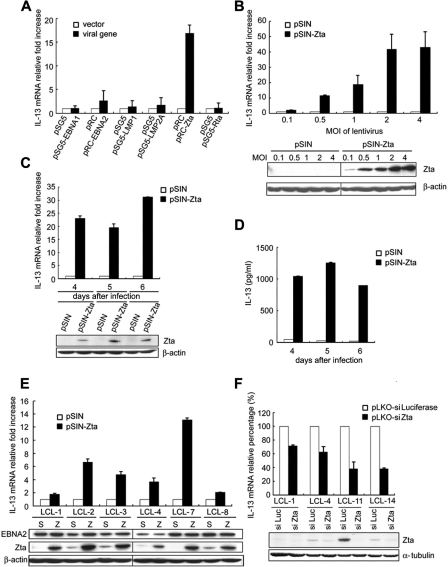
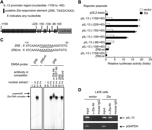
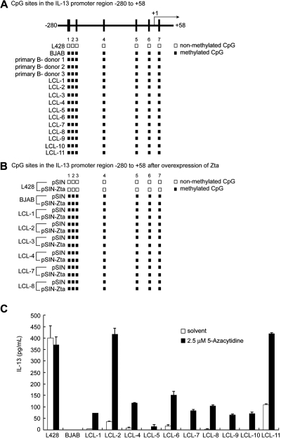
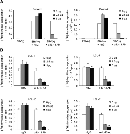
Epstein-Barr virus (EBV) infection can modify the cytokine expression profiles of host cells and determine the fate of those cells. Of note, expression of interleukin-13 (IL-13) may be detected in EBV-associated Hodgkin lymphoma and the natural killer (NK) cells of chronic active EBV-infected patients, but its biologic role and regulatory mechanisms are not understood. Using cytokine antibody arrays, we found that IL-13 production is induced in B cells early during EBV infection. Furthermore, the EBV lytic protein, Zta (also known as the BZLF-1 product), which is a transcriptional activator, was found to induce IL-13 expression following transfection. Mechanistically, induction of IL-13 expression by Zta is mediated directly through its binding to the IL-13 promoter, via a consensus AP-1 binding site. Blockade of IL-13 by antibody neutralization showed that IL-13 is required at an early stage of EBV-induced proliferation and for long-term maintenance of the growth of EBV immortalized lymphoblastoid cell lines (LCLs). Thus, Zta-induced IL-13 production facilitates B-cell proliferation and may contribute to the pathogenesis of EBV-associated lymphoproliferative disorders, such as posttransplantation lymphoproliferative disease (PTLD) and Hodgkin lymphoma.


Figures

Figure 1
Figure 1
IL-13 production is induced after EBV infection. (A) PBMCs were infected with EBV or not infected, and supernatants were collected on day 7 after infection. Expression of cytokines in supernatants was measured using the human cytokine antibody array III. Each spot on the blot represents 1 cytokine, and each cytokine was duplicated. The cytokines tested are listed in the table under the blot. (B-F) CD19-positive B cells were infected with EBV, and RNA was extracted at the times indicated. The relative fold increase of transcript expression was normalized by the amount of RNA from uninfected B cells. RNA and supernatants were pooled from 6 individual donors for 1 experiment. A representative of 2 independent experiments is shown. (B) Expression of viral latent EBNA1 and EBNA2 genes was detected by RT-Q-PCR. (C) Expression of viral lytic genes (Zta and Rta) was detected by RT-Q-PCR. (D) Expression of IL-13 transcripts was measured by RT-Q-PCR. (E) IL-13 protein secreted in cultured medium was quantified by ELISA. (F) RNA was extracted from 9 LCL cells, and the IL-13 transcripts were examined by RT-Q-PCR. The relative fold of transcript expression was normalized by the amount of RNA from primary uninfected B cells.
Figure 2
Figure 2
EBV lytic cycle progression enhances IL-13 expression. EBV negative (−) or EBV positive (+) Akata B-cell lines were treated with or without 10 μg/mL goat anti–human IgG antibody (α-hIgG Ab). RNA, supernatants, and cell lysates were harvested after 48 hours. Expression of IL-13 transcripts and protein was measured by RT-Q-PCR (A) and ELISA (B). The relative expression levels of IL-13 transcripts were normalized by the amount of IL-13 expressed in EBV(−) Akata B cells. (C) Expression of EBV latent (EBNA1) or lytic (Zta and Rta) proteins was detected by Western blot analysis; β-actin served as the internal control.
Figure 3
Figure 3
Expression of the EBV lytic protein, Zta, contributed to induction of IL-13. (A) BJAB cells were transfected with plasmids expressing EBNA1, EBNA2, LMP1, LMP2A, Zta, or Rta. RNA was extracted from each transfectant at 48 hours after transfection, and IL-13 transcripts were measured by RT-Q-PCR. (B) BJAB cells were infected with pSIN-vector or pSIN-Zta at the MOI indicated. RNA and cell lysates were harvested from each transfectant at 5 days after infection. IL-13 transcripts were measured by RT-Q-PCR (top panel), and Zta expression was detected by Western blot analysis (bottom panel; vertical lines have been inserted to indicate a repositioned gel lane). (C,D) L428 cells were infected with pSIN-Zta or pSIN-vector at an MOI of 4. Cell lysates, RNA, and supernatants were collected on days 4, 5, and 6 after infection. IL-13 transcripts were measured by RT-Q-PCR, and then relative expression of IL-13 transcripts was normalized to the amounts of IL-13 expressed in pSIN-vector transfected cells (panel C top). Zta expression was detected by Western blot analysis, and β-actin was used as an internal control (panel C bottom). Meanwhile, IL-13 protein in supernatants was quantified by ELISA (D). (E) In total, 6 LCLs cells were infected with pSIN-Zta or pSIN-vector lentivirus at an MOI of 4, and RNA and cell lysates were harvested after 7 days. Expression of IL-13 transcripts was detected by RT-Q-PCR, and the relative fold was normalized with the amounts of IL-13 mRNA in pSIN-vector lentivirus control cells (top panel). EBNA2 and Zta expression were measured by Western blot analyses, and β-actin was used as an internal control (bottom panel; vertical lines have been inserted to indicate a repositioned gel lane). (F) LCLs were infected with pLKO-siZta or pLKO-siLuciferase lentivirus, and then RNA and cell lysates were harvested on day 14 after infection. Expression of IL-13 transcripts was detected by RT-Q-PCR, and the relative fold was normalized with the amounts of IL-13 mRNA in pLKO-siLuciferase lentivirus infected cells (top panel). Zta expression was measured by Western blot analysis, and α-tubulin was used as an internal control (bottom panel).
Figure 4
Figure 4
Mechanism involved in Zta-mediated IL-13 gene expression. (A) Schematic illustration of the IL-13 promoter that drives the luciferase gene in the reporter plasmid; putative ZRE sequences are indicated. The IL-13 promoter contains 34 putative ZRE regions in the sequence from −1159 to +65. (B) HEK293T cells were transfected with Zta-expressing plasmid or vector in combination with serial 5′-deleted pIL-13 reporter plasmids and pEGFP-C1 reporter control. After 72 hours, luciferase activities from each transfectant were normalized with the GFP intensities. Zta-driven fold activation for each reporter was calculated by normalizing luciferase activities for the Zta transfectant versus that for the pGL2-basic vector control. (C) DNA sequences of the probes used in EMSA experiments. Bold lines indicate ZRE or ZREmt within the IL-13 promoter sequences from −90 to −110. Nuclear extracts were harvested at 72 hours after transfection of pRC vector or pRC-Zta in HEK293T cells. The binding of Zta with 32P-labeled ZRE (lanes 3 and 7) or ZREmt (lane 4) probes was examined by EMSA. The supershift signal represents the complex of Zta and IL-13 promoter DNA and was detected by the addition of specific anti-Zta (lane 8) or anti-GST control (lane 9) antibodies. For the competition EMSA, a 30-fold excess of wild-type ZRE (lane 10) or ZREmt (lane 11) oligonucleotides was added to the reaction mixture. (D) A ChIP experiment was carried out as described previously. L428 cells were infected with pSIN-Zta or pSIN-vector lentivirus for 5 days. DNA-protein complexes were immunoprecipitated with anti-Zta antibody or mouse IgG (negative control). IL-13 promoter DNA (pIL-13, from nt −232 to +66) and control GAPDH promoter DNA (pGAPDH, from nt −93 to +64) were detected in the immunoprecipitates by PCR. Total DNA was harvested from L428 cells and used as the input control.
Figure 5
Figure 5
Methylation patterns were examined in IL-13 promoter sequences from −280 to +58. (A) The IL-13 promoter contains 7 CpG sites at the positions indicated, between −280 and +58 relative to the transcription start site. Genomic DNA was harvested from L428 cells, BJAB cells, primary uninfected B cells, and 11 LCLs, and CpG methylation was analyzed by bisulfite sequencing. Numbers (1-7) under the sequence represents the CpG position; [□], nonmethylated CpG; [■], methylated CpG. (B) In total, 6 LCLs cells were infected with pSIN-Zta or pSIN-vector lentivirus at an MOI of 4. Genomic DNA was extracted, and CpG methylation was analyzed by bisulfite sequencing after 7 days. (C) L428 cells, BJAB cells, and a total of 10 LCLs were treated with or without 2.5 μM 5-azacytidine for 5 days. Cultured supernatants were harvested, and IL-13 expression was measured by ELISA.
Figure 6
Figure 6
IL-13 is crucial for triggering the cell growth of EBV-infected primary B cells and for maintaining the proliferation of LCLs. (A) B cells purified from 2 healthy donors were infected with EBV. Five days after infection, various doses of anti–IL-13 neutralizing antibodies (α-IL-13 Ab) or rabbit IgG were added. After 24 hours incubation, [3H]-thymidine was added, incubation was continued for a further 24 hours, and the amount of [3H]-thymidine incorporation was measured. (B) LCLs were cultured for 24 hours, and various doses of anti–IL-13 neutralizing antibodies or rabbit IgG were added. After 48 hours incubation, [3H]-thymidine was added, incubation continued for 18 hours, and the amount of [3H]-thymidine incorporation was measured.

Similar articles

Cited by

References

    1. Mogensen TH, Paludan SR. Molecular pathways in virus-induced cytokine production. Microbiol Mol Biol Rev. 2001;65:131–150. - PMC - PubMed
    1. Young LS, Rickinson AB. Epstein-Barr virus: 40 years on. Nat Rev Cancer. 2004;4:757–768. - PubMed
    1. Hislop AD, Taylor GS, Sauce D, Rickinson AB. Cellular responses to viral infection in humans: lessons from Epstein-Barr virus. Annu Rev Immunol. 2007;25:587–617. - PubMed
    1. Rickinson AB, Kieff E. Fields virology. In: Knipe DM, Howley PM, Griffin DE, Lamb MA, Martin MA, Roizman B, Straus SE, editors. Epstein-Barr Virus. 4th Ed. Vol 2. Hagerstown, MD: Lippincott Williams & Wilkin; 2001.
    1. Torkildsen O, Nyland H, Myrmel H, Myhr KM. Epstein-Barr virus reactivation and multiple sclerosis. Eur J Neurol. 2008;15:106–108. - PubMed

Publication types

MeSH terms