Review
. 2009 Sep;50(9):1721-34.
doi: 10.1194/jlr.R900011-JLR200. Epub 2009 May 5.

Bile acids: regulation of apoptosis by ursodeoxycholic acid

Affiliations
Review

Bile acids: regulation of apoptosis by ursodeoxycholic acid

Joana D Amaral et al. J Lipid Res. 2009 Sep.

Abstract

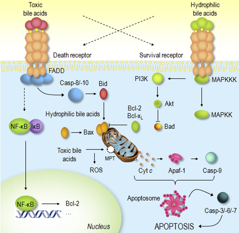
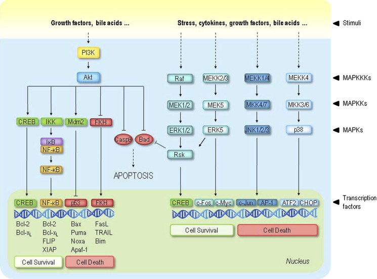
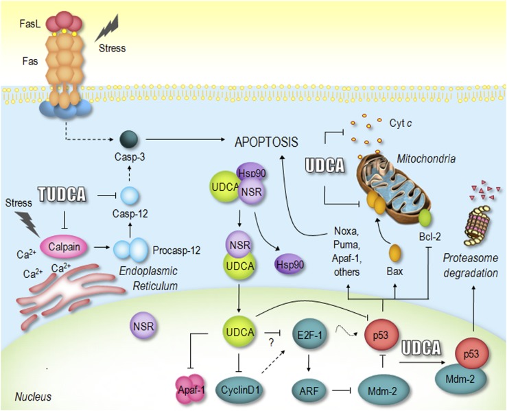
Bile acids are a group of molecular species of acidic steroids with peculiar physical-chemical and biological characteristics. At high concentrations they become toxic to mammalian cells, and their presence is pertinent in the pathogenesis of several liver diseases and colon cancer. Bile acid cytoxicity has been related to membrane damage, but also to nondetergent effects, such as oxidative stress and apoptosis. Strikingly, hydrophilic ursodeoxycholic acid (UDCA), and its taurine-conjugated form (TUDCA), show profound cytoprotective properties. Indeed, these molecules have been described as potent inhibitors of classic pathways of apoptosis, although their precise mode of action remains to be clarified. UDCA, originally used for cholesterol gallstone dissolution, is currently considered the first choice therapy for several forms of cholestatic syndromes. However, the beneficial effects of both UDCA and TUDCA have been tested in other experimental pathological conditions with deregulated levels of apoptosis, including neurological disorders, such as Alzheimer's, Parkinson's, and Huntington's diseases. Here, we review the role of bile acids in modulating the apoptosis process, emphasizing the anti-apoptotic effects of UDCA and TUDCA, as well as their potential use as novel and alternate therapeutic agents for the treatment of apoptosis-related diseases.


Figures

Fig. 1.
Fig. 1.
Schematic representation of bile acid modulation of death and survival transduction pathways. Toxic bile acids induce apoptosis by activating both ligand-dependent and -independent death receptor oligomerization. The activation of death receptors by bile acids invariably signals the mitochondrial pathway of apoptosis in type II cells, such as hepatocytes. Moreover, toxic bile acids may directly target mitochondria, either through induction of the MPT and ROS production or activation of pro-apoptotic Bcl-2 family members. Finally, hydrophobic bile acids partially activate death-receptor-dependent survival pathways, such as the NF-κB. Hydrophilic bile acids do not induce apoptosis as they simultaneously activate survival signaling pathways, such as the MAPK and PI3K, antagonizing proapototic Bcl-2 family members and preventing mitochondrial dysfunction and apoptosis. See text for more complete description. Cyt c, cytochrome c; MAPKK, mitogen-activated protein kinase kinase; MAPKKK, mitogen-activated protein kinase kinase kinase; MPT, mitochondrial permeability transition.
Fig. 2.
Fig. 2.
Schematic representation of the interplay between cellular survival signaling and apoptosis regulators. Cell survival requires the active inhibition of apoptosis, which is accomplished by reducing the expression of pro-apoptotic factors as well as promoting expression of anti-apoptotic factors. Survival pathways may be triggered by a wide variety of extracellular signals, such as growth factors, stress, proinflammatory cyto kines, and bile acids. Activation of Akt results in inhibition of pro-apoptotic factors, such as Bad and caspase-9, and activation of FKH, CREB, and NF-κB transcription factors, all involved in cell survival regulation. The MAPK pathways include ERK, JNK, and p38 cascades, all of which contain the same series of kinases. Although ERK signaling is considered mainly cytoprotective, JNK and p38 are referred to primarily as stress-activated proteins. The effect of MAPK pathways on survival is mediated, at least partly, by activation of ribosomal S6 kinase (RSK) family members. Much like Akt, RSKs inactivate Bad and activate CREB, ultimately leading to cell survival.
Fig. 3.
Fig. 3.
Proposed mechanisms of UDCA and TUDCA inhibition of apoptosis. UDCA negatively modulates the mitochondrial pathway by inhibiting Bax translocation, ROS formation, cytochrome c release, and caspase-3 activation. UDCA can also interfere with the death receptor pathway, inhibiting caspase-3 activation. Moreover, TUDCA inhibits apoptosis associated with ER stress by modulating intracellular calcium levels and inhibiting calpain and caspase-12 activation. Importantly, UDCA interacts with NSR, leading to NSR/hsp90 dissociation and nuclear translocation of the UDCA/NSR complex. Once in the nucleus, UDCA modulates the E2F-1/p53/Bax pathway, thus preventing apoptosis. Finally, UDCA downregulates cyclin D1 and Apaf-1, further inhibiting the mitochondrial apoptotic cascade. See text for more complete description. Cyt c, cytochrome c; Hsp90, heat shock protein 90.

References

    1. Bayerdorffer E., Mannes G. A., Richter W. O., Ochsenkuhn T., Wiebecke B., Kopcke W., Paumgartner G. 1993. Increased serum deoxycholic acid levels in men with colorectal adenomas. Gastroenterology. 104: 145–151. - PubMed
    1. Patel T., Gores G. J. 1995. Apoptosis and hepatobiliary disease. Hepatology. 21: 1725–1741. - PubMed
    1. Hofmann A. F., Roda A. 1984. Physicochemical properties of bile acids and their relationship to biological properties: an overview of the problem. J. Lipid Res. 25: 1477–1489. - PubMed
    1. Babu P., Sangeetha N., Maitra U. 2006. Supramolecular chemistry of bile acids derivatives: formation of gels. Macromol. Symp. 241: 60–67.
    1. Russell D. W., Setchell K. D. 1992. Bile acid biosynthesis. Biochemistry. 31: 4737–4749. - PubMed

MeSH terms

LinkOut - more resources