DNA/amphiphilic block copolymer nanospheres promote low-dose DNA vaccination
- PMID: 19417740
- PMCID: PMC2835232
- DOI: 10.1038/mt.2009.84
DNA/amphiphilic block copolymer nanospheres promote low-dose DNA vaccination
Abstract
Intramuscular (i.m.) DNA vaccination induces strong cellular immune responses in the mouse, but only at DNA doses that cannot be achieved in humans. Because antigen expression is weak after naked DNA injection, we screened five nonionic block copolymers of poly(ethyleneoxide)-poly(propyleneoxide) (PEO-PPO) for their ability to enhance DNA vaccination using a beta-galactosidase (betaGal) encoding plasmid, pCMV-betaGal, as immunogen. At a high DNA dose, formulation with the tetrafunctional block copolymers 304 (molecular weight [MW] 1,650) and 704 (MW 5,500) and the triblock copolymer Lutrol (MW 8,600) increased betaGal-specific interferon-gamma enzyme-linked immunosorbent spot (ELISPOT) responses 2-2.5-fold. More importantly, 704 allowed significant reductions in the dose of antigen-encoding plasmid. A single injection of 2 microg pCMV-betaGal with 704 gave humoral and ELISPOT responses equivalent to those obtained with 100 microg naked DNA and conferred protection in tumor vaccination models. However, 704 had no adjuvant properties for betaGal protein, and immune responses were only elicited by low doses of pCMV-betaGal formulated with 704 if noncoding carrier DNA was added to maintain total DNA dose at 20 microg. Overall, these results show that formulation with 704 and carrier DNA can reduce the dose of antigen-encoding plasmid by at least 50-fold.
Figures

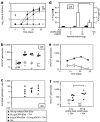
 ) or formulated with 3% Lutrol (Lut, □), 0.05% PE6400 (PE, ⋄), 5% 304 (*), 0.25% 704 (▵), or 0.1% 904 (○). n = 7 for all groups except PE6400 n = 6. (b) Antigen expression. Two mice per group were killed at day 7 after the first DNA injection, and βGal expression in situ was monitored by coloration with X-Gal. One representative TA muscle of four is shown for each group. (c) Humoral response. Geometric mean titers (GMTs) are shown for each group. Arrows indicate DNA injection at day 0 and boost at day 21. For clarity, sera at day 0 with no detectable βGal-specific IgG are presented at a titer of 1/25. (d) Class I–restricted cellular response. Splenocytes were prepared at day 42, stimulated overnight with the ICPMYARV peptide, and the number of IFNγ spot-forming cells (SFCs) was determined. Each symbol represents the result from one mouse. Significant differences between groups are indicated. *P < 0.05; **P < 0.01 by analysis of variance and Tukey's least significant difference post hoc test.
 , ⋄), 0.2 µg (•, ○), pCMV-βGal formulated with 0.25% 704 in the absence (a,c) or the presence (b,d) of pUC19 plasmid to maintain a total DNA dose of 20 µg per mouse. (a,b) Humoral response. Geometric mean titers are shown for each group ± standard deviation. Arrows indicate DNA injection at day 0 and boost at day 21. Figures on the right indicate the pCMV-βGal plasmid dose for each group. For clarity, sera with no detectable βGal-specific IgG are presented at a titer of 1/25. (c,d) Class I–restricted cellular response. Splenocytes were prepared at day 42, stimulated overnight with the βGal ICPMYARV peptide (▴,
  , •) or control peptide (▵, ⋄, ○), and the number of IFNγ SFCs was determined. Each symbol represents the result from one mouse. Data from the group injected with 20 µg pCMV-βGal are duplicated in panels a and c, and b and d. Significant differences between groups are indicated. *P < 0.05, **P < 0.01 by analysis of variance and Tukey's least significant difference post hoc test. SFC, spot-forming cell.
 ), 2 µg pCMV-βGal + 18 µg pQE30 plasmid formulated with 0.25% 704 (•), or 0.25% 704 alone (▪), and different parameters of the immune response were measured. (a) Humoral response. Geometric mean titers are shown for each group ± standard deviation. The arrow indicates DNA injection at day 0. Figures on the right indicate the pCMV-βGal plasmid dose for each group. For clarity, sera with no detectable βGal-specific IgG are presented at a titer of 1/25. Wide deviations at day 14 and day 21 in the group vaccinated with 2 µg pCMV-βGal + 18 µg pQE30 formulated with 704 are due to two mice that only seroconverted by day 28. (b) Class I–restricted cellular response. Splenocytes were prepared at day 28, stimulated overnight with the βGal ICPMYARV peptide (▴,
  , •, ▪), or control peptide (▵, ⋄, ○, □), and the number of IFNγ SFCs was determined. Each symbol represents the result from one mouse. (c) Isotype profile of βGal-specific antibodies. βGal-specific IgG1 and IgG2c were titered in sera at day 28 after a single DNA vaccination, and the IgG2c/IgG1 ratio is shown. (d) T-helper response. Splenocytes were prepared at day 28, stimulated for 72 hours with recombinant βGal protein, and IFNγ and IL-4 in the supernatants were determined by ELISA. *P < 0.05 by the Wilcoxon test. (e) Time course of the class I–restricted response. Splenocytes were prepared at 7, 14, 21, and 28 days after a single vaccination with 2 µg pCMV-βGal formulated with 0.25% 704 without carrier DNA, then stimulated overnight with the βGal ICPMYARV peptide (•) or control peptide (○), and the number of IFNγ SFCs was determined. (f) Groups of mice (n = 6) were injected i.m. with 2 µg or 20 µg pCMV-βGal formulated with 704 as indicated. Splenocytes were prepared at day 28, stimulated overnight with the βGal ICPMYARV peptide (•,
  ) or control peptide (○, ⋄), and the number of IFNγ SFCs was determined. Each symbol represents the result from one mouse. *P < 0.05 by the Wilcoxon test. IL-4, interleukin-4; SFC, spot-forming cell.
 ), or 2 µg pCMV-βGal + 18 µg pQE30 formulated with 0.25% 704 (704, ▴). Two weeks after vaccination, mice were challenged subcutaneously with 5 × 10⁶ Hepa1.6 cells stably expressing βGal. Kaplan-Meier analysis of uncontrolled tumor growth (tumor size >250 mm3) in the three groups is shown. Significant differences between groups are indicated. *P < 0.05 by log-rank test. (b–d) Orthotopic tumor model. Groups of AFP-βGal mice were injected i.m. with 0.25% 704 alone (•, n = 6), 100 µg pCMV-βGal (▴, n = 6), or 2 µg pCMV-βGal + 18 µg pQE30 formulated with 0.25% 704 (
 , n = 6). A booster vaccination was given at day 21, and then at day 28, mice were injected via the portal vein with 2 × 10⁶ HepaβGal cells. Mice were killed 3 weeks later at day 49. (b) Images of livers from two mice in each group. (c) Liver weight as a percentage of body weight. (d) Specific βGal activity in liver extracts. PBS, phosphate-buffered saline.
 ), or 20 µg pCMV-Ova formulated with 0.25% 704 (▴). Arrows indicate DNA injection at day 0 and boost at day 21. Heparinized blood was sampled once per week, and Ova-specific CD8+ cells were identified by staining with H2-Kb:SIINFEKL pentamers. Data points represent median values for each group. Significant differences between groups at the time of peak response (day 35) are indicated. *P < 0.05 by analysis of variance and Tukey's least significant difference test. (b) Groups of C57BL/6 mice (n = 8) were vaccinated i.m. either with 0.25% 704 alone (•), 100 µg naked pCMV-Ova DNA (▪), or 2 µg pCMV-Ova formulated with 704 and 18 µg pQE30 (▴). A booster vaccination was given at day 21, and 7 days later, mice were challenged by subcutaneous injection of 2.5 × 10⁵ B16-Ova cells. Kaplan-Meier analysis of uncontrolled tumor growth (tumor size >250 mm3) in the three groups is shown. Significant differences between groups are indicated. **P < 0.01 by log-rank test.
 , n = 5), or formulated with 0.25% 704 (▴, n = 5) or subcutaneous 100 µg recombinant βGal protein in IFA (▪, n = 3). (a) Humoral response. Geometric mean titers are shown for each group ± standard deviation. Significantly higher titers after vaccination with βGal protein in IFA are indicated. **P < 0.01 by analysis of variance and Tukey's least significant difference test. (b) Class I–restricted cellular response. Splenocytes were prepared at day 28, stimulated overnight with the βGal ICPMYARV peptide (
 , ▴, ▪) or control peptide (⋄, ▵, □), and the number of IFNγ SFCs was determined. Each symbol represents the result from one mouse. (c) Isotype profile of βGal-specific antibodies. βGal-specific IgG1 and IgG2c were titered in sera at day 28 after a single vaccination, and the IgG2c/IgG1 ratio is shown. IFA, incomplete Freund's adjuvant; SFC, spot-forming cell.
References
-
- Ulmer JB, Donnelly JJ, Parker SE, Rhodes GH, Felgner PL, Dwarki VJ, et al. Heterologous protection against influenza by injection of DNA encoding a viral protein. Science. 1993;259:1745–1749. - PubMed
-
- Donnelly JJ, Ulmer JB, Shiver JW., and , Liu MA. DNA vaccines. Annu Rev Immunol. 1997;15:617–648. - PubMed
-
- Boyer JD, Cohen AD, Vogt S, Schumann K, Nath B, Ahn L, et al. Vaccination of seronegative volunteers with a human immunodeficiency virus type 1 env/rev DNA vaccine induces antigen-specific proliferation and lymphocyte production of β-chemokines. J Infect Dis. 2000;181:476–483. - PubMed
Publication types
MeSH terms
Substances
LinkOut - more resources
Full Text Sources
Other Literature Sources
Molecular Biology Databases

