

doi: 10.1038/icb.2009.32.
Epub 2009 May 5.
Leukocyte adhesion deficiency syndrome: a controversy solved
Affiliations
- PMID: 19417769
- PMCID: PMC4464832
- DOI: 10.1038/icb.2009.32
Item in Clipboard
Leukocyte adhesion deficiency syndrome: a controversy solved
Immunol Cell Biol.
2009 Aug-Sep.
No abstract available
Figures
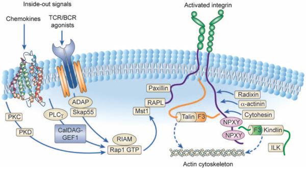
Integrin inside–out signaling. The figure outlines the key signaling pathways that have been implicated in integrin inside–out signaling downstream of chemokine and B- or T-cell receptor stimulation. In their inactive state, integrins adopt a bent conformation that is unable to bind ligand at high affinity. Initiation of most signaling pathways that lead to integrin activation involve the recruitment of Rap1 GTPase. Rap1 activation is controlled by upstream GEFs, in particular CalDAG-GEF1, which has been implicated in regulating cell adhesion and migration. Following Rap1 activation, its effectors RIAM and RapL form a complex with the cytoplasmic tails of integrins. This leads to a critical change in the association of talin with the integrin cytoplasmic tail, and binding of a number of other proteins to the integrin tail, including paxillin, radixin, cytohesin, α-actinin and the kindlins. Altogether, these proteins facilitate separation of the cytoplasmic tails of the integrins, leading to full conformational changes required for high-affinity ligand binding and coupling of the integrin to the actin cytoskeleton. The precise sequence of events from Rap1 activation to integrin tail separation and protein associations are unclear; however, this overall theme is likely common between the β1-, β2- and β3-integrins.
References
-
- Kuijpers TW, van de Vijver E, Weterman MA, de Boer M, Tool AT, van den Berg TK, et al. LAD-1/variant syndrome is caused by mutations in FERMT3. Blood. (e-pub ahead of print 8 December 2008; doi:10.1182/blood-2008-10-182154) - PubMed
-
- Etzioni A. Leukocyte adhesion deficiencies: molecular basis, clinical findings, and therapeutic options. Adv Exp Med Biol. 2007;601:51–60. - PubMed
MeSH terms
Substances
Grants and funding
LinkOut - more resources
Full Text Sources
Other Literature Sources
