. 2009 Aug;26(8):1369-78.
doi: 10.1089/neu.2008-0870.

Temporal and spatial dynamics of peroxynitrite-induced oxidative damage after spinal cord contusion injury

Affiliations

Temporal and spatial dynamics of peroxynitrite-induced oxidative damage after spinal cord contusion injury

Kimberly M Carrico et al. J Neurotrauma. 2009 Aug.

Abstract

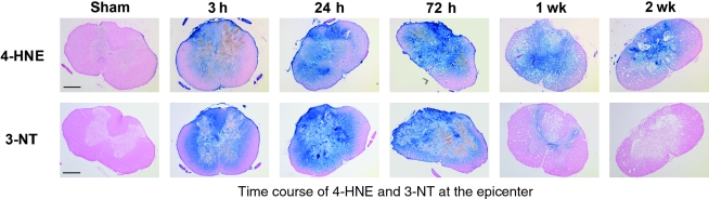
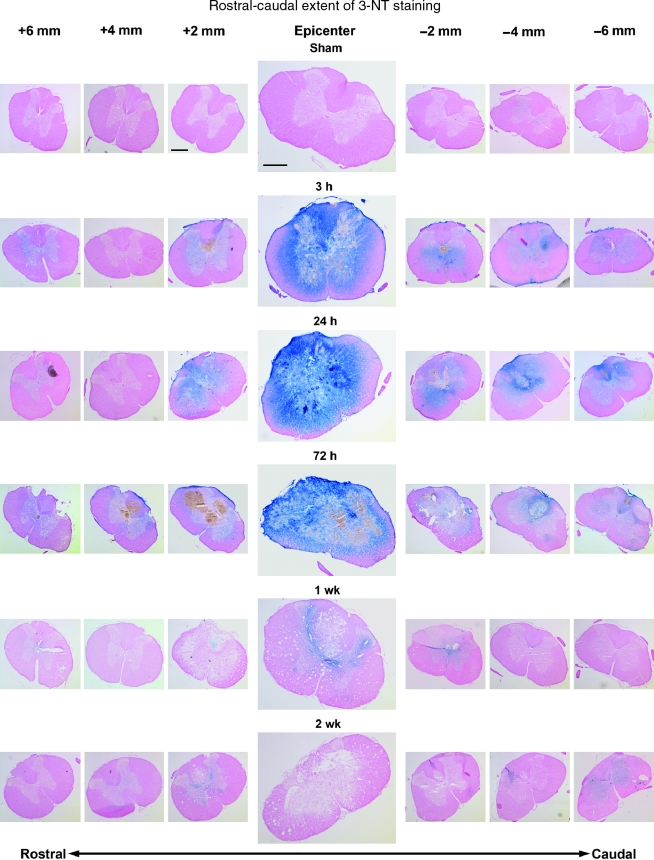
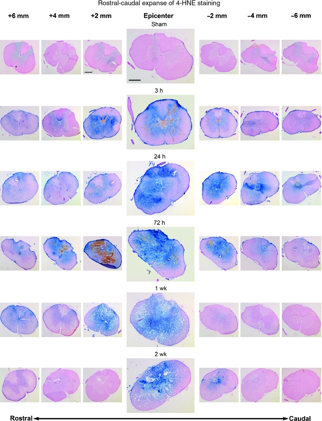
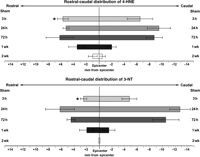
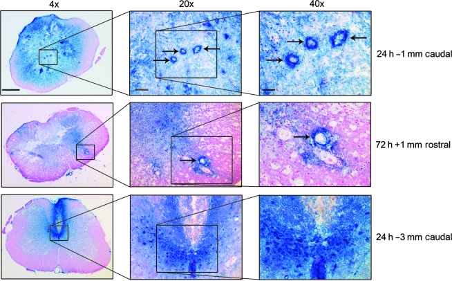
The reactive nitrogen species peroxynitrite (PN) has been suggested to be an important mediator of the secondary oxidative damage that occurs following acute spinal cord injury (SCI). The PN decomposition products nitrogen dioxide (*NO(2)), hydroxyl radical (*OH), and carbonate radical (*CO(3)) are highly reactive with cellular lipids and proteins. In this immunohistochemical study, we examined the temporal (3, 24, and 72 h, and 1 and 2 weeks) and spatial relationships of PN-mediated oxidative damage in the contusion-injured rat thoracic spinal cord (IH device, 200 kdyn, T10) using 3-nitrotyrosine (3-NT), a marker for protein nitration by PN-derived *NO(2) and 4-hydroxynonenal (4-HNE), an indicator of lipid peroxidation (LP) initiated by any of the PN radicals. Minimal 3-NT or 4-HNE immunostaining was seen in sham, non-injured spinal cords. In contrast, both markers showed a substantial increase at 3 h post-injury at the epicenter, that extended throughout the gray matter and into the surrounding white matter. At 24 and 72 h, the oxidative damage expanded circumferentially to involve all but a small rim of white matter tissue at the injury site, and longitudinally as much as 6-9 mm in the rostral and caudal directions. The staining was observed in neuronal soma, axons, and microvessels. At all time points except 3 h, there was no significant difference in the mean rostral or caudal extent of 3-NT and 4-HNE staining. By 1, and more so at 2 weeks, the longitudinal extent of the oxidative damage staining was greatly decreased. The spatial and temporal overlap of 3-NT and 4-HNE staining supports the concept that PN is involved in both damage produced by lipid peroxidation and protein nitration, and that antioxidant agents that target PN or PN-derived radicals should be effective neuroprotectants for acute SCI if administered during the first post-injury hours.


Figures

FIG. 1.
FIG. 1.
Representative examples showing the post-traumatic time course of oxidative damage as revealed by 3-NT and 4-HNE immunostaining. Images are representative coronal sections at the epicenter (T10) at 3, 24, and 72 h and at 1 and 2 weeks post-injury. Adjacent sections from the same animal were stained for 3-NT and 4-HNE. All sections were counterstained with nuclear fast red. The sham animal showed minimal staining for both markers. By 3 h post-injury, staining was substantially elevated, encompassing all of the gray matter and extending into the white matter. It peaked for 3-NT and 4-HNE around 24 to 72 h post-injury, with only a small rim of white matter remaining unstained. By 1 week, 3-NT staining had nearly disappeared, while the elevation of 4-HNE persisted throughout the gray matter until at least 2 weeks post-injury (scale bar = 500 μm).
FIG. 2.
FIG. 2.
Rostral-caudal extent of 4-HNE staining at all time-points as shown by immunohistochemistry. All sections were counterstained with nuclear fast red. Images were taken every 2 mm starting 6 mm rostral to the epicenter and ending 6 mm caudal to the epicenter. At each time point, the images were taken from only one animal. By 3 h post-injury, staining was elevated above sham levels at the epicenter, with this elevation present at least 6 mm in both the rostral and caudal directions. This staining pattern persisted up to 72 h post-injury, with 4-HNE indicated at least as much as 6 mm from the epicenter in both directions. At 1 week post-injury, staining was still intense 2 mm rostral to the epicenter, but had abated somewhat by 4 mm, although it remained above sham levels out to 6 mm rostrally. Very little staining was seen at 1 week post-injury in the caudal direction. By 2 weeks post-injury, staining was still present at the epicenter, but was not seen more than 2 mm in either direction. Compare Figure 2 with Figure 3, which shows protein nitration-related 3-NT staining of the adjacent sections (scale bar = 500 μm).
FIG. 3.
FIG. 3.
Rostral-caudal extent of 3-NT staining at all time-points as shown by immunohistochemistry. All sections were counterstained with nuclear fast red. Images were taken every 2 mm starting 6 mm rostral to the epicenter, and ending 6 mm caudal to the epicenter. At each time-point, the images were taken from only one animal. By 3 h post-injury, staining was elevated above sham levels at the epicenter and for several millimeters in both the rostral and caudal directions. The largest spread of staining was present between 24 and 72 h post-injury, with 3-NT apparent at least 6 mm from the epicenter in either direction. By 1 week post-injury, staining had largely abated and was found sparingly and only in sections within 2 mm of the epicenter. At 2 weeks post-injury staining had returned to near-sham levels even at the epicenter. (Compare Figure 3 with Figure 2, which shows lipid peroxidative-related 4-HNE staining of the adjacent sections [scale bar = 500 μm]).
FIG. 4.
FIG. 4.
Graphic summary of the rostral-caudal distribution of staining for both 4-HNE and 3-NT over various time-points in the injured rat spinal cord (values = mean ± SEM for 4 animals/time-point). The extent of staining for 4-HNE and 3-NT are expressed in millimeters from the epicenter (0) in both the rostral (+) and caudal (–) directions. See methods section for details on measurements. The top panel shows the extent of 4-HNE staining, with the peak of its distribution between 24 and 72 h post-injury and some staining found up to 1 mm from the epicenter as late as 2 weeks post-injury. The bottom panel shows the extent of 3-NT staining, with the peak again occurring between 24 and 72 h post-injury. At 1 week post-injury, some staining was present up to 2 mm from the epicenter, but by 2 weeks post-injury, staining was found only in the epicenter sections (*p < 0.05 versus 4-HNE at the same time-point by paired t-test).
FIG. 5.
FIG. 5.
Higher-power views of 3-NT oxidative damage in the injured spinal cord. The top row shows a 24-h post-injury spinal cross-section at 1 mm caudal to the epicenter, and shows intense oxidative damage staining of gray matter microvessels (arrows), as well as surrounding parenchymal elements. The middle row displays a 72-h post-injury cross-section 1 mm rostral to the epicenter, which again shows an example of microvascular staining (arrow). The bottom row shows an example at 24 h that was taken 3 mm caudal to the epicenter, in which intense 3-NT staining of presumptive dorsal commissural axons and cell bodies is apparent (4× scale bar = 500 μm; 20× scale bar = 200 μm; 40× scale bar = 100 μm).

References

    1. Aksenova M. Butterfield D.A. Zhang S.X. Underwood M. Geddes J.W. Increased protein oxidation and decreased creatine kinase BB expression and activity after spinal cord contusion injury. J. Neurotrauma. 2002;19:491–502. - PubMed
    1. Baldwin S.A. Broderick R. Osbourne D. Waeg G. Blades D.A. Scheff S.W. The presence of 4-hydroxynonenal/protein complex as an indicator of oxidative stress after experimental spinal cord contusion in a rat model. J. Neurosurg. 1998;88:874–883. - PubMed
    1. Bao F. DeWitt D.S. Prough D.S. Liu D. Peroxynitrite generated in the rat spinal cord induces oxidation and nitration of proteins: reduction by Mn (III) tetrakis (4-benzoic acid) porphyrin. J. Neurosci. Res. 2003;71:220–227. - PubMed
    1. Beckman J.S. Beckman T.W. Chen J. Marshall P.A. Freeman B.A. Apparent hydroxyl radical production by peroxynitrite: implications for endothelial injury from nitric oxide and superoxide. Proc. Natl. Acad. Sci. U.S.A. 1990;87:1620–1624. - PMC - PubMed
    1. Beckman J.S. Koppenol W.H. Nitric oxide, superoxide, and peroxynitrite: the good, the bad, and ugly. Am. J. Physiol. 1996;271:C1424–C1427. - PubMed

Publication types