. 2009 Nov;24(11):1869-78.
doi: 10.1359/jbmr.090512.

Oxygen tension regulates the expression of ANK (progressive ankylosis) in an HIF-1-dependent manner in growth plate chondrocytes

Affiliations

Oxygen tension regulates the expression of ANK (progressive ankylosis) in an HIF-1-dependent manner in growth plate chondrocytes

Raihana Zaka et al. J Bone Miner Res. 2009 Nov.

Abstract

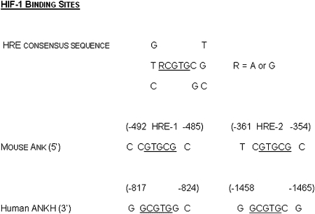
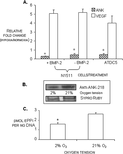
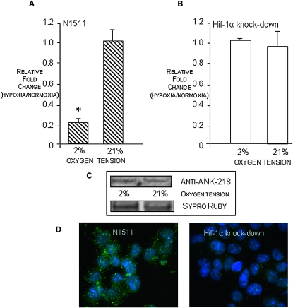
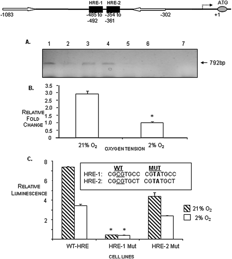
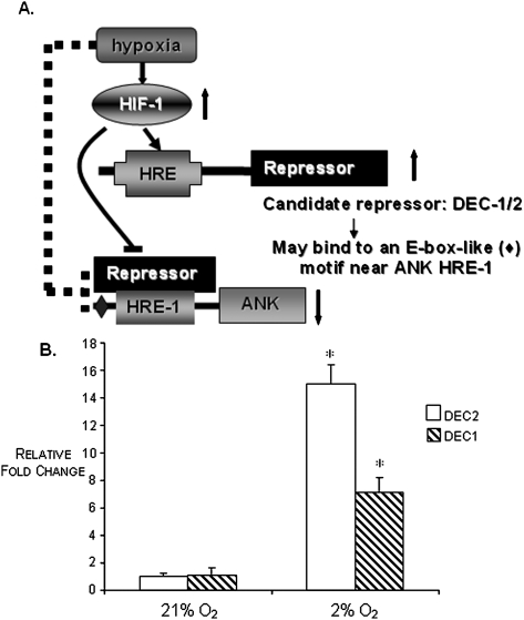
The proximal promoter region of ANK, a gene that codes for a protein that regulates the transport of inorganic pyrophosphate, contains two hypoxia responsive elements (HREs); therefore, we studied the expression and function of ANK at different oxygen tensions. ATDC5 and N1511 clonal chondrocytic cells were cultured in either hypoxia (2% O(2)) or normoxia (21% O(2)). Transcript and protein levels of ANK were depressed in hypoxic conditions, as were levels of extracellular pyrophosphate (ePPi). To determine whether HIF-1 was involved in the oxemic response, Hif-1alpha knockdown cells were exposed to varying oxygen conditions and ANK expression was assessed. Knockdown of Hif-1alpha resulted in low levels of expression of ANK in hypoxia and normoxia. Chromatin immunoprecipitation (ChIP) assays explored the binding of Hif-1alpha to ANK HREs and showed that Hif-1alpha is able to bind to the HREs of ANK more avidly in normoxia than in hypoxia. Furthermore, functional studies of Hif-1alpha activity using luciferase reporter assays of wildtype and mutagenized HREs showed that only HRE-1 binds Hif-1alpha in normoxia. Expression of ANK in growth plate and articular cartilage was low in hypoxic regions of the tissues, and higher levels of ANK expression were observed in the synovium and meniscus in regions that have a normally higher oxygen tension. The data suggest that ANK expression and function in vitro and in vivo are repressed in hypoxic environments and that the effect is regulated by HIF-1.


Figures

FIG. 1
FIG. 1
HIF-1 consensus binding site. Underlined nucleotides represent core consensus element; putative HIF-1 binding sites and their positions in the proximal promoter regions of human and mouse ANKH/Ank genes.
FIG. 2
FIG. 2
(A) Relative expression of ANK and VEGF transcripts in normoxia vs. hypoxia (24-h treatment) as determined by qRT-PCR in ATDC5 and N1511 cells. Transcript levels for ANK are <1, indicating that ANK expression is repressed in hypoxia. Conversely, expression levels for VEGF are >1, indicating that VEGF is upregulated in hypoxia. Relative transcript levels of ANK in N1511 cells at immaturity (−BMP-2) and at nonmineralizing hypertrophy (+BMP-2), as well as in immature ATDC5 cells is shown. *p < 0.05 for fold change for ANK in hypoxia vs. normoxia; n = 9. (B) Western blot analysis of ANK protein expression in mature N1511 cells in hypoxic (2% O2) and normoxic (21% O2) conditions. Sypro Ruby staining of the mirror image blot depicts relative protein loading. (C) Extracellular PPi levels in N1511 cells in normoxia (21% O2) and hypoxia (2% O2) at the proliferative phase of their differentiation. Repression of ANK expression in hypoxia results in statistically significant decrease in the elaboration of ePPi. *p < 0.05, n = 6.
FIG. 3
FIG. 3
(A and B) Real-time PCR of ANK transcript levels in hypoxia vs. normoxia in N1511and Hif-1α knockdown cells. Transcript levels <1 indicate repression of ANK expression in hypoxia; transcript levels >1 indicate stimulation of expression in hypoxia. In contrast to hypoxic repression of ANK expression in N1511 cells, knockdown of Hif-1α results in equivalent expression of ANK in normoxia and hypoxia. (C) Western blot analysis of ANK protein expression in mature Hif-1α knockdown cells in hypoxic (2% O2) and normoxic (21% O2) conditions. Sypro Ruby staining of the mirror image blot depicts relative protein loading. Note low levels of expression of ANK in hypoxia and normoxia, showing relative loss of ANK expression in normoxia (21% O2) in cells in which Hif-1α is knocked down. (D) Immunocytochemical visualization of ANK expression in mature N1511 cells and Hif-1α knockdown cells grown in normoxic conditions showing high expression of ANK in N1511 cells and lack of significant ANK expression in cells in which Hif-1α has been knocked down.
FIG. 4
FIG. 4
(A) ChIP assay depicting binding of Hif-1α to the ANK HREs. Hif-1α–immunoprecipitated DNA–protein complexes were disrupted, and the recovered DNA was PCR-amplified with primers (white arrows in depiction of the position of the ANK proximal promoter HREs, HRE-1 and HRE-2, above ChIP assay results) surrounding the HRE elements in the ANK promoter. PCR products (792 bp) were resolved on a 2% agarose gel. Lane 1, IP with anti-Hif-1α antibody in normoxia; lane 2, IP with anti-Hif-1α antibody in hypoxia; lane 3, positive control: input DNA binding to anti-Hif-1α in normoxia; lane 4, positive control: input DNA binding to anti-Hif-1α in hypoxia; lane 5, negative control: no antibody in normoxia; lane 6, negative control: no antibody in hypoxia; lane 7, water control. Note that binding of the Hif-1α antibody to the promoter PCR fragment is repressed in hypoxia (lane 2). (B) qPCR quantitation of the recovery of the Hif-1α–binding PCR fragments in normoxia vs. hypoxia as depicted on the gel. (C) Site-directed mutagenesis studies of the ANK HREs. HRE-1 and HRE-2 were mutagenized as shown, cloned into a luciferase reporter plasmid, and used to transfect COS-7 cells under normoxic conditions. Expression of the ANK promoter harboring wildtype (WT) HRE sequences resulted in relatively high expression of ANK in normoxia compared with hypoxia. When HRE-1 was mutated, the expression of the luciferase reporter construct was dramatically decreased. When the HRE-2 site was mutated, expression of the corresponding luciferase construct was only slightly decreased. These results suggest that HRE-1 functions as the site primarily used for HIF-1 mediation of ANK expression in response to oxygen. *p < 0.05 compared with WT, n = 6.
FIG. 5
FIG. 5
Immunohistochemical studies of ANK expression in the mouse growth plate and articular cartilage. (A) ANK is primarily expressed in the hypertrophic and calcification zones of the growth plate, and its expression is particularly strong in oxygen-rich areas of the growth plate undergoing vascular invasion. (B) ANK expression in the superficial zone of articular cartilage from 5-day postnatal mice corresponds to relatively high levels of oxygen from synovial fluid compared with the proliferative zone of articular cartilage where severe hypoxia results in very low ANK expression. (C) Immunohistochemical staining of ANK in meniscus shows high levels of expression; also note high levels of ANK in the superficial zone of the femoral condyle and tibial plateau in a mouse knee joint from a P7 animal. Autofluorescing anuclear RBCs in vessel walls emphasize high levels of ANK staining in these areas as well (white arrows). (D) H&E and (E) anti-ANK staining of human synovial tissue, showing high levels of expression of ANK in synoviocytes and in other regions of the tissue (F, fatty globules). (F) qRT-PCR showing very low levels of ANK expression in hypoxia in primary normal human articular chondrocytes. Values >1 indicate a relative increase in expression in hypoxia; values <1 indicate a relative decrease in expression in hypoxia. Data show that the expression of ANK is repressed in hypoxia, whereas the expression of VEGF is upregulated in hypoxia. *p < 0.05 for fold change in ANK expression in hypoxia vs. normoxia; n = 3. (G) qRT-PCR showing the relative expression of ANK in osteocytic MLO-A5 cells in normoxia and hypoxia. Note very high levels of ANK expression in normoxic conditions at times of mineralization (days 5–7) and repression of ANK expression in hypoxia at these same times. Results depict two separate experiments with qRT-PCR analyses performed in triplicate for each experiment.
FIG. 6
FIG. 6
(A) Model depicting the repression of ANK expression in hypoxia. We hypothesize that elevated expression of HIF-1 in hypoxia upregulates the expression of some repressor gene whose protein product is able to repress the expression of ANK in hypoxic conditions. A possible candidate gene(s) for this repressor is DEC1 and/or DEC2. (B) Relative expression of DEC1 and DEC2 in N1511 cells in hypoxia and normoxia showing that these transcripts are highly expressed in hypoxic conditions in the chondrocytic cells used in these studies. n = 3; *p < 0.05 with respect to expression of DEC1 and DEC2 in hypoxia vs. normoxia.

References

    1. Ho AM, Johnson MD, Kingsley DM. Role of the mouse ank gene in control of tissue calcification and arthritis. Science. 2000;289:265–270. - PubMed
    1. Zaka R, Williams CJ. Role of the progressive ankylosis gene in cartilage mineralization. Curr Opin Rheumatol. 2006;18:181–186. - PubMed
    1. Sohn P, Crowley M, Slattery E, Serra R. Developmental and TGFβ-mediated regulation of ANK mRNA expression in cartilage and bone. Osteoarthrit Cartilage. 2002;10:482–490. - PubMed
    1. Johnson K, Terkeltaub R. Upregulated ank expression in osteoarthritis can promote both chondrocyte MMP-13 expression and calcification via chondrocyte extracellular PPi excess. Osteoarthrit Cartilage. 2004;12:321–335. - PubMed
    1. Wang W, Xu J, Du B, Kirsch T. Role of the progressive ankylosis gene (ank) in cartilage mineralization. Mol Cell Biol. 2005;25:312–323. - PMC - PubMed

Publication types

MeSH terms