. 2009 May;11(3):256-69.
doi: 10.1111/j.1399-5618.2009.00675.x.

Valproate activates the Notch3/c-FLIP signaling cascade: a strategy to attenuate white matter hyperintensities in bipolar disorder in late life?

Affiliations

Valproate activates the Notch3/c-FLIP signaling cascade: a strategy to attenuate white matter hyperintensities in bipolar disorder in late life?

Peixiong Yuan et al. Bipolar Disord. 2009 May.

Abstract

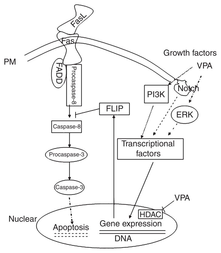
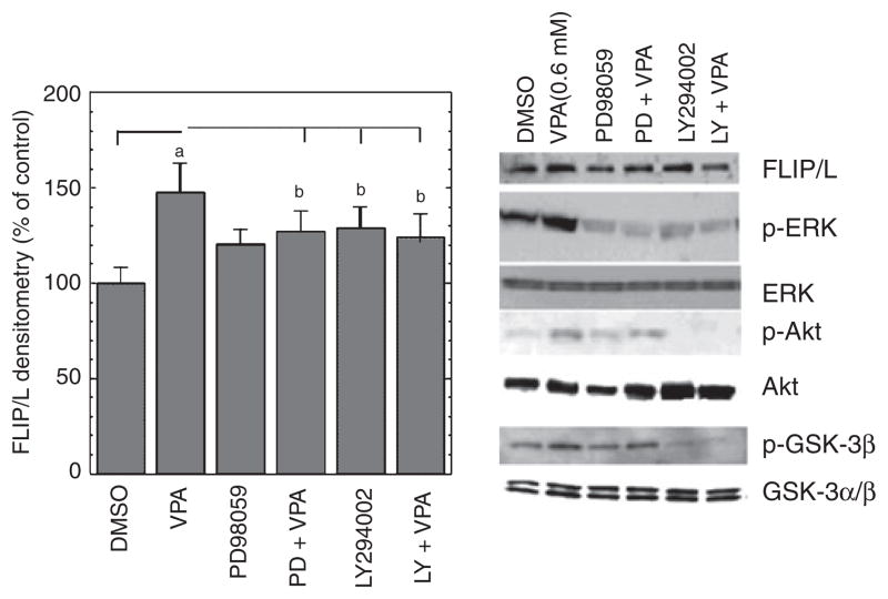
Objectives: Increased prevalence of deep white matter hyperintensities (DWMHs) has been consistently observed in patients with geriatric depression and bipolar disorder. DMWHs are associated with chronicity, disability, and poor quality of life. They are thought to be ischemic in their etiology and may be related to the underlying pathophysiology of mood disorders in the elderly. Notably, these lesions strikingly resemble radiological findings related to the cerebral autosomal dominant arteriopathy with subcortical infarcts and leukoencephelopathy (CADASIL) syndrome. CADASIL arises from mutations in Notch3, resulting in impaired signaling via cellular Fas-associated death domain-like interleukin-1-beta-converting enzyme-inhibitory protein (c-FLIP) through an extracellular signal-regulated kinase (ERK)-dependent pathway. These signaling abnormalities have been postulated to underlie the progressive degeneration of vascular smooth muscle cells (VSMC). This study investigates the possibility that the anticonvulsant valproate (VPA), which robustly activates the ERK mitogen-activated protein kinase (MAPK) cascade, may exert cytoprotective effects on VSMC through the Notch3/c-FLIP pathway.

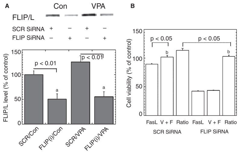
Methods: Human VSMC were treated with therapeutic concentrations of VPA subchronically. c-FLIP was knocked down via small interfering ribonucleic acid transfection. Cell survival, apoptosis, and protein levels were measured.

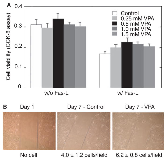
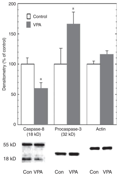
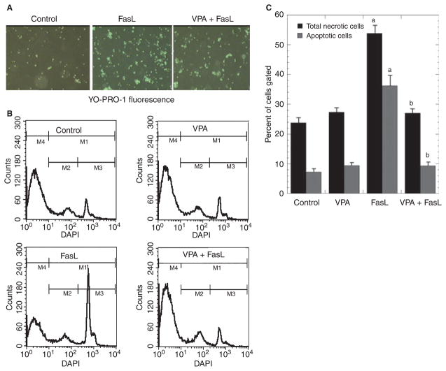
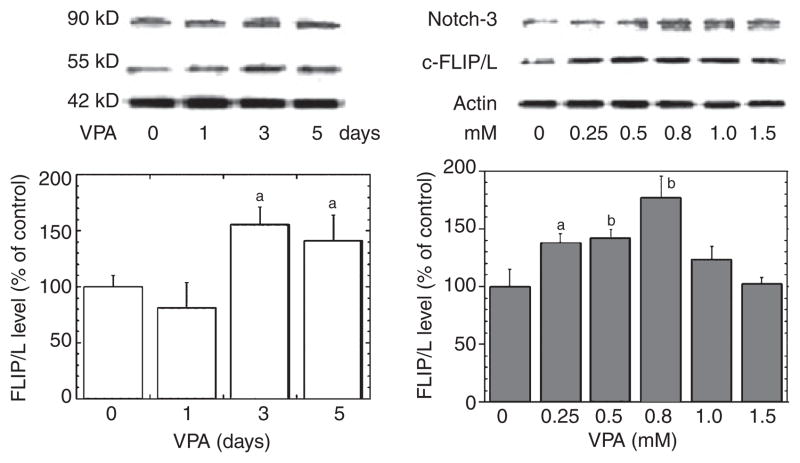
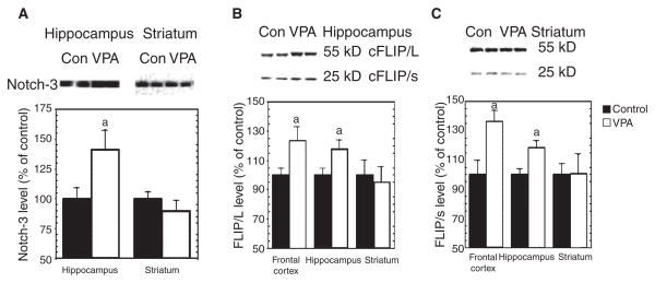
Results: VPA increased c-FLIP levels dose- and time-dependently and promoted VSMC survival in response to Fas ligand-induced apoptosis in VSMC. The anti-apoptotic effect of VPA was abolished by c-FLIP knockdown. VPA also produced similar in vivo effects in rat brain.

Conclusions: These results raise the intriguing possibility that VPA may be a novel therapeutic agent for the treatment of CADASIL and related disorders. They also suggest that VPA might decrease the liability of patients with late-life mood disorders to develop DWMHs.


Figures

Fig. 1
Fig. 1
Schematic representation of valproate’s intracellular cytoprotective mechanisms. Fas ligand (FasL)/Fas interactions cause effector caspase activation. FasL-mediated apoptosis can be blocked by the inhibition of caspase-8 protease through cellular Fas-associated death domain-like interleukin- 1-beta-converting enzyme-inhibitory protein (c-FLIP). Valproate (VPA) activates mitogen-activated protein kinase (MAPK)/extracellular signal-regulated kinase (ERK) and PI- 3K pathways, which upregulate Notch3, c-FLIP, and other proteins (Bcl-2, BDNF, etc.) via transcriptional factors. VPA also regulates gene expression by inhibiting histone deacetylase (HDAC). Hatched lines represent membrane; dotted lines represent indirect effects. FADD = Fas-associated death domain- containing protein.
Fig. 2
Fig. 2
(A) Valproate (VPA) promotes vascular smooth muscle cell (VSMC) survival under Fas-ligand (FasL) challenge. Human VSMC were subcultured in 96-well plates, pretreated with (or without) VPA at the indicated concentrations in serum-free medium for two days, and then exposed to FasL (25 ng/ml) in the serum-free medium with VPA for 24 hours. Cell viability was measured with Cell Counting Kit-8 (CCK-8) as described in Materials and methods. Data represent means ± SEM, n = 6, p < 0.05. (B) Representative photomicrographs of cells in each group show that VPA increased VSMC motility in serum-free medium. VSMC were cultured in six-well plates; the cell monolayer was disrupted and cells were treated with or without VPA (0.8 mM) in serum-free medium. Migration of cells into the wound was quantified under microscopy. The numbers below the panels represent the number of cells that migrated into the wound area. Results are representative of at least three separate experiments.
Fig. 3
Fig. 3
Valproate (VPA) inhibited caspase-8 and caspase-3 activation. Human vascular smooth muscle cells (VSMC) were treated with VPA (0.8 mM) in serum-free medium for three days, and Fas-ligand (25 ng/ml) was added to the medium during the last day of treatment. Immunoblot analyses were performed with anti-caspase-8, anti-caspase-3, and anti-β-actin antibodies. Pro-caspase and/or cleaved caspase bands were quantified with Kodak Image Station. VPA significantly inhibited caspase-8 and caspase-3 cleavage (n = 6). ap < 0.05.
Fig. 4
Fig. 4
Valproate (VPA) inhibits Fas-ligand (FasL)-induced vascular smooth muscle cells (VSMC) apoptosis. Human VSMC were pretreated with or without VPA (0.8 mM) for two days, and challenged with Fas-ligand (25 ng/ml) for 12 hours. (A) Representative photographs of VSMC stained with YO-PRO-1 fluoresence. (B) Fluorescence-activated cell sorting (FACS) analyses after staining with DAPI (4′,6-diamidino-2-phenylindole dihydrochloride) to detect apoptotic cells. (C) Data represent three independent experiments. FasL significantly induced apoptotic cells (ap < 0.01 versus control); pretreatment with VPA (two days) significantly blocked FasL’s proapoptotic effect in VSMC (bp < 0.01, VPA + FasL versus FasL).
Fig. 5
Fig. 5
Valproate (VPA) increases Notch3 and cellular Fas-associated death domain-like interleukin-1-beta-converting enzyme-inhibitory protein (c-FLIP) levels dose- and time-dependently in vascular smooth muscle cells (VSMC). Human VSMC were cultured in growth media close to confluence. The media were then replaced with serum-free media, and the cells were treated with or without VPA at concentrations for the time period as indicated (same endpoint). Immunoblotting of Notch3, c-FLIP, or β-actin was conducted. VPA concentration (0~1.5 mM for 48 h) and time (1, 3, or 5 days at 0.8 mM) dependently increased Notch3 and c-FLIP levels. Bar graph densitometric results representing mean ± SEM of three or more sets of samples immunoblotted in duplicate. ap < 0.05; bp < 0.01 compared with control cells.
Fig. 6
Fig. 6
Cellular Fas-associated death domain-like interleukin-1-beta-converting enzyme-inhibitory protein (c-FLIP) small interfering ribonucleic acid (siRNA) abolishes valproate’s (VPA) cytoprotective effects. (A) Transfection with c-FLIP siRNA plasmid knocked down the expression of c-FLIP/L in human vascular smooth muscle cells. The single blot is representative of three separate experiments. ap < 0.01. (B) Cell viability determined with Cell Counting Kit-8 showed that c-FLIP siRNA transfection suppressed cell viability and partially eliminated VPA’s cytoprotective effect. Data are mean ± SEM and represent three separate experiments, bp < 0.05. Con = control medium without VPA; SCR = scramble siRNA plasmid, which served as negative control.
Fig. 7
Fig. 7
Valproate (VPA) activates extracellular signal-regulated kinase (ERK) and phosphoinositide-3 kinase (PI-3K) signal pathways in human vascular smooth muscle cells (VSMC), and specific inhibitors of MEK and PI-3K abolish VPA’s upregulation of c-FLIP. Human VSMC were cultured in growth media close to confluence. The media were then replaced with serum-free media, and the cells were treated with VPA (0.8 mM) in the absence or presence of indicated inhibitors for two days. Immunoblotting was conducted as described in Materials and Methods. MEK inhibitor PD98059 (50 μM) and PI-3K inhibitor LY294002 (20 μM) attenuated VPA-induced increases in c-FLIP/L, phospho-ERK1/2 (P-p44/42), phospho-Akt, and phospho-GSK-3β, ap < 0.01, bp < 0.05, compared with cells treated with dimethy sulfoxide (DMSO, final concentration 0.1%) alone. Total ERK1/2 (p44/42), Akt, and GSK-3 α/β levels did not change significantly.
Fig. 8
Fig. 8
Valproate (VPA) activates Notch3 signaling in vivo. Adult male Wistar rats received VPA chow for four weeks. Protein extracts from frontal cortex, hippocampus, or striatum were separated on SDS-PAGE, and then immunoblotted with anti-Notch3 or anti-cellular Fas-associated death domain-like interleukin-1-beta-converting enzyme-inhibitory protein (c-FLIP) antibody. The resultant bands were quantified and standardized by bands of β-actin. (A) VPA significantly increased cleaved Notch3 protein levels in hippocampus [t(22) = 2.51, p = 0.02], but not striatum. (B) VPA significantly increased c-FLIP/L and c-FLIP/s levels in rat frontal cortex and hippocampus (ap < 0.05 versus control), but not in striatum. (C) Representative blot with frontal cortex samples was similar to that of hippocampal samples.

References

    1. Videbech P. MRI findings in patients with affective disorder: a meta-analysis. Acta Psychiatr Scand. 1997;96:157–168. - PubMed
    1. Altshuler LL, Curran JG, Hauser P, Mintz J, Denico K, Post R. T-2 hyperintensities in bipolar disorder – magnetic-resonance- imaging comparison and literature metaanalysis. Am J Psychiatry. 1995;152:1139–1144. - PubMed
    1. Thomas AJ, Perry R, Kalaria RN, Oakley A, McMeekin W, O’Brien JT. Pathologies and pathological mechanisms for white matter hyperintensities in depression. Ann N Y Acad Sci. 2002;977:333–339. - PubMed
    1. Thomas AJ, O’Brien JT, Davis S, et al. Ischemic basis for deep white matter hyperintensities in major depression – a neuropathological study. Arch Gen Psychiatry. 2002;59:785–792. - PubMed
    1. Ruchoux MM, Guerouaou D, Vandenhaute B, Pruvo JP, Vermersch P, Leys D. Systemic vascular smooth muscle cell impairment in cerebral autosomal dominant arteriopathy with subcortical infarcts and leukoencephalopathy. Acta Neuropathol (Berl) 1995;89:500–512. - PubMed

Publication types

MeSH terms