. 2009 May 6:2:9.
doi: 10.1186/1756-6606-2-9.

Sexual attraction enhances glutamate transmission in mammalian anterior cingulate cortex

Affiliations

Sexual attraction enhances glutamate transmission in mammalian anterior cingulate cortex

Long-Jun Wu et al. Mol Brain. .

Abstract

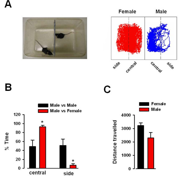
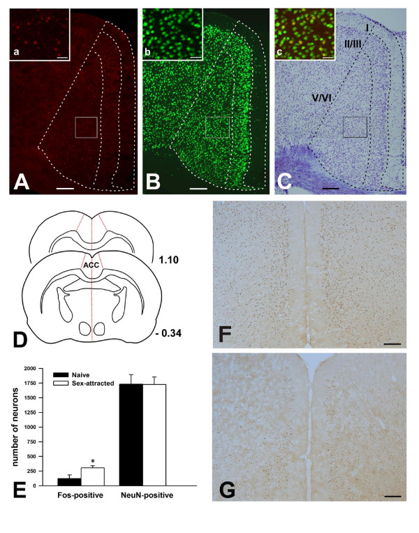
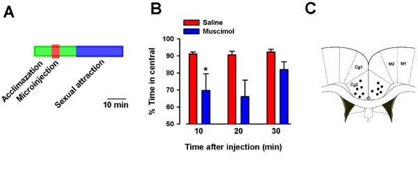
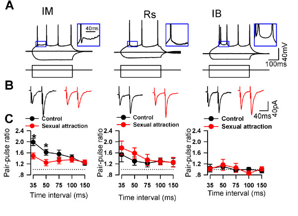
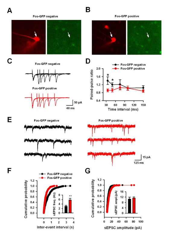
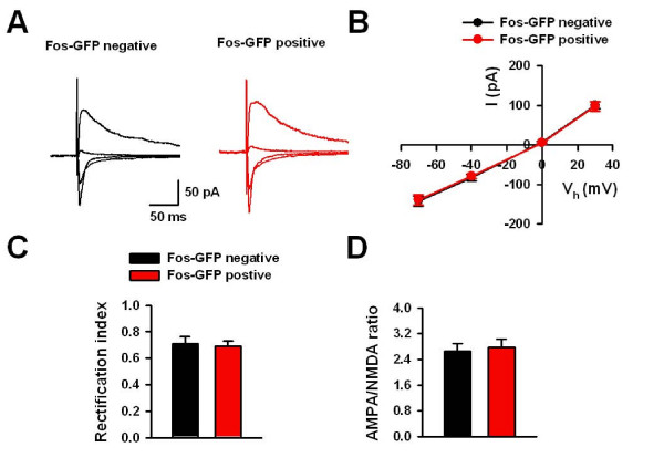
Functional human brain imaging studies have indicated the essential role of cortical regions, such as the anterior cingulate cortex (ACC), in romantic love and sex. However, the neurobiological basis of how the ACC neurons are activated and engaged in sexual attraction remains unknown. Using transgenic mice in which the expression of green fluorescent protein (GFP) is controlled by the promoter of the activity-dependent gene c-fos, we found that ACC pyramidal neurons are activated by sexual attraction. The presynaptic glutamate release to the activated neurons is increased and pharmacological inhibition of neuronal activities in the ACC reduced the interest of male mice to female mice. Our results present direct evidence of the critical role of the ACC in sexual attraction, and long-term increases in glutamate mediated excitatory transmission may contribute to sexual attraction between male and female mice.


Figures

Figure 1
Figure 1
Behavioral test for sexual attraction. (A) Photograph and diagram of the sexual attraction test. The two boxes are further divided into side and central zones as indicated by the dashed line. Representative traces showing the movement of the male and female mice for 30 min. Left = female (red); right = male (blue). (B) Male mice (n = 5) spent significantly more time in the central zone than in the side zone (male vs. female) (n = 5), while there is no difference in the time spent in either zone in male vs. male test. * P = 0.007. (C) There is no difference in the total distance traveled between male and female mice in the sexual attraction test.
Figure 2
Figure 2
Sexual attraction activated Fos expression in the ACC neurons in male mice. (A) Fos immunostaining in the ACC after sexual attraction. The framed area is magnified in (a). (B) NeuN immunostaining in the ACC. The framed area is magnified in (b). (C) The same section as shown by (A, B), was counterstained by Nissl technique; demarcation for neuronal layers in the ACC. The inset (c) is the overlapping of (a) and (b). Yellow color in (c) represents the Fos/NeuN double-labeled neurons. (D) Diagram showing the ACC from Bregma -0.34 to 1.1, where the Fos-positive and NeuN positive cells were counted. (E) Bar graph showing the significant increase of Fos neurons in layer II/III and ACC after sexual attraction test compared to naïve mice. (F and G) The representative slices with Fos staining between sex-attracted group (F) and naïve mice (G). The increased Fos expression was found in layer II/III and layer V/VI. Bar = 150 μm in A – C, F and G; and 35 μm in a – c.
Figure 3
Figure 3
The ACC is required for sexual attraction. (A) Experimental design for the behavioral experiment after musiimol injection. (B) Local injection of muscimol in the ACC significantly reduced sexual attraction in male mice 10 minutes after injection. The sexual attraction was recovered within 30 minutes. (C) Location of injection sites from all animals included in the study.
Figure 4
Figure 4
PPF was changed in the ACC after sexual attraction. (A) Sample traces show the three types of pyramidal neurons in layer II/III of ACC. IM, intermediate neurons; RS, regular spiking neurons; IB, intrinsic bursting neurons. (B) PPF of IM neurons (upper) was decreased after sexual attraction in the pathway of Layer V-Layer II/III, while no change has been detected on other two kinds of pyramidal neurons. (C) No interaction between two pathways induced by layer V and layer I stimulations. Time interval between two stimulations was 55 ms. (D) PPF induced by S1 (layer I stimulation) did not change after sexual attraction.
Figure 5
Figure 5
The excitability of pyramidal neurons was not changed after sexual attraction. (A) Action potentials were induced by current injection from 50 pA to 200 pA with 25 pA step. The number of spikes was counted at different current injections to measure the excitability of neurons. There was no change in the excitability of IM neurons between two groups. (B) No change in the number action potentials in RS neurons between two groups. (C) No change in the number action potentials in IB neurons between two groups.
Figure 6
Figure 6
PPF in the pathway of Layer I-Layer II/III were not changed after sexual attraction. (A) No interaction between two pathways induced by layer V and layer I stimulations. Time interval between two stimulations was 55 ms. (B, C and D) PPF induced by S1 (layer I stimulation) did not change after sexual attraction.
Figure 7
Figure 7
GFP is co-stained with Fos protein and NeuN in the ACC of transgenic mice. (A) The GFP-positive cells in the ACC after sexual attraction are overlapped with Fos-positive cells. The framed area was used for magnification in a-c. (B) Some of GFP-positive cells in the ACC after sexual attraction are overlapped with NeuN-positive cells. The framed area was used for magnification in d-f. Bar = 200 μm in (A, B), 30 μm in (a – c) and 20 μm in (d – f).
Figure 8
Figure 8
Increased presynaptic glutamate release to Fos-GFP positive neurons after sexual attraction. (A and B) Example of Fos-GFP negative (A) and positive (B) cells from layer II/III of the ACC that was targeted for whole-cell recording dilated with Alexa fluor 594. (C) Typical traces showing paired-pulse stimulations with interval of 35, 50, 75,100, 150 ms in Fos-GFP negative and positive neurons. (D) Decreased paired-pulse facilitation in Fos-GFP positive neurons at interval 35 ms or 50 ms compared with those in Fos-GFP negative neurons. (E) Representative traces showing mEPSC recordings in Fos-GFP negative and positive neurons. (F) Cumulative probability and pooled data (inset) showing the increased mEPSC frequency in Fos-GFP positive neurons than that in Fos-GFP negative neurons. (G) Cumulative probability and pooled data (inset) showing that there is no difference in mEPSC amplitude between Fos-GFP positive and negative neurons.
Figure 9
Figure 9
The postsynaptic responses in Fos-GFP positive neurons after sexual attraction. (A) Typical traces of EPSCs at holding potentials of -70, -40, 0 and 30 mV in Fos-GFP negative and positive neurons. (B) The pooled data showing the normal I–V responses in Fos-GFP positive neurons. (C and D) The rectification index (C) and AMPA/NMDA ratio (D) for EPSCs was similar in Fos-GFP positive and negative neurons.

Similar articles

Cited by

References

    1. Fisher HE, Aron A, Brown LL. Romantic love: a mammalian brain system for mate choice. Philos Trans R Soc Lond B Biol Sci. 2006;361:2173–2186. doi: 10.1098/rstb.2006.1938. - DOI - PMC - PubMed
    1. Boehm U, Zou Z, Buck LB. Feedback loops link odor and pheromone signaling with reproduction. Cell. 2005;123:683–695. doi: 10.1016/j.cell.2005.09.027. - DOI - PubMed
    1. Lin DY, Zhang SZ, Block E, Katz LC. Encoding social signals in the mouse main olfactory bulb. Nature. 2005;434:470–477. doi: 10.1038/nature03414. - DOI - PubMed
    1. Luo M, Fee MS, Katz LC. Encoding pheromonal signals in the accessory olfactory bulb of behaving mice. Science. 2003;299:1196–1201. doi: 10.1126/science.1082133. - DOI - PubMed
    1. Stowers L, Holy TE, Meister M, Dulac C, Koentges G. Loss of sex discrimination and male-male aggression in mice deficient for TRP2. Science. 2002;295:1493–1500. doi: 10.1126/science.1069259. - DOI - PubMed

Publication types

MeSH terms

LinkOut - more resources