. 2009 Jul 3;284(27):18515-24.
doi: 10.1074/jbc.M109.006676. Epub 2009 May 6.

Up-regulation of miR-21 by HER2/neu signaling promotes cell invasion

Affiliations

Up-regulation of miR-21 by HER2/neu signaling promotes cell invasion

Tzu-Hsuan Huang et al. J Biol Chem. .

Abstract

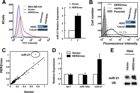
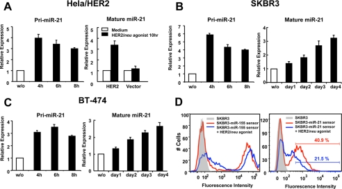
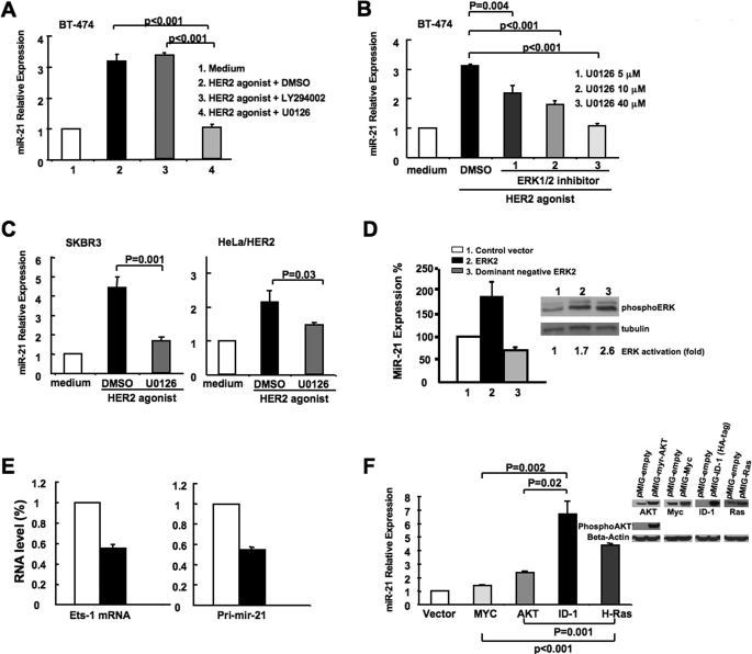
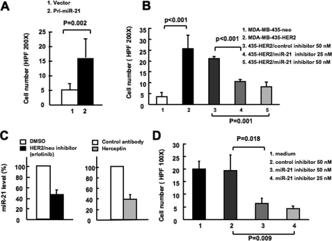
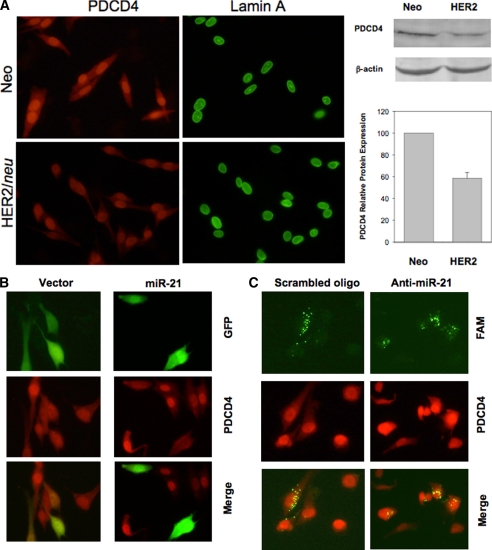
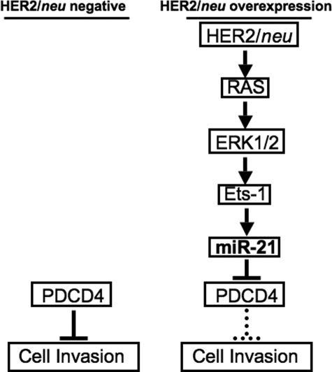
The cell surface receptor tyrosine kinase HER2/neu enhances tumor metastasis. Recent studies suggest that deregulated microRNA (miRNA) expression promotes invasion and metastasis of cancer cells; we therefore explored the possibility that HER2/neu signaling induces the expression of specific miRNAs involved in this process. We identified a putative oncogenic miRNA, miR-21, whose expression is correlated with HER2/neu up-regulation and is functionally involved in HER2/neu-induced cell invasion. We show that miR-21 is up-regulated via the MAPK (ERK1/2) pathway upon stimulation of HER2/neu signaling in breast cancer cells, and overexpression of other ERK1/2 activators such as RASV12 or ID-1 is sufficient to induce miR-21 up-regulation in HER2/neu-negative breast cancer cells. Furthermore, the metastasis suppressor protein PDCD4 (programmed cell death 4) is down-regulated by miR-21 in breast cancer cells expressing HER2/neu. Our data reveal a mechanism for HER2/neu-induced cancer cell invasion via miRNA deregulation. In addition, our results identify miR-21 as a potential therapeutic target for the prevention of breast cancer invasion and metastasis.


Figures

FIGURE 1.
FIGURE 1.
Ectopic expression of HER2/neu induces miR-21 up-regulation. A, MDA-MB-435 cells transfected with neo vector (blue line) or MDA-MB-435 transfected with HER2/neu-expressing vector (red line) were stained with fluorescein isothiocyanate (FITC)-conjugated anti-human HER2/neu antibody (left panel), and the miR-21 expression level in the MDA-MB-435 cells was assessed by using qRT-PCR (right panel). The cell lysates were separated by SDS-PAGE and analyzed by Western blot using anti-HER2/neu antibody or anti-tubulin antibody. B, HER2/neu expression in HeLa cells transfected with different vectors. HeLa cell line (gray histogram), HeLa cell line transfected with pEBS7 empty vector (dashed line), or HeLa cell line transfected with pEBS7-HER2/neu (black line) were stained with fluorescein isothiocyanate-conjugated anti-human HER2/neu antibody. The cell lysates were separated by SDS-PAGE and analyzed by Western blot using anti-HER2/neu antibody or anti-tubulin antibody. C, miRNA microarray analysis of the effect of HER2/neu overexpression. RNA from HeLa cells transfected with pEBS7 and pEBS7-HER2/neu were labeled with different fluorophores and competitively hybridized on microarrays. Each spot in the scatter plot represents the normalized mean fluorescence for one microRNA probe from two dye swapped arrays. D and E, RNA used in B was analyzed by qRT-PCR to assay expression of miR-21 (D) and in a separate experiment by Northern blot analysis under the same conditions (E). The mean and standard error from triplicate experiments are indicated.
FIGURE 2.
FIGURE 2.
HER2/neu signaling induced miR-21 up-regulation with distinct kinetics in different cancer cell lines. Real time PCR analysis of miR-21 pri-cursor (pri-miR-21) and mature miR-21 expression in HeLa-HER2/neu or HeLa-vector cells (A), SKBR3 cells (B), and BT-474 cells (C) stimulated with 100 nm HER2/neu agonist at varying time points. D, SKBR3 cells infected with lentivirus carrying a mCherry gene with two complementary miR-21 or miR-155 binding sites in the 3′-untranslated region (mCherry miR-21 sensor or mCherry miR-155 sensor, respectively) were stimulated with 100 nm HER2/neu agonist for 4 days, and the fluorescent signal was detected by flow cytometry. The mean and standard error from triplicate experiments are indicated.
FIGURE 3.
FIGURE 3.
HER2/neu regulates miR-21 expression via the MAPK (ERK1/2) pathway. A, BT-474 cells were treated with 100 nm HER2/neu agonist in the presence of 40 μm Me2SO, LY294002 or U0126. After 4 days, RNA was collected, and miR-21 expression was analyzed by qRT-PCR. B, BT-474 cells were treated with 100 nm HER2/neu agonist in the presence of 5, 10 and 40 μm U0126, and miR-21 expression was assessed as described for A. C, SKBR3 and HeLa-HER2/neu cells were treated with 100 nm HER2/neu agonist in the presence of Me2SO or 40 μm U0126, and miR-21 expression was determined as described for A. D, 293T cells were transfected with either control vector, wild type ERK2 or dominant negative ERK2. After 2 days, RNA was collected from each sample, and the mature miR-21 level was analyzed by real time PCR. The cell lysates were separated by SDS-PAGE and analyzed by Western blot using anti-phospho-ERK antibody or anti-tubulin antibody. The intensity of phospho-ERK was normalized with the intensity of tubulin to obtain the fold activation for ERK. E, BT-474 cells were transfected with an expression plasmid encoding both GFP and a Ets-1-targeting small hairpin RNA or both GFP and a scrambled small hairpin RNA sequence. After 7 days, green fluorescent-positive cells were sorted, and Ets-1 and pri-miR-21 transcript levels were determined by real time PCR. The values for the control samples were taken as reference. F, RNA was collected from MCF10A cells infected with recombinant retrovirus expressing a control vector (pMIG-empty), activated H-Ras (G12V), activated myristoylated AKT, Myc, and ID-1-hemagglutinin tag. miR-21 expression was analyzed by qRT-PCR. The mean and standard error from triplicate experiments are indicated. The cell lysates were separated by SDS-PAGE and analyzed by Western blot using the respective antibodies (anti-phospho-AKT, anti-Myc, anti-hemagglutinin tag, and anti-RAS) to demonstrate overexpression of the corresponding proteins.
FIGURE 4.
FIGURE 4.
miR-21 is crucial for HER2/neu induced cancer cell invasion. A, MDA-MB-435 cells were infected with lentivirus carrying a miR-21 pri-cursor (pri-miR-21) or control vector, and cell invasion was assessed by using a Matrigel transwell assay. B, MDA-MB-435/HER2/neu cells were transfected with miR-21 inhibitor or control inhibitor. After 24 h, cell invasion was assessed by using Matrigel transwell assay. C, in the right panel, BT-474 cells were treated with either isotype control antibody or Herceptin antibody for 7 days. In the left panel, BT-474 cells were treated with either Me2SO or erlotinib for 2 day. The mature miR- 21 expression was detected by qRT-PCR. D, BT-474 cells were transfected with miR-21 inhibitor or control inhibitor. After 48 h, cell invasion was assessed using Matrigel transwell assay. The mean and standard error from triplicate experiments are indicated.
FIGURE 5.
FIGURE 5.
HER2/neu down-regulates PDCD4 expression via miR-21. A, detection of PDCD4 expression in MDA-MB-435/HER2/neu and MDA-MB-435/Neo cells by immunofluorescence microscopy. Nuclear structure protein lamin A represented a positive control in this experiment. B, miR-21 down-regulates PDCD4 in MDA-MB-435/Neo cells. C, anti-miR-21 increased PDCD4 level in MDA-MB-435/HER2/neu. In B and C, cells were first transfected with either miR-21 expression vector or carboxyfluorescein (FAM)-labeled anti-miR-21 and then immunostained with PDCD4 antibody, as described under “Experimental Procedures.”
FIGURE 6.
FIGURE 6.
Regulation of cell invasion via miR-21 in HER2/neu overexpression cancer cells. In the normal cells without overexpression of HER2/neu or other oncogenes associated with Ras pathway, the level of endogenous PDCD4 restricts the cell invasiveness. However, in the cancer cells with HER2/neu overexpressing, HER2/neu activates Ets-1 via the Ras/ERK1/2 pathway and consequently initiates the miR-21 transcription. miR-21 inhibits the translation of metastasis suppressor protein PDCD4 and/or other unidentified targets to promote the invasiveness of the HER2/neu-overexpressing cancer cells.

References

    1. Slamon D. J., Godolphin W., Jones L. A., Holt J. A., Wong S. G., Keith D. E., Levin W. J., Stuart S. G., Udove J., Ullrich A., et al. ( 1989) Science 244, 707– 712 - PubMed
    1. Yu D., Hung M. C. ( 2000) Oncogene 19, 6115– 6121 - PubMed
    1. Tan M., Yao J., Yu D. ( 1997) Cancer Res. 57, 1199– 1205 - PubMed
    1. Guy C. T., Webster M. A., Schaller M., Parsons T. J., Cardiff R. D., Muller W. J. ( 1992) Proc. Natl. Acad. Sci. U.S.A. 89, 10578– 10582 - PMC - PubMed
    1. Subbaramaiah K., Norton L., Gerald W., Dannenberg A. J. ( 2002) J. Biol. Chem. 277, 18649– 18657 - PubMed

Publication types

MeSH terms