. 2009 Jul 10;284(28):18664-73.
doi: 10.1074/jbc.M109.004895. Epub 2009 May 5.

Single molecule analysis of a red fluorescent RecA protein reveals a defect in nucleoprotein filament nucleation that relates to its reduced biological functions

Affiliations

Single molecule analysis of a red fluorescent RecA protein reveals a defect in nucleoprotein filament nucleation that relates to its reduced biological functions

Naofumi Handa et al. J Biol Chem. .

Abstract

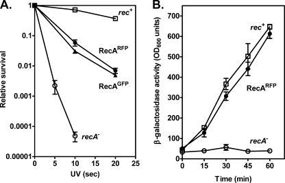
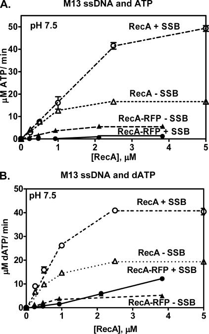
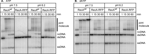
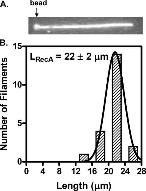
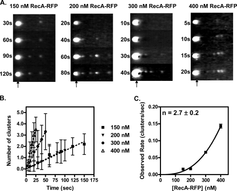
Fluorescent fusion proteins are exceedingly useful for monitoring protein localization in situ or visualizing protein behavior at the single molecule level. Unfortunately, some proteins are rendered inactive by the fusion. To circumvent this problem, we fused a hyperactive RecA protein (RecA803 protein) to monomeric red fluorescent protein (mRFP1) to produce a functional protein (RecA-RFP) that is suitable for in vivo and in vitro analysis. In vivo, the RecA-RFP partially restores UV resistance, conjugational recombination, and SOS induction to recA(-) cells. In vitro, the purified RecA-RFP protein forms a nucleoprotein filament whose k(cat) for single-stranded DNA-dependent ATPase activity is reduced approximately 3-fold relative to wild-type protein, and which is largely inhibited by single-stranded DNA-binding protein. However, RecA protein is also a dATPase; dATP supports RecA-RFP nucleoprotein filament formation in the presence of single-stranded DNA-binding protein. Furthermore, as for the wild-type protein, the activities of RecA-RFP are further enhanced by shifting the pH to 6.2. As a consequence, RecA-RFP is proficient for DNA strand exchange with dATP or at lower pH. Finally, using single molecule visualization, RecA-RFP was seen to assemble into a continuous filament on duplex DNA, and to extend the DNA approximately 1.7-fold. Consistent with its attenuated activities, RecA-RFP nucleates onto double-stranded DNA approximately 3-fold more slowly than the wild-type protein, but still requires approximately 3 monomers to form the rate-limited nucleus needed for filament assembly. Thus, RecA-RFP reveals that its attenuated biological functions correlate with a reduced frequency of nucleoprotein filament nucleation at the single molecule level.


Figures

FIGURE 1.
FIGURE 1.
Fluorescent proteins fused to RecA partially suppress the UV sensitivity and SOS induction defects of a recA strain. A, fresh log-phase cultures were diluted, plated onto L plates, and irradiated at various doses of UV light. Open squares, AB1157 (wild-type); open circles, BIK733 (ΔrecA) with the pBR322 empty vector; closed circles, BIK733 with pNG1 (recA803-mrfp1); closed triangles, BIK733 with pSJS1379 (recA-gfp). B, cells were assayed (in duplicate) for β-galactosidase activity originating from a fusion to the sulA gene at the indicated times. The isogenic strains are derivatives of DM4000 (39); open squares, DM4000 (recA+); open circles, SS2060 (Δ(recA)::kan); and closed circles, SS2065 (recA803-mrfp1). The data are the mean ± S.E. of at least three independent experiments.
FIGURE 2.
FIGURE 2.
Absorption, excitation, and fluorescence emission spectra of RecA-RFP protein. A, absorption spectrum of RecA-RFP protein (4 μm) in storage buffer. B, fluorescence spectra of RecA-RFP (215 nm) in storage buffer. The excitation spectrum was obtained by measuring the fluorescence at 608 nm, and emission spectrum was as obtained by exciting at 582 nm. Each spectrum was normalized to the peak value in each scan.
FIGURE 3.
FIGURE 3.
Purified RecA-RFP protein possesses ssDNA-dependent ATP and dATP hydrolysis activities. A, ATPase activity in the presence of poly(dT). Open triangles, wild-type RecA protein; closed triangles, RecA-RFP protein. B, dATPase activity in the presence of poly(dT). Open triangles, wild-type RecA protein; closed triangles, RecA-RFP protein. Data are mean ± S.D.
FIGURE 4.
FIGURE 4.
The M13 ssDNA-dependent ATPase activity of RecA-RFP protein is inhibited by SSB protein at pH 7.5. A, M13 ssDNA-dependent ATPase activity in the presence or absence of SSB protein (1 μm). Open triangles, wild-type RecA protein without SSB; open circles, wild-type RecA protein with SSB protein; closed triangles, RecA-RFP without SSB; closed circles, RecA-RFP with SSB protein. B, M13 ssDNA-dependent dATPase activity in the presence or absence of SSB protein (1 μm). Symbols are the same as in panel A. Data are mean ± S.D.
FIGURE 5.
FIGURE 5.
The M13 ssDNA-dependent ATP hydrolysis of RecA-RFP protein is not inhibited by SSB protein at pH 6.2. A, M13 ssDNA-dependent ATPase activity in the presence or absence of SSB protein (1 μm). Open triangles, wild-type RecA without SSB; open circles, wild-type RecA with SSB protein; closed triangles, RecA-RFP without SSB protein; closed circles, RecA-RFP with SSB protein. B, M13 ssDNA-dependent dATPase activity in the presence or absence of SSB protein (1 μm). C, M13 ssDNA-dependent ATPase activity in the presence or absence of SSB protein (0.45 μm). D, M13 ssDNA-dependent dATPase activity in the presence or absence of SSB protein (0.45 μm). Symbols in panels B–D are the same as in panel A. Data are mean ± S.D.
FIGURE 6.
FIGURE 6.
The RecA-RFP promotes DNA strand exchange in the presence of ATP or dATP at pH 6.2. DNA strand exchange was carried out with 5′-end labeled linear dsDNA, circular ssDNA, and either wild-type or the fluorescent RecA protein at either pH 7.5 or 6.2. A, DNA strand exchange in the presence of ATP. B, DNA strand exchange in the presence of dATP. The bands are labeled as follows: joint molecule, homologously paired joint molecule intermediate; ncDNA, nicked circular dsDNA product; dsDNA, linear dsDNA substrate; ssDNA, the displaced ssDNA product of complete DNA strand exchange.
FIGURE 7.
FIGURE 7.
RecA-RFP protein assembles on single molecules of dsDNA to form extended nucleoprotein filaments. A, an individual RecA-RFP protein filament formed on a single λ DNA molecule, imaged in the observation channel after a 5-min incubation with RecA-RFP protein (1 μm) at pH 6.2 in the presence of ATPγS (1 mm). The trapped bead is indicated by the arrow. The direction of flow is from left to right. The size of this image is 28 × 4.2 μm. B, length distribution of RecA-RFP nucleoprotein filaments (n = 21). The distribution was fit to a Gaussian function, which yielded a mean length (LRecA) = 22 ± 2 μm.
FIGURE 8.
FIGURE 8.
Visualization of the kinetics of RecA-RFP protein nucleation on dsDNA. A, representative video frames from recordings of nucleation obtained at four different RecA-RFP protein concentrations in nucleation buffer. Flow is right to left. Each vertical strip represents the same DNA molecule repeatedly dipped into the RecA-RFP protein solution in channel 3 for the incubation times indicated. The trapped bead position is indicated by an arrow. B, clusters of RecA-RFP protein were scored and the average values were plotted as a function of time for the following concentrations of RecA-RFP protein: squares, 150 nm; inverted triangles, 200 nm; circles, 300 nm; triangles, 400 nm. C, the observed rate of cluster formation plotted as a function of RecA-RFP protein concentration. The line represents the fit to a power function, kobs = 1.2 × 10⁻⁸ [RecA]2.7, which yielded the value of 2.7 ± 0.2 for the exponent. Error bars represent the standard deviation; where the error bars are not visible, the standard deviation is smaller than the symbols.

References

    1. Chudakov D. M., Lukyanov S., Lukyanov K. A. (2005) Trends Biotechnol. 23, 605–613 - PubMed
    1. Nie S., Chiu D. T., Zare R. N. (1994) Science 266, 1018–1021 - PubMed
    1. Funatsu T., Harada Y., Tokunaga M., Saito K., Yanagida T. (1995) Nature 374, 555–559 - PubMed
    1. Noji H., Yasuda R., Yoshida M., Kinosita K., Jr. (1997) Nature 386, 299–302 - PubMed
    1. Ha T., Ting A. Y., Liang J., Caldwell W. B., Deniz A. A., Chemla D. S., Schultz P. G., Weiss S. (1999) Proc. Natl. Acad. Sci. U.S.A. 96, 893–898 - PMC - PubMed

Publication types