. 2009 Jul 10;284(28):18833-9.
doi: 10.1074/jbc.M109.012385. Epub 2009 May 6.

Syk tyrosine 317 negatively regulates osteoclast function via the ubiquitin-protein isopeptide ligase activity of Cbl

Affiliations

Syk tyrosine 317 negatively regulates osteoclast function via the ubiquitin-protein isopeptide ligase activity of Cbl

Wei Zou et al. J Biol Chem. .

Abstract

Cytoskeletal organization of the osteoclast (OC), which is central to the capacity of the cell to resorb bone, is induced by occupancy of the alphavbeta3 integrin or the macrophage colony-stimulating factor (M-CSF) receptor c-Fms. In both circumstances, the tyrosine kinase Syk is an essential signaling intermediary. We demonstrate that Cbl negatively regulates OC function by interacting with Syk(Y317). Expression of nonphosphorylatable Syk(Y317F) in primary Syk(-/-) OCs enhances M-CSF- and alphavbeta3-induced phosphorylation of the cytoskeleton-organizing molecules, SLP76, Vav3, and PLCgamma2, to levels greater than wild type, thereby accelerating the resorptive capacity of the cell. Syk(Y317) suppresses cytoskeletal organization and function while binding the ubiquitin-protein isopeptide ligase Cbl. Consequently, Syk(Y317F) abolishes M-CSF- and integrin-stimulated Syk ubiquitination. Thus, Cbl/Syk(Y317) association negatively regulates OC function and therefore is essential for maintenance of skeletal homeostasis.


Figures

FIGURE 1.
FIGURE 1.
SykY317F mutation does not affect OC differentiation. Syk−/− BMMs were transduced with either WT Syk, SykY317F, or empty vector (Vect). A, Syk expression by transduced Syk−/− BMMs was determined by immunoblot. B, BMMs were cultured with M-CSF (30 ng/ml) and increasing amounts of RANKL (RL) for 6 days after which the cells were stained for TRAP activity. Spread OCs (arrows), which are absent in vector-transduced cells, appear at lower RANKL doses and are more abundant in those expressing SykY317F than the WT construct. C, similar intensity of TRAP activity (red reaction product) in wells containing OCs generated from SykY317F and WT BMMs. D, histological quantification of the numbers of OCs/well (more than three nuclei per cell) generated from SykY317F and WT BMMs generated in the presence of RANKL (100 ng/ml) and M-CSF (30 ng/ml). E, quantification of the numbers of spread OCs/well generated from SykY317F and WT BMMs. Inset shows characteristic spread and nonspread OCs, respectively. (***, p < 0.001.) F, transduced Syk−/− BMMs were cultured with RANKL (100 ng/ml) and M-CSF (30 ng/ml), with time. OC differentiation markers were determined by immunoblot. Actin serves as loading control. CathK, cathepsin K. G and H, transduced Syk−/− BMMs, cultured in 50 ng/ml M-CSF for 3 days, were serum- and cytokine-starved overnight. The cells were then exposed to either 100 ng/ml M-CSF (G) or 100 ng/ml RANKL (H) with time. Signaling molecules were identified by immunoblotting. Actin serves as a loading control.
FIGURE 2.
FIGURE 2.
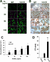
SykY317F enhances OC function. In parallel with the experiments depicted in Fig. 1, Syk−/− BMMs, transduced with either WT Syk or SykY317F, were cultured with M-CSF (30 ng/ml) and increasing amounts of RANKL for 6 days on bone slices. A, actin ring formation was determined by immunofluorescence following fluorescein isothiocyanate-phalloidin staining. B, after 6 days, OCs were removed and the bone slices stained with horseradish peroxidase-labeled wheat germ agglutinin to visualize resorption lacunae. C, histomorphometric analysis of pit area/field. D, medium was collected after 6 days in M-CSF and RANKL (100 ng/ml) and assayed for CTx concentration. *, p < 0.01; **, p < 0.001.
FIGURE 3.
FIGURE 3.
SykY317F does not alter Syk kinase activity. Syk−/− BMMs, transduced with either WT Syk or SykY317F, were cultured with RANKL and M-CSF for 3 days. A, cells were then lifted and either maintained in suspension (S) or plated on vitronectin (A) for 30 min. Phosphorylated tyrosine (p-Y) in Syk immunoprecipitates (IP) was determined by immunoblot. B, cells were treated as in A. Total tyrosine-phosphorylated proteins and phosphorylated SrcY416 were determined by immunoblot. C, cells were treated with or without M-CSF (100 ng/ml) for 5 min. Phosphorylated tyrosine in Syk immunoprecipitates was determined by immunoblot. D, cells were treated as in C. Total tyrosine-phosphorylated proteins were determined by immunoblot. (Numbers represent densitometric analysis relative to suspension, or 0 min of M-CSF, in WT cells.) E, cells were then lifted and replated on vitronectin for 30 min. Cells were lysed and incubated for reaction for 30, 60, and 90 min. The kinase activity was tested using fluorescence enzyme-linked immunosorbent assay reader. Numbers represent average of three time points. RFU, relative fluorescent units.
FIGURE 4.
FIGURE 4.
SykY317F super-activates cytoskeleton-organizing signaling molecules. Syk−/− BMMs, transduced with either WT Syk or SykY317F, were cultured with RANKL and M-CSF for 3 days. A, cells were lifted and either maintained in suspension (S) or plated on vitronectin (A) for 30 min. Phosphorylated tyrosine (p-Y) in Vav3 immunoprecipitates (IP) was determined by immunoblot. B, cells were serum- and cytokine-starved and treated with M-CSF (100 ng/ml) with time. Phosphorylated tyrosine in Vav3 immunoprecipitates was determined by immunoblot. C, cells were treated as in A. Phosphorylated tyrosine in PLCγ2 immunoprecipitates was determined by immunoblot. D, cells were serum- and cytokine-starved and treated with M-CSF (100 ng/ml) for 5 min. Phosphorylated tyrosine in PLCγ2 immunoprecipitates was determined by immunoblot. E, cells were treated as in A. Phosphorylated tyrosine in SLP-76 immunoprecipitates was determined by immunoblot. F, serum- and cytokine-starved cells were exposed to M-CSF (100 ng/ml) for 5 min. Phosphorylated tyrosine in SLP76 immunoprecipitates was determined by immunoblot. (Numbers represent densitometric analysis relative to suspension or 0 min of M-CSF in WT cells.)
FIGURE 5.
FIGURE 5.
SykY317F abolishes Syk-Cbl association in OCs. A, WT BMMs were cultured with RANKL and M-CSF for 3 days. The cells were then lifted and either maintained in suspension (S) or plated on vitronectin (A) for 30 min. Expression of phosphorylated SykY317 and total Syk was determined by immunoblot. Actin serves as loading control. B–D, Syk−/− BMMs, transduced with either WT Syk or SykY317F, were cultured with RANKL and M-CSF for 3 days. B, cells were then lifted and either maintained in suspension (S) or plated on vitronectin (A) for 30 min. Cbl immunoprecipitates (IP) were probed by immunoblot for Syk. IgG immunoprecipitate serves as negative control. C, cells were treated as in B. Phosphorylated tyrosine (p-Y) in Cbl immunoprecipitates (IP) was determined by immunoblot. D, cells were treated with M-CSF (100 ng/ml) for 5 min. Phosphorylated tyrosine in Cbl immunoprecipitates was determined by immunoblot. E, cells were treated as in B. Phosphorylated Cbl in total cell lysates was determined by immunoblot using a specific phospho-CblY774 antibody.
FIGURE 6.
FIGURE 6.
SykY317F inhibits Syk ubiquitination and degradation. Syk−/− BMMs, transduced with either WT Syk or SykY317F, were cultured with RANKL and M-CSF for 3 days. A, cells were pretreated with proteasome inhibitor MG-132 (10 μm) for 1 h and then stimulated with M-CSF with time. Syk immunoprecipitates (IP) were immunoblotted for ubiquitin (Ub). B, cells, lifted and maintained in suspension in the presence MG-132 (10 μm) for 1 h, were retained in suspension (S) or plated to vitronectin (A) for 3 h. Syk immunoprecipitates (IP) were immunoblotted for ubiquitin. C, cytokine- and serum-starved cells were pretreated with the protein synthesis inhibitor cycloheximide (20 μg/ml). After 20 min, M-CSF was added and Syk immunoblotted with time. Actin serves as loading control. Numbers represent densitometric analysis related to 0 h in each group. p-Y, phosphorylated tyrosine.

References

    1. Teitelbaum S. L. (2000) Science 289, 1504–1508 - PubMed
    1. Teitelbaum S. L., Ross F. P. (2003) Nat. Rev. Genet. 4, 638–649 - PubMed
    1. Kumar C. C. (1998) Oncogene 17, 1365–1373 - PubMed
    1. McHugh K. P., Hodivala-Dilke K., Zheng M. H., Namba N., Lam J., Novack D., Feng X., Ross F. P., Hynes R. O., Teitelbaum S. L. (2000) J. Clin. Invest. 105, 433–440 - PMC - PubMed
    1. Pixley F. J., Stanley E. R. (2004) Trends Cell Biol. 14, 628–638 - PubMed

Publication types

MeSH terms