

Case Reports
doi: 10.1056/NEJMoa0900082.
STIM1 mutation associated with a syndrome of immunodeficiency and autoimmunity
Affiliations
- PMID: 19420366
- PMCID: PMC2851618
- DOI: 10.1056/NEJMoa0900082
Item in Clipboard
Case Reports
STIM1 mutation associated with a syndrome of immunodeficiency and autoimmunity
N Engl J Med.
.
Abstract
A mutation in ORAI1, the gene encoding the pore-forming subunit of the Ca(2+)-release-activated Ca(2+) (CRAC) channel, abrogates the store-operated entry of Ca(2+) into cells and impairs lymphocyte activation. Stromal interaction molecule 1 (STIM1) in the endoplasmic reticulum activates ORAI1-CRAC channels. We report on three siblings from one kindred with a clinical syndrome of immunodeficiency, hepatosplenomegaly, autoimmune hemolytic anemia, thrombocytopenia, muscular hypotonia, and defective enamel dentition. Two of these patients have a homozygous nonsense mutation in STIM1 that abrogates expression of STIM1 and Ca(2+) influx.
2009 Massachusetts Medical Society
Figures
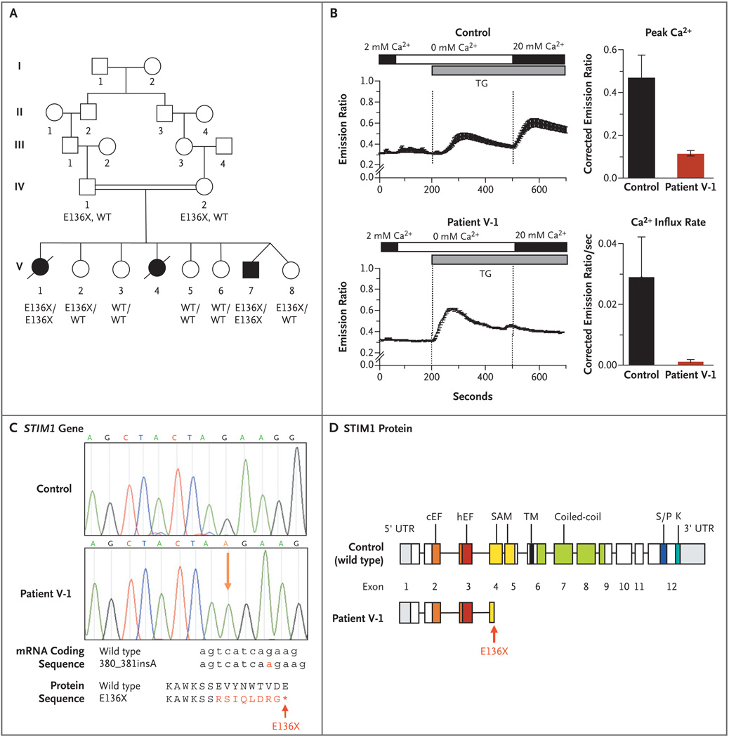
Panel A shows the pedigree of the proband with combined immunodeficiency disease (Patient V-1) and her affected siblings (Patients V-4 and V-7). The STIM1 genotypes of patients and family members from whom DNA was available for sequence analysis (all of whom were in generation IV or V) are listed under their symbols; no DNA was available for Patient V-4. WT denotes wild type. Squares indicate male family members, circles female family members, solid symbols patients, open symbols unaffected persons, slashes deceased persons, and double horizontal lines consanguinity. Panel B shows intracellular calcium levels measured in fibroblasts from Patient V-1 and from a control, showing a reduction of Ca2+ influx in fibroblasts from Patient V-1. Depletion of Ca2+ stores was induced with thapsigargin (TG) in Ca2+-free Ringer’s solution, followed by perfusion with Ringer’s solution containing 20 mM Ca2+ to allow for Ca2+ influx. The resultant intracellular Ca2+ levels were measured as the ratio of fluorescence emissions at 510 nm of Fura-2, a fluorescent dye that binds calcium, after excitation at 340 nm and 380 nm, respectively. Between 40 and 80 cells per experiment were analyzed; results of one representative experiment are shown in the plots on the left side, in which the horizontal bars indicate the durations of perfusion with Ca2+-containing (black) and Ca2+-free (white) Ringer’s solution and the duration of stimulation with thapsigargin (gray). The plots to the right show the average peak Ca2+ levels and initial Ca2+ influx rates after the perfusion of 20 mM Ca2+ (corrected for the baseline values, which were those before the initial depletion of the Ca2+ stores) from four independent experiments. I bars represent the standard error. Panel C shows the results of sequence analysis of genomic STIM1 DNA obtained from Patient V-1 and from a control, revealing an insertion of a single adenine in STIM1 exon 3, between positions 380 and 381 of the coding sequence (380_381insA; NM_003156) resulting in a frame shift and premature termination codon (X) at position 136 of the protein (E136X; NP_003147). The structures of the STIM1 protein domains are superimposed on the sequence of STIM1 exons (modified from Williams et al.15) in Panel D, showing the location of the E136X truncation mutation in STIM1 exon 3, resulting in a truncated STIM1 protein in the N-terminal end of its sterile alpha-motif (SAM) domain. The abbreviation cEF denotes canonical EF-hand domain, hEF hidden EF-hand domain (as recently identified by Stathopulos et al.16), K lysine-rich domain, S/P serine–proline-rich domain, TM transmembrane domain, and UTR untranslated region.
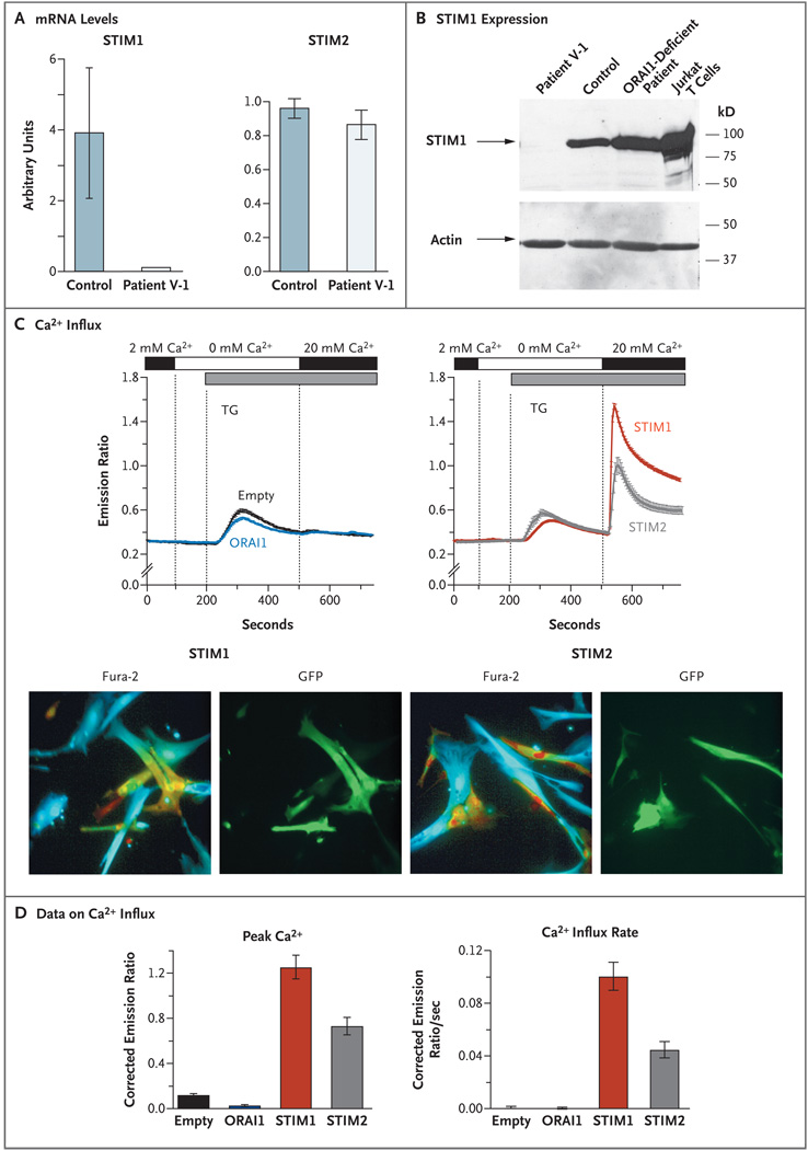
Panel A shows the STIM1 messenger RNA (mRNA) levels in fibroblasts from controls and Patient V-1. Quantitative real-time PCR was performed on complementary DNA (cDNA) from Patient V-1’s fibroblasts and from control fibroblast cell lines (seven lines for the STIM1 experiments and one line for the STIM2 experiments), amplified with the use of primers specific for STIM1, STIM2, and the gene encoding glyceraldehyde-3-phos-phate dehydrogenase (GAPDH). Threshold cycles (CT) for STIM1 and STIM2 expression, normalized to those of GAPDH (ΔCT), are plotted as 0.5ΔCT×104 (arbitrary units). The experiments were run in triplicate; results from one representative experiment are shown. The I bars indicate the standard error. Panel B shows the STIM1 protein expression, as detected on Western blotting, in fibroblasts from Patient V-1, a control, and a previously described patient with ORAI1 deficiency (due to an R91W mutation), as well as Jurkat T cells that were known to express STIM1 (derived from a patient with T-cell acute lymphoblastic leukemia). Total cell lysates were separated by means of sodium dodecyl sulfate–polyacrylamidegel electrophoresis and incubated with an affinity-purified antibody against the C-terminal domain of STIM1. Actin expression (as measured after incubating separated cell lysates with anti-actin antibody) is shown as a control, indicating equal loading. In Panel C (top), store-operated Ca2+ entry in fibroblasts from Patient V-1 is shown to be restored after transduction with bicistronic retroviral vectors expressing full-length STIM1 or STIM2 (right) but not after transduction with an empty vector (no insert) or a vector expressing ORAI1 (left). All vectors contained an internal ribosome entry site green fluorescent protein (GFP) cassette for the detection of transduced cells. Ca2+ measurements were obtained, and are presented, as described in Figure 1B. Between 20 and 40 cells per experiment were analyzed. Panel C (bottom) shows representative results for fibroblasts from Patient V-1 transduced with STIM1 or STIM2 vectors promoting bicistronic GFP expression. The Fura-2 images show pseudocolored intracellular Ca2+ levels: blue represents low Ca2+, green intermediate Ca2+, and yellow–red high Ca2+. The GFP images show cells expressing GFP from an internal ribosome entry site, indicative of simultaneous STIM1 or STIM2 expression. In contrast to nontransduced cells (not visible in the GFP images), transduced cells (green) show high intracellular Ca2+ levels. Panel D shows the average (from between three and five experiments) peak Ca2+ level and initial rate of Ca2+ influx in fibroblasts from Patient V-1 containing empty vectors or vectors expressing ORAI1, STIM1, or STIM2. The data shown were measured after the addition of 20 mM of Ca2+ at 500 seconds. In Panel D, the I bars indicate the standard error.
References
-
- Fischer A. Human primary immunodeficiency diseases. Immunity. 2007;27:835–845. - PubMed
-
- Feske S. Calcium signalling in lymphocyte activation and disease. Nat Rev Immunol. 2007;7:690–702. - PubMed
-
- Lewis RS. The molecular choreography of a store-operated calcium channel. Nature. 2007;446:284–287. - PubMed
Publication types
MeSH terms
Substances
Grants and funding
LinkOut - more resources
Full Text Sources
Other Literature Sources
Medical
Molecular Biology Databases
Miscellaneous