Review
. 2009 Jun;7(6):411-23.
doi: 10.1038/nrmicro2130.

Making the cut: central roles of intramembrane proteolysis in pathogenic microorganisms

Affiliations
Review

Making the cut: central roles of intramembrane proteolysis in pathogenic microorganisms

Sinisa Urban. Nat Rev Microbiol. 2009 Jun.

Abstract

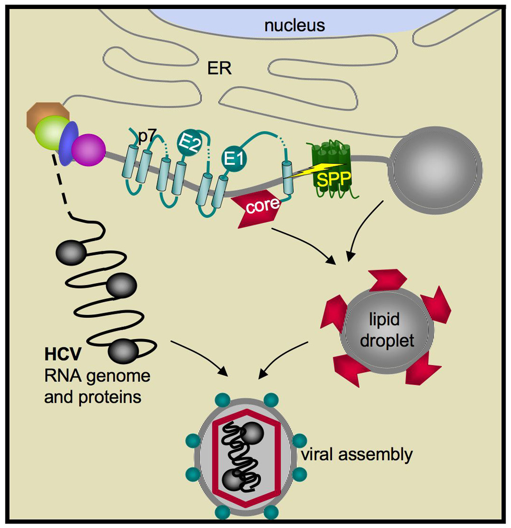
Proteolysis in cellular membranes to liberate effector domains from their transmembrane anchors is a well-studied regulatory mechanism in animal biology and disease. By contrast, the function of intramembrane proteases in unicellular organisms has received little attention. Recent progress has now established that intramembrane proteases execute pivotal roles in a range of pathogens, from regulating Mycobacterium tuberculosis envelope composition, cholera toxin production, bacterial adherence and conjugation, to malaria parasite invasion, fungal virulence, immune evasion by parasitic amoebae and hepatitis C virus assembly. These advances raise the exciting possibility that intramembrane proteases may serve as targets for combating a wide range of infectious diseases. This Review focuses on summarizing the advances, evaluating the limitations and highlighting the promise of this newly emerging field.


Figures

Figure 1
Figure 1. The three families of intramembrane proteases
Graphical representation of the general topology of each intramembrane protease (cytoplasm is down). Active site residues are shown in yellow and the zinc ion of S2P in green. Generic substrates are diagramed in black, with a lightning bolt depicting the proteolytic event, arrow for direction of domain release, and activated effector as a star in black and yellow. The core six transmembrane segments of rhomboid are in blue, with an additional transmembrane segment and cytosolic domain that are present in many homologues depicted in red and brown, respectively. The conserved three transmembrane core of S2P is depicted in purple, with variable transmembrane segments and extramembrane domains (frequently a PDZ domain) of some S2P members shown in orange. The domains present in E. coli, human, and Methanocladococcus jannaschii S2Ps are delineated below the topology diagram. SPP is shown in green (note that presenilins also have nine transmembrane segments but the opposite membrane orientation). In the bottom left panel, the crystal structures of membrane core fragments of prokaryotic rhomboid and S2P are shown (colors and orientation correspond to topology diagram). Bottom right illustrates the conservation of each intramembrane protease in different forms of life. Presenilins are conserved only in multicellular organisms (not shown).
Figure 2
Figure 2. S2P circuits and bacterial virulence: variations on a theme
The E. coli extracytoplasmic function (top left panel) serves as a paradigm for the S2P circuit in bacterial pathogenesis. An input signal (yellow box and arrow), in this case envelope stress, activates the site-1 protease DegS and relief of inhibition by RseB, allowing cleavage of the anti-sigmaE factor RseA at site-1 (top bolt). Intramembrane proteolysis by RseP ensues at site-2 (bottom bolt), resulting in cytoplasmic (bottom) release, degradation of RseA, and liberation of σE to activate the output response, in this case transcription of stress response genes (black arrow). The other S2P circuits involved in regulating virulence of various bacterial pathogens (detailed in the text) are depicted in the same way for the sake of comparison. Components that have been identified are named, while missing components are labeled with a question mark.
Figure 3
Figure 3. Dis-similarity in signalling by rhomboid proteases in Drosophila and a bacterium
In Drosophila cells (left), the EGF precursor Spitz is transported by Star from the ER to the Golgi apparatus, where intramembrane proteolysis by Rhomboid-1 ensures. The processed Spitz is then secreted from the cell, ready to activate EGF receptor signalling in neighboring cells. In this context, rhomboid directly processes the signalling molecule to activate it from a latent form. Conversely in a bacterium (right), the signal itself is unknown and does not require rhomboid processing. Instead, the rhomboid protease AarA cleaves the TatA component of the twin arginine transporter, presumably to facilitate export of the unknown quorum-sensing signal. Notably, the mechanisms are fundamentally different because AarA does not act on the signal itself. Removal of the N terminal extension of TatA by AarA is shown to be required for TatA oligomerzation to form the export pore (along with other subunits), although this is only one model and other possibilities exist.
Figure 4
Figure 4. Function of rhomboid proteases in two different protozoan parasites
An example of parasitism of a human red blood cell by two different protozoan pathogens is shown. In the top panel, a Plasmodium merozoite invading the red blood cell using rhomboid enzymes PfROM4 (green) and PfROM1 (red, initially in mononeme) to breakdown the moving junction (formed between parasite adhesins and host receptors) at the end of invasion. The steps and roles of rhomboid enzymes are detailed in the text, but lead to malaria in the host. The invasion mechanism and role of rhomboid is similar in Toxoplasma. Bottom, an Entamoeba histolytica trophozoite is in the process of internalizing a red blood cell via phagocytosis. The rhomboid EhROM1 (green) relocalizes from the secretory pathway to the phagosome, colocalizing with its putative substrate the Gal/GalNAc lectin (blue). On the right, the amoeba translocates antibody-bound surface proteins to the cap, a membraneous vesicle-like structure that it will jettison to evade immune system attack. EhROM1 colocalizes to the neck of the cap during the capping process. Penetration of the intestine by E. histolytica and evasion of the immune system can lead to life-threatening abcesses.
Figure 5
Figure 5. S2P mediates adaptation to hypoxia during fungal dissemination
Outside the body, Cryptococcus (drawn in blue throughout) experiences a highly aerobic environment, but encounters hypoxia upon inhalation and dissemination into host tissues. Hypoxia is thought to lower sterol production, since biosynthesis requires oxygen. As in human cells, lower sterol levels are thought to result in release of the Scp1:Sre1 complex (SCAP:SREBP in humans) from the ER to the Golgi apparatus (depicted on the right), followed by sequential cleavage by an unidentified Cryptococcal site-1 protease (S1P in humans) and Stp1 (S2P in humans). The site-2 cleavage releases the Sre1 transcription factor domain from the membrane, allowing it to enter the nucleus and activate biosynthetic and hypoxia response genes. In humans, SREBP does not participate in a hypoxic response, but if Stp1 activity is blocked by mutation, Cryptococcus is unable to adapt to hypoxic conditions and cannot survive in host tissues.
Figure 6
Figure 6. SPP processes core protein for hepatitis C virus assembly
The structural HCV components E1 and E2 envelope proteins, core, and p7 are shown inserted into the ER membrane and already processed by signal peptidase. SPP is subsequently responsible for cleaving the first signal peptide, which following signal peptidase cleavage is attached to the C terminus of core protein. SPP cleavage (depicted by the yellow bolt) is required for core trafficking to lipid droplets (right) and recruitment of viral genomic RNA and factors (left) for capsid assembly. Note the SPP is not encoded by the virus, but rather is an ER-resident protein of the host cell.

Similar articles

Cited by

References

    1. Brown MS, Ye J, Rawson RB, Goldstein JL. Regulated intramembrane proteolysis: a control mechanism conserved from bacteria to humans. Cell. 2000;100:391–398. - PubMed
    1. Wolfe MS, Kopan R. Intramembrane proteolysis: theme and variations. Science. 2004;305:1119–1123. - PubMed
    1. Urban S. Rhomboid proteins: conserved membrane proteases with divergent biological functions. Genes Dev. 2006;20:3054–3068. - PubMed
    1. Koonin EV, et al. The rhomboids: a nearly ubiquitous family of intramembrane serine proteases that probably evolved by multiple ancient horizontal gene transfers. Genome Biol. 2003;4:R19. - PMC - PubMed
    1. Lemberg MK, Freeman M. Functional and evolutionary implications of enhanced genomic analysis of rhomboid intramembrane proteases. Genome Res. 2007;17:1634–1646. - PMC - PubMed

Publication types

MeSH terms