Glutathione and catalase suppress TGFbeta-induced cataract-related changes in cultured rat lenses and lens epithelial explants
- PMID: 19421408
- PMCID: PMC2676196
Glutathione and catalase suppress TGFbeta-induced cataract-related changes in cultured rat lenses and lens epithelial explants
Abstract
Purpose: The damaging effects of oxidative stress and transforming growth factor-beta (TGFbeta)-induced transdifferentiation of lens epithelial cells have both been implicated independently in the etiology of cataract. The aim of this study was to investigate whether the presence of antioxidant systems in the lens influences the ability of lens epithelial cells to respond to TGFbeta.
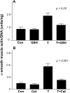
Methods: Whole lenses from young rats were cultured with or without TGFbeta in the presence or absence of reduced glutathione (GSH). Lens epithelial explants from weanling rats were used to investigate the effects of GSH and catalase on TGFbeta-induced cataract-related changes. Lenses were monitored for opacification for three to four days, photographed, and then processed for routine histology. Explants were assessed by phase contrast microscopy, enzyme-linked immunosorbent assay (ELISA) of alpha-smooth muscle actin (alphaSMA), and/or immunolocalization of alphaSMA and Pax6, markers for transdifferentiation and normal lens epithelial phenotype, respectively.
Results: In cultured lenses, GSH strongly suppressed TGFbeta-induced opacification and subcapsular plaque formation. In explants, both GSH and catalase suppressed changes typically associated with TGFbeta-induced transdifferentiation including wrinkling of the lens capsule, cell-surface blebbing, apoptotic cell loss, induction of alphaSMA, and loss of Pax6 expression.
Conclusions: This study suggests that antioxidant systems present in the normal lens, which protect the epithelium against the damaging effects of reactive oxygen species, may also serve to protect it against the potentially cataractogenic effects of TGFbeta. Taken together with other recent studies, it also raises the possibility that TGFbeta may induce cataract-related changes in lens epithelial cells via release of hydrogen peroxide.
Figures


Similar articles
-
Aberrant lens fiber differentiation in anterior subcapsular cataract formation: a process dependent on reduced levels of Pax6.Invest Ophthalmol Vis Sci. 2004 Jun;45(6):1946-53. doi: 10.1167/iovs.03-1206. Invest Ophthalmol Vis Sci. 2004. PMID: 15161862
-
Posterior capsule opacification-like changes in rat lens explants cultured with TGFbeta and FGF: effects of cell coverage and regional differences.Exp Eye Res. 2006 Apr;82(4):693-9. doi: 10.1016/j.exer.2005.09.008. Epub 2005 Dec 15. Exp Eye Res. 2006. PMID: 16359663
-
FGF-2 counteracts loss of TGFbeta affected cells from rat lens explants: implications for PCO (after cataract).Mol Vis. 2004 Jul 22;10:521-32. Mol Vis. 2004. PMID: 15303087
-
Transforming growth factor-beta-induced epithelial-mesenchymal transition in the lens: a model for cataract formation.Cells Tissues Organs. 2005;179(1-2):43-55. doi: 10.1159/000084508. Cells Tissues Organs. 2005. PMID: 15942192 Review.
-
Fibrosis in the lens. Sprouty regulation of TGFβ-signaling prevents lens EMT leading to cataract.Exp Eye Res. 2016 Jan;142:92-101. doi: 10.1016/j.exer.2015.02.004. Epub 2015 May 21. Exp Eye Res. 2016. PMID: 26003864 Free PMC article. Review.
Cited by
-
Understanding cataract development in axial myopia: The contribution of oxidative stress and related pathways.Redox Biol. 2025 Mar;80:103495. doi: 10.1016/j.redox.2025.103495. Epub 2025 Jan 10. Redox Biol. 2025. PMID: 39813957 Free PMC article. Review.
-
Tonic ErbB signaling underlies TGFβ-induced activation of ERK and is required for lens cell epithelial to myofibroblast transition.Mol Biol Cell. 2024 Mar 1;35(3):ar35. doi: 10.1091/mbc.E23-07-0294. Epub 2024 Jan 3. Mol Biol Cell. 2024. PMID: 38170570 Free PMC article.
-
Exploring Angiotensin II and Oxidative Stress in Radiation-Induced Cataract Formation: Potential for Therapeutic Intervention.Antioxidants (Basel). 2024 Oct 8;13(10):1207. doi: 10.3390/antiox13101207. Antioxidants (Basel). 2024. PMID: 39456460 Free PMC article. Review.
-
Oxidative Stress and Cataract Formation: Evaluating the Efficacy of Antioxidant Therapies.Biomolecules. 2024 Aug 25;14(9):1055. doi: 10.3390/biom14091055. Biomolecules. 2024. PMID: 39334822 Free PMC article. Review.
-
Fisetin attenuates hydrogen peroxide-induced cell damage by scavenging reactive oxygen species and activating protective functions of cellular glutathione system.In Vitro Cell Dev Biol Anim. 2014 Jan;50(1):66-74. doi: 10.1007/s11626-013-9681-6. Epub 2013 Aug 27. In Vitro Cell Dev Biol Anim. 2014. PMID: 23982916
References
-
- Congdon N, Vingerling JR, Klein BE, West S, Friedman DS, Kempen J, O'Colmain B, Wu SY, Taylor HR. Prevalence of cataract and pseudophakia/aphakia among adults in the United States. Arch Ophthalmol. 2004;122:487–94. - PubMed
-
- Tabin G, Chen M, Espandar L. Cataract surgery for the developing world. Curr Opin Ophthalmol. 2008;19:55–9. - PubMed
-
- West S. Epidemiology of cataract: accomplishments over 25 years and future directions. Ophthalmic Epidemiol. 2007;14:173–8. - PubMed
-
- Hutnik CM, Nichols BD. Cataracts in systemic diseases and syndromes. Curr Opin Ophthalmol. 1999;10:22–8. - PubMed
Publication types
MeSH terms
Substances
LinkOut - more resources
Full Text Sources
Medical
