. 2009 Sep;36(9):1495-509.
doi: 10.1007/s00259-009-1142-9. Epub 2009 May 7.

Targeting murine heart and brain: visualisation conditions for multi-pinhole SPECT with (99m)Tc- and (123)I-labelled probes

Affiliations

Targeting murine heart and brain: visualisation conditions for multi-pinhole SPECT with (99m)Tc- and (123)I-labelled probes

M Pissarek et al. Eur J Nucl Med Mol Imaging. 2009 Sep.

Abstract

Purpose: The study serves to optimise conditions for multi-pinhole SPECT small animal imaging of (123)I- and (99m)Tc-labelled radiopharmaceuticals with different distributions in murine heart and brain and to investigate detection and dose range thresholds for verification of differences in tracer uptake.

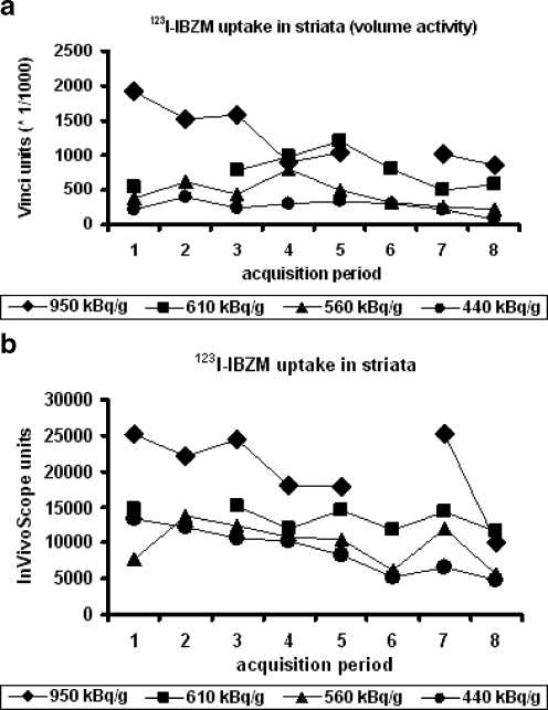
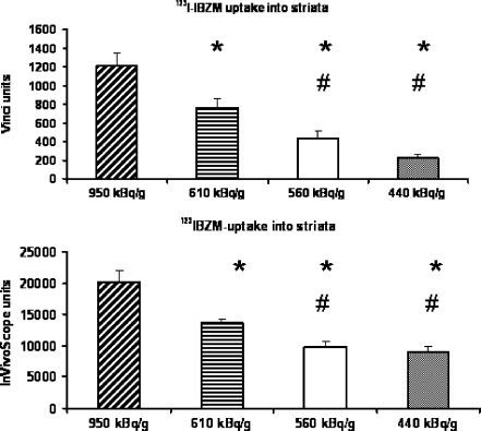
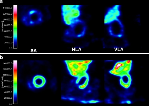
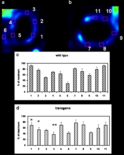
Methods: A Triad 88/Trionix system with three 6-pinhole collimators was used for investigation of dose requirements for imaging of the dopamine D(2) receptor ligand [(123)I]IBZM and the cerebral perfusion tracer [(99m)Tc]HMPAO (1.2-0.4 MBq/g body weight) in healthy mice. The fatty acid [(123)I]IPPA (0.94 +/- 0.05 MBq/g body weight) and the perfusion tracer [(99m)Tc]sestamibi (3.8 +/- 0.45 MBq/g body weight) were applied to cardiomyopathic mice overexpressing the prostaglandin EP(3) receptor.

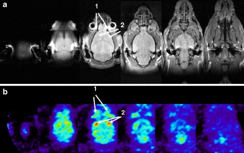
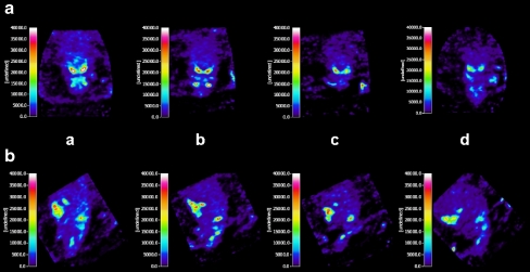
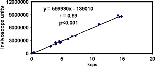
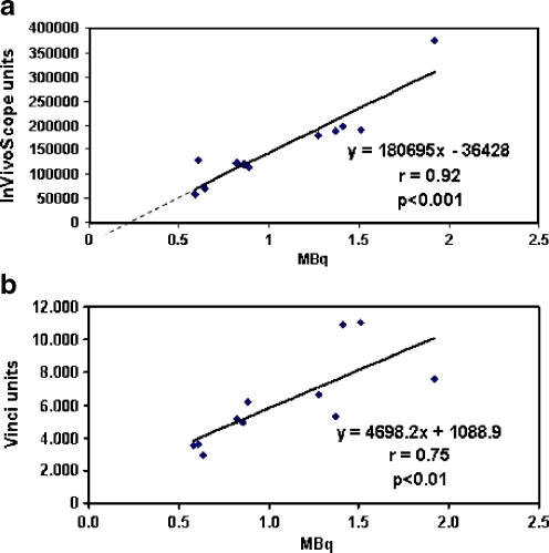
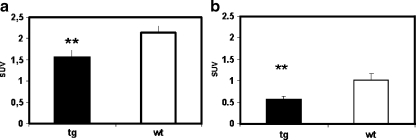
Results: In vivo imaging and in vitro data revealed 45 kBq total cerebral uptake and 201 kBq cardiac uptake as thresholds for visualisation of striatal [(123)I]IBZM and of cardiac [(99m)Tc]sestamibi using 100 and 150 s acquisition time, respectively. Alterations of maximal cerebral uptake of [(123)I]IBZM by >20% (116 kBq) were verified with the prerequisite of 50% striatal of total uptake. The labelling with [(99m)Tc]sestamibi revealed a 30% lower uptake in cardiomyopathic hearts compared to wild types. [(123)I]IPPA uptake could be visualised at activity doses of 0.8 MBq/g body weight.

Conclusion: Multi-pinhole SPECT enables detection of alterations of the cerebral uptake of (123)I- and (99m)Tc-labelled tracers in an appropriate dose range in murine models targeting physiological processes in brain and heart. The thresholds of detection for differences in the tracer uptake determined under the conditions of our experiments well reflect distinctions in molar activity and uptake characteristics of the tracers.


Figures

Fig. 1
Fig. 1
MR and [123I]IBZM SPECT images of murine brains. a Transversal MR images were obtained with an echo time of TE 14 ms and a repetition time of TR 2,000 ms. Measurement was performed using a field of view of 32 × 19.2 mm and a matrix of 512 × 256. Slices of 0.7 mm thickness were scanned with 0.7-mm gaps between the registered planes. b Transversal multi-pinhole SPECT images of a brain obtained following processing by means of Vinci 2.3.1; 0.61 MBq [123I]IBZM/g body weight were applied. SPECT measurement was performed using acquisition times of 100 s and 10 steps of rotation. 1 position of the harderian glands, 2 position of the striata
Fig. 2
Fig. 2
Transversal and sagittal slices of murine brains with different uptake of [123I]IBZM. Brains of four mice are shown following application of 0.95 (a), 0.61 (b), 0.56 (c) and 0.44 (d) MBq/g during the fifth acquisition period (A transversal slices, B sagittal slices). MPS was performed using acquisition times of 100 s per scan, 10 steps of rotation and a radius of rotation of 37 mm. Body weights of the mice were 42 g (a), 34 g (b), 34 g (c) and 50 g (d). Maximum intensity projections of a have been shown in [30]
Fig. 3
Fig. 3
Time course of striatal uptake of [123I]IBZM. Uptake of [123I]IBZM is shown after application of 0.44, 0.56, 0.61 and 0.95 MBq/g body weight in mice. Imaging conditions were used as described in the legend of Fig. 2. The numbers of the x-axis correspond to the consecutive acquisition periods 1 to 8. a Time course analysed using the Vinci 2.3.1. processing tool. b Time course analysed using the InVivoScope processing tool
Fig. 4
Fig. 4
[123I]IBZM uptake into murine striata. Mean ± SEM was calculated for each animal from 12 data points (right and left striata) involving six acquisition periods, when the complete set of intensity data at the four activity doses was available. *p < 0.05 vs 950 kBq/g body weight. #p < 0.05 vs 610 kBq/g body weight. ANOVA followed by the Student-Newman-Keuls test was used for statistical comparison. See Fig. 2 for further description of the imaging conditions
Fig. 5
Fig. 5
In vitro uptake of [123I]IBZM versus in vivo imaging data obtained with different processing tools. a Correlation between striatal uptake of [123I]IBZM in vivo analysed using the InVivoScope processing tool with the total cerebral activities observed. In vivo data reflect the activity detected in right and left striata of the four animals analysed also in Figs. 2–4. Conditions of the measurements are in accordance with that described in the legend of Fig. 2. The offset for y (in vivo intensity) =0 was determined as x0 45 kBq and regarded as the border of detection under the acquisition conditions chosen (100 s acquisition time and 10 steps of rotation). Higher acquisition times should diminish the detection threshold. b, c Correlation between data of striatal accumulation of [123I]IBZM obtained using the Vinci 2.3.1. and InVivoScope processing tools during acquisition periods 3 (b) and 8 (c). Right and left striata of four animals were involved in the calculations
Fig. 6
Fig. 6
Approach to estimation of activities for equipotent visualisation of 123I-labelled receptor ligands in different species according to Eq. 1. The figure shows activities proposed for the application to different species to obtain a visualisation equipotent to that observed in mice. The numbers 1 to 6 of the x-axis correspond to the conditions listed in the table below the diagram. BRwt1 brain weight of the first species, BRwt2 brain weight of the second species, BDwt2 body weight of the second species. The lines connect the doses per animal resulting in visualisation levels (v) corresponding to the visualisation quality expected in mice following application of 0.5 MBq [123I]IBZM/g body weight (v1), 1 MBq/g body weight (v2), 2 MBq/g body weight (v3) and 3 MBq/g body weight (v4)
Fig. 7
Fig. 7
Uptake of [99mTc]HMPAO in the murine brain. [99mTc]HMPAO uptake after application of 0.47 (A) and 1.16 MBq/g body weight (B). The body weight of the animals was 36 g. Acquisition time of 140 s and 10 steps of rotation were used for imaging. 1 thyroid gland, 2 salivary gland, 3 lacrimal gland and ducts. Planes of the sections: a coronal, b transversal, c sagittal. Sections Aa and Ba were measured at prone position of the animals, however, are shown here in supine position
Fig. 8
Fig. 8
Multi-pinhole SPECT of murine heart (wild-type) after application of [123I]IPPA. Images were obtained using the Vinci 2.3.1. processing tool during three consecutive acquisition periods following application of 0.89 MBq/g [123I]IPPA into a B6C3F1(EP3) wild-type mouse with 22 g body weight. a Short axis view. b Horizontal long axis view. c Vertical long axis view. 1 liver, 2 heart
Fig. 9
Fig. 9
MR and SPECT images of cardiomyopathic and wild-type hearts. ad Short axis view of end-diastolic (a and c) and end-systolic hearts (b and d) of B6CBF1.Tg(EP3)5 mice (a and b) and a wild-type littermate (c and d). e, f Short axis view of a cardiomyopathic (e) and wild-type (f) heart labelled with [99mTc]sestamibi. g, h Short axis view of a cardiomyopathic (g) and a wild-type (h) heart labelled with [123I]IPPA
Fig. 10
Fig. 10
Images of cardiomyopathic and wild-type hearts: short axis and horizontal and vertical long axis views after application of [99mTc]sestamibi. a A cardiomyopathic heart with cardiac overexpression of the EP3 receptor. Body weight of the mouse was 22 g and heart weight 187 mg. SA short axis view, HLA horizontal long axis view, VLA vertical long axis view. b A wild-type mouse with a body weight of 24 g and 138 mg heart weight. Measurements were performed using 150 s acquisition time and 10 steps of rotation after application of 3.8 MBq/g body weight [99mTc]sestamibi
Fig. 11
Fig. 11
Yield obtained per detector during measurements of cardiac uptake of [99mTc]sestamibi. Data of 14 mean values of measurements in phase 1 and phase 2 and data of four ex vivo heart measurements (the four lowest values depicted) are correlated with arbitrary units obtained using the InVivoScope data processing tool. Kcps reflecting the average of counts displayed on the screen during all acquisition periods (150 s and 10 steps of rotation) are correlated with the results of the evaluation by means of the InVivoScope data processing tool. The regression line demonstrates the linearity of the multi-pinhole device
Fig. 12
Fig. 12
Correlation of cardiac accumulation of [99mTc]sestamibi in vitro and in vivo. a Comparison of in vivo data obtained using the InVivoScope data processing tool with in vitro activities in transgenic and wild-type animals. b Comparison of in vivo data obtained using the Vinci 2.3.1. data processing tool with in vitro activities in transgenic and wild-type animals
Fig. 13
Fig. 13
SUV in B6C3F1-Tg(EP3) and wild-type mice in vitro and in vivo. Comparison of [99mTc]sestamibi uptake in the heart: in vitro (a) and in vivo (b). [99mTc]sestamibi accumulations in five B6C3F1-Tg(EP3) mice and their wild-type littermates were used for calculation of cardiac SUV. Means ± SEM are shown. Asterisks indicate p < 0.005 calculated for the two-tailed paired Student’s t test. Calculation of standard uptake in vitro was performed using activities and weights determined in excised hearts. Calculation of in vivo data was performed using activities determined using the regression line in Fig. 12a referring to the apparent heart volume calculated by the InVivoScope tool. SUV in vitro is given without dimension and SUV in vivo has the dimension arbitrary units (InVivoScope)/MBq
Fig. 14
Fig. 14
Comparison of regional cardiac accumulation of [99mTc]sestamibi in cardiomyopathic and wild-type mice. a Horizontal long axis view. b Vertical long axis view. ROI: 1 lateral basal, 2 lateral apical, 3 apical, 4 septal apical, 5 septal basal, 6 right ventricle, 7 anterior basal, 8 anterior apical, 9 apical, 10 inferior apical, 11 inferior basal. Means ± SEM are shown. Asterisks indicate differences between the respective ROIs between cardiomyopathic hearts and the hearts of the wild-type littermates with p < 0.05; 5–11 data points observed in the acquisition periods 1–3 were used for the calculation of each mean value

Similar articles

References

    1. {'text': '', 'ref_index': 1, 'ids': [{'type': 'DOI', 'value': '10.1007/s00259-002-0805-6', 'is_inner': False, 'url': 'https://doi.org/10.1007/s00259-002-0805-6'}, {'type': 'PubMed', 'value': '12111135', 'is_inner': True, 'url': 'https://pubmed.ncbi.nlm.nih.gov/12111135/'}]}
    2. Beekman FJ, McElroy DP, Berger F, Gambhir SS, Hoffman EJ, Cherry SR. Towards in vivo nuclear microscopy: iodine-125 imaging in mice using micro-pinholes. Eur J Nucl Med Mol Imaging 2002;29:933–8. - PubMed
    1. {'text': '', 'ref_index': 1, 'ids': [{'type': 'DOI', 'value': '10.1109/TNS.2003.812437', 'is_inner': False, 'url': 'https://doi.org/10.1109/tns.2003.812437'}]}
    2. Schramm NU, Ebel G, Engeland U, Schurrat T, Béhé M, Behr TM. High-resolution SPECT using multipinhole collimation. IEEE Trans Nucl Sci 2003;50:315–20.
    1. {'text': '', 'ref_index': 1, 'ids': [{'type': 'DOI', 'value': '10.1016/S0735-1097(03)00716-2', 'is_inner': False, 'url': 'https://doi.org/10.1016/s0735-1097(03)00716-2'}, {'type': 'PubMed', 'value': '12906991', 'is_inner': True, 'url': 'https://pubmed.ncbi.nlm.nih.gov/12906991/'}]}
    2. Wu MC, Gao DW, Sievers RE, Lee RJ, Hasegawa BH, Dae MW. Pinhole single-photon emission computed tomography for myocardial perfusion imaging of mice. J Am Coll Cardiol 2003;42:576–82. - PubMed
    1. {'text': '', 'ref_index': 1, 'ids': [{'type': 'DOI', 'value': '10.1109/TMI.2005.843782', 'is_inner': False, 'url': 'https://doi.org/10.1109/tmi.2005.843782'}, {'type': 'PubMed', 'value': '16011315', 'is_inner': True, 'url': 'https://pubmed.ncbi.nlm.nih.gov/16011315/'}]}
    2. Goertzen AL, Jones DW, Seidel J, Li K, Green MV. First results from the high-resolution mouseSPECT annular scintillation camera. IEEE Trans Med Imaging 2005;24:863–7. - PubMed
    1. {'text': '', 'ref_index': 1, 'ids': [{'type': 'DOI', 'value': '10.1002/art.21732', 'is_inner': False, 'url': 'https://doi.org/10.1002/art.21732'}, {'type': 'PubMed', 'value': '16572444', 'is_inner': True, 'url': 'https://pubmed.ncbi.nlm.nih.gov/16572444/'}]}
    2. Ostendorf B, Scherer A, Wirrwar A, Hoppin JW, Lackas C, Schramm NU, et al. High-resolution multipinhole single-photon-emission computed tomography in experimental and human arthritis. Arthritis Rheum 2006;54:1096–104. - PubMed

MeSH terms