. 2009 Nov;104(6):621-9.
doi: 10.1007/s00395-009-0031-5. Epub 2009 May 7.

Transcription profiling of HCN-channel isotypes throughout mouse cardiac development

Affiliations

Transcription profiling of HCN-channel isotypes throughout mouse cardiac development

Patrick A Schweizer et al. Basic Res Cardiol. 2009 Nov.

Abstract

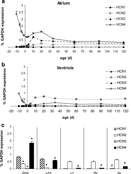
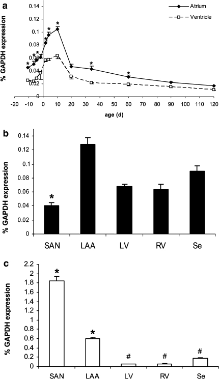
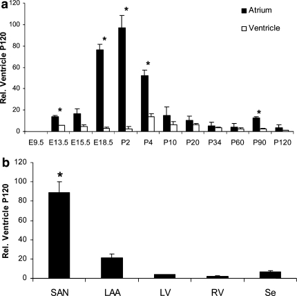
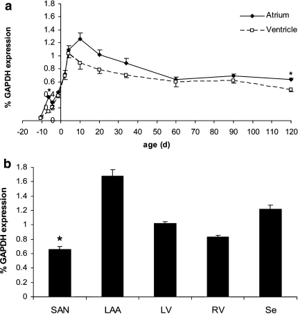
Hyperpolarization-activated ion channels, encoded by four mammalian genes (HCN1-4), contribute in an important way to the cardiac pacemaker current I(f). Here, we describe the transcription profiles of the four HCN genes, the NRSF, KCNE2 and Kir2.1 genes from embryonic stage E9.5 dpc to postnatal day 120 in the mouse. Embryonic atrium and ventricle revealed abundant HCN4 transcription but other HCN transcripts were almost absent. Towards birth, HCN4 was downregulated in the atrium and almost vanished from the ventricle. After birth, however, HCN isotype transcription changed remarkably, showing increased levels of HCN1, HCN2 and HCN4 in the atrium and of HCN2 and HCN4 in the ventricle. HCN3 showed highest transcription at early embryonic stages and was hardly detectable thereafter. At postnatal day 10, HCN4 was highest in the sinoatrial node, being twofold higher than HCN1 and fivefold higher than HCN2. In the atrium, HCN4 was similar to HCN1 and sevenfold higher than HCN2. In the ventricle, in contrast, HCN2 was sixfold higher than HCN4, while HCN1 was absent. Subsequently all HCN isotype transcripts declined to lower adult levels, while ratios of HCN isotypes remained stable. In conclusion, substantial changes of HCN isotype transcription throughout cardiac development suggest that a regulated pattern of HCN isotypes is required to establish and ensure a stable heart rhythm. Furthermore, constantly low HCN transcription in adult myocardium may be required to prevent atrial and ventricular arrhythmogenesis.


Figures

Fig. 1
Fig. 1
Transcription profiling of HCN isotypes throughout mouse cardiac development. a Atrial HCN isotype transcription. In the early embryonic atrium, HCN4 transcripts are the sole source of If current. HCN1-3 is absent. From birth to P34, HCN transcription is reorganized and achieves constant adult levels at P60. b Ventricular HCN isotype transcription. Only HCN4 transcripts appear in the early embryonic ventricle. Postnatally, HCN transcription is changed to predominant HCN2 and moderate HCN4 levels while HCN1 and HCN3 transcripts are virtually missing. c Regional differences at P10. In the sinoatrial node (SAN), HCN4 is expressed at a significantly higher level than in the whole atrial tissue (P < 0.05) and clearly dominates HCN1 and HCN2 transcription. In the left atrial auricle (LAA), HCN4 is reduced to one-third of the SAN level, being expressed significantly lower than in whole atrium (P < 0.05). In the left (LV) and right (RV) ventricle and in the interventricular septum (Se) HCN2 clearly dominates HCN4 while HCN1 like HCN3 is negligible. Interestingly, HCN4 is significantly higher in the Se and significantly lower in LV and RV than in the whole ventricular tissue, respectively. Statistical analysis: *P < 0.05, SAN and LAA, respectively, are compared pairwise with whole atrium at P10. #P < 0.05, LV, RV and Se, respectively, are compared pairwise with whole ventricle at P10 (n = 3)
Fig. 2
Fig. 2
Comparative analysis of atrial and ventricular HCN isotype transcription in relation to their ventricular levels at P120. a HCN1 transcripts, low at embryonic myocardium, rise in the atrium after birth, reaching a maximum level at P10 that slowly declines to adult levels. In the ventricle, HCN1 transcription is negligible. b HCN2 transcripts, almost absent from the embryonic myocardium, appear postnatally with ventricular levels always higher than atrial levels. c HCN3 transcription, although very low at all, shows highest levels in the atrium and at early embryonic stages that gradually decrease to adult levels. d HCN4 transcription is highest at E9.5 dpc, then declines towards birth. A moderate rise after birth falls to stable adult levels after P20. Atrial HCN4 levels always dominate ventricular levels. Statistical analysis: *P < 0.05, atrium versus ventricle compared (n = 3)
Fig. 3
Fig. 3
Cardiac NRSF transcription. a Although higher in atrium than in ventricle, NRSF transcription reaches a maximum level at P10 and declines thereafter to adult levels. Statistical analysis: *P < 0.05, atrium versus ventricle compared. b At P10, NRSF level is significantly lower in the sinoatrial node (SAN) in comparison to whole atrial tissue. Intermediate levels are detected in the right ventricle (RV), left ventricle (LV) and interventricular septum (Se). c Correspondingly, minor HCN4 levels in LAA, RV, LV and Se indicate inverse relations to NRSF. Statistical analysis: *P < 0.05, SAN and LAA, respectively, are compared pairwise to whole atrium at P10. #P < 0.05, LV, RV and Se, respectively, are compared pairwise to whole ventricle at P10 (n = 3)
Fig. 4
Fig. 4
KCNE2 transcription related to ventricular level at P120. a At embryonic stage E9.5 dpc, KCNE2 is undetectable in atrium and ventricle. KCNE2 transcription, first detected at E13.5 dpc, rises in the atrium to attain maximum level at P2 but remains low in the ventricle. After P2, atrial KCNE2 declines rapidly to adult levels in atrium and ventricle. Statistical analysis: *P < 0.05, atrium versus ventricle compared. b At P10, the majority of KCNE2 appears in the sinoatrial node (SAN), levels are significantly higher than in the whole atrium, intermediate amounts in the left atrial auricle (LAA), and low levels in the right (RV) and left ventricle (LV) and the interventricular septum (Se). Statistical analysis: *P < 0.05, SAN and LAA, respectively, are compared pairwise to whole atrium at P10. #P < 0.05, LV, RV and Se, respectively, are compared pairwise to whole ventricle at P10 (n = 3)
Fig. 5
Fig. 5
Cardiac Kir2.1 transcription. a From barely detectable levels at E9.5 dpc, Kir2.1 levels rise similarly in atrium and ventricle reaching maximum shortly after birth and slowly decline to adult levels. Statistical analysis: *P < 0.05, atrium versus ventricle compared. b Kir2.1 transcription is lowest in the sinoatrial node (SAN), highest in the left atrial auricle (LAA) and shows intermediate levels in the right (RV) and left ventricle (LV) and interventricular septum (Se). Statistical analysis: *P < 0.05, SAN and LAA, respectively, are compared pairwise to whole atrium at P10. #P < 0.05, LV, RV and Se, respectively, are compared pairwise to whole ventricle at P10 (n = 3)

References

    1. {'text': '', 'ref_index': 1, 'ids': [{'type': 'DOI', 'value': '10.1113/jphysiol.2002.027698', 'is_inner': False, 'url': 'https://doi.org/10.1113/jphysiol.2002.027698'}, {'type': 'PMC', 'value': 'PMC2342966', 'is_inner': False, 'url': 'https://pmc.ncbi.nlm.nih.gov/articles/PMC2342966/'}, {'type': 'PubMed', 'value': '12702747', 'is_inner': True, 'url': 'https://pubmed.ncbi.nlm.nih.gov/12702747/'}]}
    2. Altomare C, Terragni B, Brioschi C, Milanesi R, Pagliuca C, Viscomi C, Moroni A, Baruscotti M, DiFrancesco D (2003) Heteromeric HCN1-HCN4 channels: a comparison with native pacemaker channels from the rabbit sinoatrial node. J Physiol 549:347–359 - PMC - PubMed
    1. {'text': '', 'ref_index': 1, 'ids': [{'type': 'DOI', 'value': '10.1096/fj.02-0889com', 'is_inner': False, 'url': 'https://doi.org/10.1096/fj.02-0889com'}, {'type': 'PubMed', 'value': '12958166', 'is_inner': True, 'url': 'https://pubmed.ncbi.nlm.nih.gov/12958166/'}]}
    2. Borlak J, Thum T (2003) Hallmarks of ion channel gene expression in end-stage heart failure. FASEB J 17:1592–1608 - PubMed
    1. {'text': '', 'ref_index': 1, 'ids': [{'type': 'PubMed', 'value': '15365256', 'is_inner': True, 'url': 'https://pubmed.ncbi.nlm.nih.gov/15365256/'}]}
    2. Chun KRJ, Koenen M, Katus HA, Zehelein J (2004) Expression of the IKr components KCNH2 (rERG) and KCNE2 (rMiRP1) during late rat heart development. Exp Mol Med 36:367–371 - PubMed
    1. {'text': '', 'ref_index': 1, 'ids': [{'type': 'DOI', 'value': '10.1161/CIRCULATIONAHA.106.616011', 'is_inner': False, 'url': 'https://doi.org/10.1161/circulationaha.106.616011'}, {'type': 'PubMed', 'value': '17420362', 'is_inner': True, 'url': 'https://pubmed.ncbi.nlm.nih.gov/17420362/'}]}
    2. Dobrzynski H, Boyett MR, Anderson RH (2007) New insights into pacemaker activity promoting understanding of sick sinus syndrome. Circulation 115:1921–1932 - PubMed
    1. None
    2. DiFrancesco D (1996) The hyperpolarization-activated (If) current: autonomic regulation and the control of pacing. In: Morad M, Ebashi S, Trautwein W, Kurachi Y (eds) Molecular physiology and pharmacology of cardiac ion channels and transporters. Kluwer Academic Publishers, Dordrecht, pp 31–37

Publication types

MeSH terms

Substances