

Solution structure of the DNA binding domain of AraC protein
- PMID: 19422057
- PMCID: PMC2745637
- DOI: 10.1002/prot.22431
Solution structure of the DNA binding domain of AraC protein
Abstract
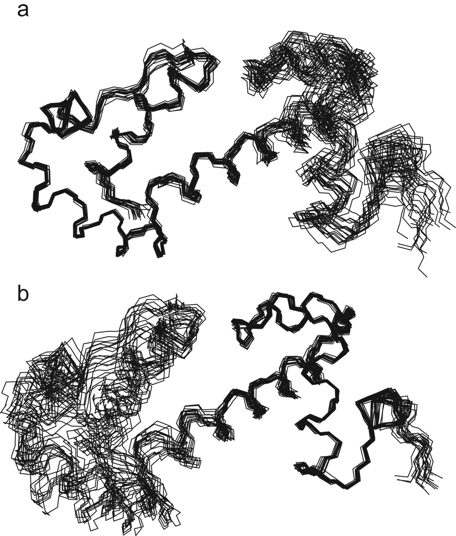
We report the solution structure of the DNA binding domain of the Escherichia coli regulatory protein AraC determined in the absence of DNA. The 20 lowest energy structures, determined on the basis of 1507 unambiguous nuclear Overhauser restraints and 180 angle restraints, are well resolved with a pair wise backbone root mean square deviation of 0.7 A. The protein, free of DNA, is well folded in solution and contains seven helices arranged in two semi-independent sub domains, each containing one helix-turn-helix DNA binding motif, joined by a 19 residue central helix. This solution structure is discussed in the context of extensive biochemical and physiological data on AraC and with respect to the DNA-bound structures of the MarA and Rob homologs.
Figures


Similar articles
-
A new and unexpected domain-domain interaction in the AraC protein.Proteins. 2012 May;80(5):1465-75. doi: 10.1002/prot.24044. Epub 2012 Mar 1. Proteins. 2012. PMID: 22383259
-
Active role of the interdomain linker of AraC.J Bacteriol. 2011 Oct;193(20):5737-46. doi: 10.1128/JB.05339-11. Epub 2011 Aug 12. J Bacteriol. 2011. PMID: 21840981 Free PMC article.
-
Computational predictions of the mutant behavior of AraC.J Mol Biol. 2010 May 7;398(3):462-70. doi: 10.1016/j.jmb.2010.03.021. Epub 2010 Mar 23. J Mol Biol. 2010. PMID: 20338183 Free PMC article.
-
AraC protein, regulation of the l-arabinose operon in Escherichia coli, and the light switch mechanism of AraC action.FEMS Microbiol Rev. 2010 Sep;34(5):779-96. doi: 10.1111/j.1574-6976.2010.00226.x. Epub 2010 Apr 8. FEMS Microbiol Rev. 2010. PMID: 20491933 Review.
-
AraC protein: a love-hate relationship.Bioessays. 2003 Mar;25(3):274-82. doi: 10.1002/bies.10237. Bioessays. 2003. PMID: 12596232 Review.
Cited by
-
Structure of Vibrio cholerae ToxT reveals a mechanism for fatty acid regulation of virulence genes.Proc Natl Acad Sci U S A. 2010 Feb 16;107(7):2860-5. doi: 10.1073/pnas.0915021107. Epub 2010 Feb 1. Proc Natl Acad Sci U S A. 2010. PMID: 20133655 Free PMC article.
-
Gene amplifications cause high-level resistance against albicidin in gram-negative bacteria.PLoS Biol. 2023 Aug 10;21(8):e3002186. doi: 10.1371/journal.pbio.3002186. eCollection 2023 Aug. PLoS Biol. 2023. PMID: 37561817 Free PMC article.
-
Structural and Molecular Mechanism of CdpR Involved in Quorum-Sensing and Bacterial Virulence in Pseudomonas aeruginosa.PLoS Biol. 2016 Apr 27;14(4):e1002449. doi: 10.1371/journal.pbio.1002449. eCollection 2016 Apr. PLoS Biol. 2016. PMID: 27119725 Free PMC article.
-
A DNA-assisted binding assay for weak protein-protein interactions.J Mol Biol. 2009 Dec 18;394(5):805-14. doi: 10.1016/j.jmb.2009.09.064. Epub 2009 Oct 6. J Mol Biol. 2009. PMID: 19815018 Free PMC article.
-
Computer-based annotation of putative AraC/XylS-family transcription factors of known structure but unknown function.J Biomed Biotechnol. 2012;2012:103132. doi: 10.1155/2012/103132. Epub 2012 Mar 13. J Biomed Biotechnol. 2012. PMID: 22505803 Free PMC article.
References
-
- Soisson S, MacDougall-Shackleton B, Schleif R, Wolberger C. Structural Basis for Ligand-Regulated Oligomerization of AraC. Science. 1997;276:421–425. - PubMed
Publication types
MeSH terms
Substances
Associated data
- Actions
Grants and funding
LinkOut - more resources
Full Text Sources
Molecular Biology Databases
