Comparative Study
. 2009 Sep;158(1):339-53.
doi: 10.1111/j.1476-5381.2009.00172.x. Epub 2009 May 5.

Characterization of P2Y receptor subtypes functionally expressed on neonatal rat cardiac myofibroblasts

Affiliations
Comparative Study

Characterization of P2Y receptor subtypes functionally expressed on neonatal rat cardiac myofibroblasts

Amarnath Talasila et al. Br J Pharmacol. 2009 Sep.

Abstract

Background and purpose: Little is known about P2Y receptors in cardiac fibroblasts, which represent the predominant cell type in the heart and differentiate into myofibroblasts under certain conditions. Therefore, we have characterized the phenotype of the cells and the different P2Y receptors at the expression and functional levels in neonatal rat non-cardiomyocytes.

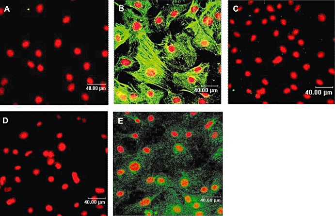
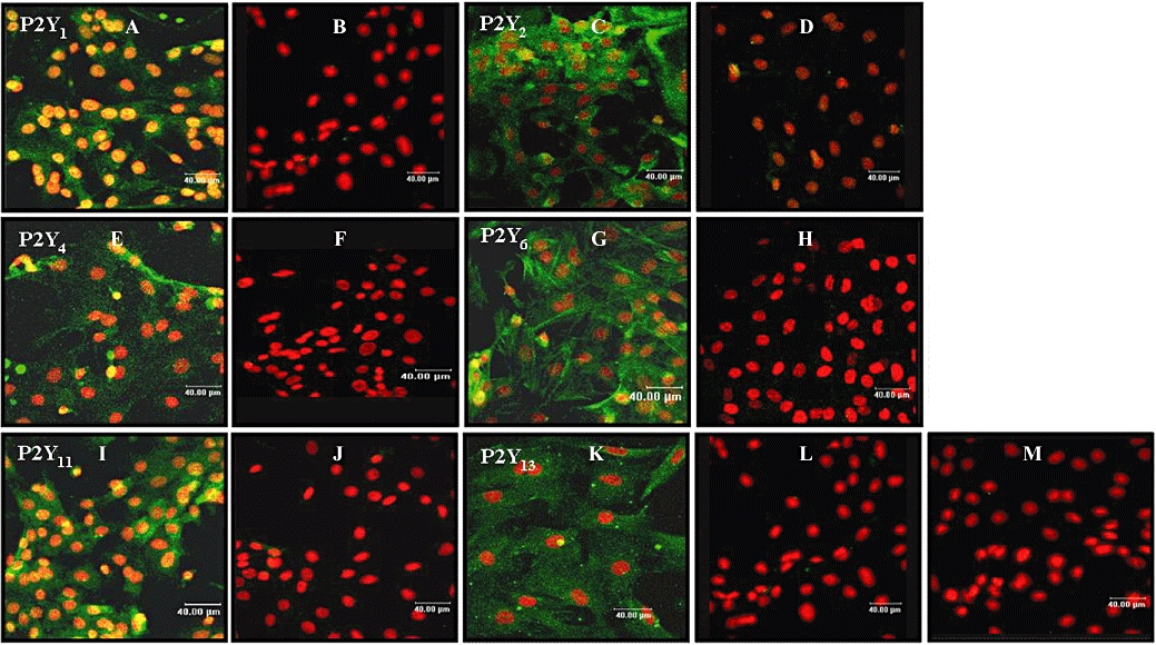
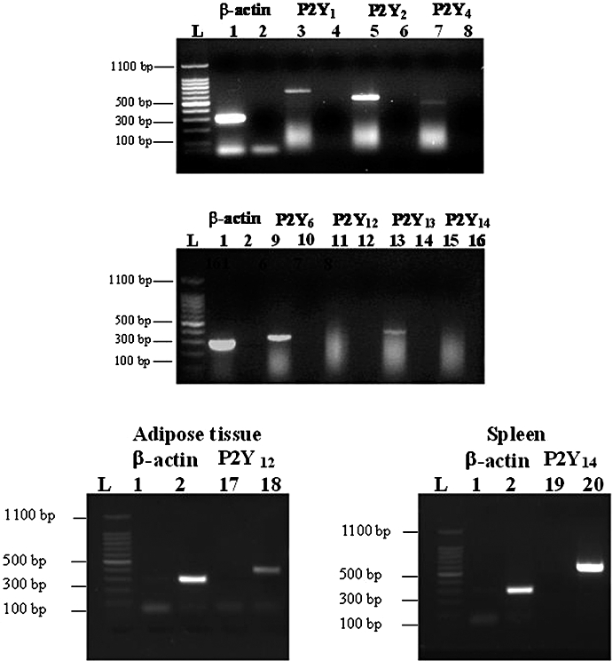
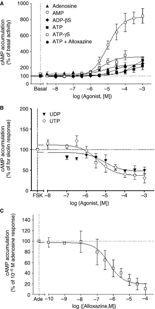
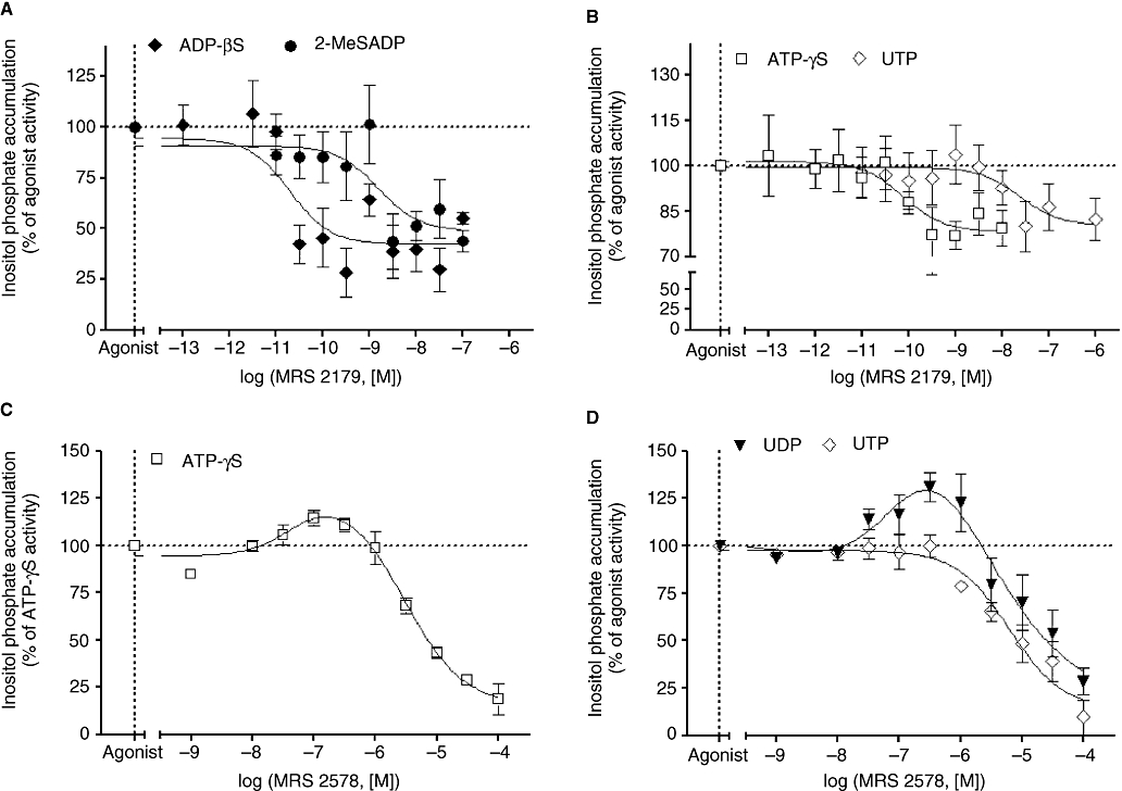
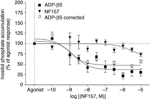
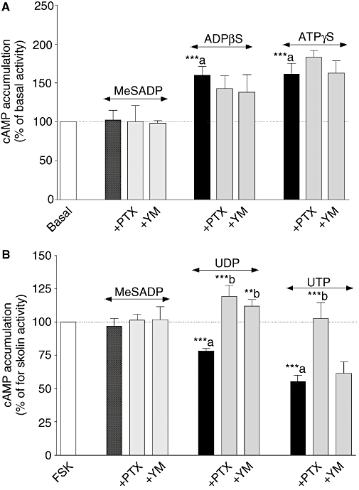
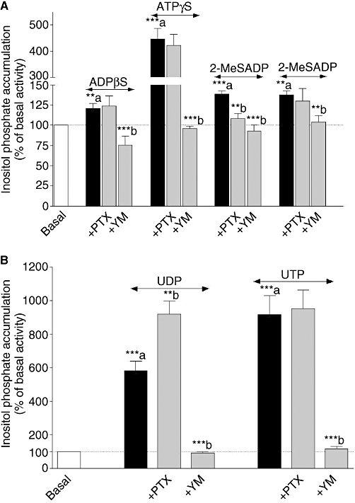
Experimental approach: Non-cardiomyocyte phenotype was determined by confocal microscopy by using discoidin domain receptor 2, alpha-actin and desmin antibodies. P2Y receptor expression was investigated by reverse transcription-polymerase chain reaction and immunocytochemistry, and receptor function by cAMP and inositol phosphate (IP) accumulation induced by adenine or uracil nucleotides in the presence or absence of selective antagonists of P2Y(1) (MRS 2179, 2-deoxy-N(6)-methyl adenosine 3',5'-diphosphate diammonium salt), P2Y(6) (MRS 2578) and P2Y(11) (NF 157, 8,8'-[carbonylbis[imino-3,1-phenylenecarbonylimino(4-fluoro-3,1-phenylene)carbonylimino]]bis-1,3,5-naphthalene trisulphonic acid hexasodium salt) receptors. G(i/o) and G(q/11) pathways were evaluated by using Pertussis toxin and YM-254890 respectively.

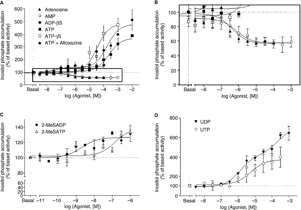
Key results: The cells (>95%) were alpha-actin and discoidin domain receptor 2-positive and desmin-negative. P2Y(1), P2Y(2), P2Y(4), P2Y(6) were detected by reverse transcription-polymerase chain reaction and immunocytochemistry, and P2Y(11)-like receptors at protein level. All di- or tri-phosphate nucleotides stimulated IP production in an YM-254890-sensitive manner. AMP, ADPbetaS, ATP and ATPgammaS increased cAMP accumulation, whereas UDP and UTP inhibited cAMP response, which was abolished by Pertussis toxin. MRS 2179 and NF 157 inhibited ADPbetaS-induced IP production. MRS 2578 blocked UDP- and UTP-mediated IP responses.

Conclusion and implications: P2Y(1)-, P2Y(2)-, P2Y(4)-, P2Y(6)-, P2Y(11)-like receptors were co-expressed and induced function through G(q/11) protein coupling in myofibroblasts. Furthermore, P2Y(2) and P2Y(4) receptor subtypes were also coupled to G(i/o). The G(s) response to adenine nucleotides suggests a possible expression of a new P2Y receptor subtype.


Figures

Figure 1
Figure 1
Phenotypic characterization of neonatal rat non-cardiomyocyte cell culture by immunocytochemistry. Immunocytochemistry by confocal microscopy was performed as described under Materials and Methods using specific α-actin, desmin and discoidin domain receptor 2 (DDR2) antibodies (green). Nuclei were stained with propidium iodide (red). Panels (B, C and E) represent confocal images of α-actin, desmin and DDR2 respectively. Controls were performed in the absence of the primary antibody and in the presence of the secondary rabbit anti-mouse (panel A) and donkey anti-goat (panel D). Images presented are from one experiment and representative of three.
Figure 3
Figure 3
Expression of P2Y receptors in neonatal rat cardiac myofibroblasts by immunocytochemistry. Immunocytochemistry by confocal microscopy was performed as described under Materials and Methods by using specific P2Y receptor antibodies (green). Nuclei were stained with propidium iodide (red). Panels (A-B, C-D, E-F, G-H, I-J and K-L) represent confocal images of P2Y1, P2Y2, P2Y4, P2Y6, P2Y11 and P2Y13 respectively. Controls were performed in the presence of the immunogenic peptide for each receptor (panels B, D, F, H, J and L) or in the absence of the primary antibody (panel M). Images presented are from one experiment and representative of 4.
Figure 2
Figure 2
Expression of P2Y receptor mRNA obtained from neonatal rat cardiac myofibroblasts. Total RNA was prepared and reverse transcription-polymerase chain reaction was carried out as described under Materials and Methods; 1.5% agarose gel electrophoresis represent mRNA coding for β-actin (lanes 1–2), P2Y1 (lanes 3–4), P2Y2 (lanes 5–6), P2Y4 (lanes 7–8), P2Y6 (lanes 9–10), P2Y12 (lanes 11–12, 17–18 in brown adipose tissue), P2Y13 (lanes 13–14) and P2Y14 (lanes 15–16 and 19–20 in spleen). Lanes 2, 4, 6, 8, 10, 12, 14, 16 in myofibroblasts, lanes 1 and 17 in adipose tissue and lanes 1 and 19 in spleen correspond to the primer control without cDNA and lane L to the ladder. Gel images presented are from one experiment and representative of seven independent experiments for myofibroblasts and three for brown adipose tissue and spleen.
Figure 5
Figure 5
Effect of adenine and uracil nucleotides on cAMP accumulation and effect of alloxazine on adenosine-induced cAMP accumulation in neonatal rat cardiac myofibroblasts. cAMP accumulation was measured as described in Methods. Cardiac myofibroblasts were stimulated with adenosine, AMP, ADPβS (adenosine 5′-[β-thio]diphosphate), ATP, ATP + alloxazine and ATPγS (adenosine 5′-[γ-thio] triphosphate) in panel (A), and UDP and UTP in panel (B). Cardiac myofibroblasts were incubated with the indicated concentrations of alloxazine for 30 min before stimulating with 10 µmol·L−1 adenosine (panel C). Data in panel (A) are expressed as percentage of basal cAMP accumulation (100%) and represent the mean ± SE of three to six independent experiments each performed in duplicate. Data in panel (B) are expressed as the percentage of 1.5 µmol·L−1 forskolin response (100%) and represent the mean ± SE of three to seven independent experiments each performed in duplicate. Data in panel (C) are expressed as the percentage of the adenosine-induced cAMP accumulation in the absence of antagonist (100%) and represent the mean ± SE of three independent experiments performed in duplicate.
Figure 4
Figure 4
Effect of adenine and uracil nucleotides on inositol phosphate accumulation (IP) in neonatal rat cardiac myofibroblasts. IP accumulation was measured as described under Materials and Methods. Cardiac myofibroblasts were stimulated with adenosine, AMP, ADPβS (adenosine 5′-[β-thio]diphosphate), ATP, ATP + alloxazine and ATPγS (adenosine 5′-[γ-thio] triphosphate) in panel (A), 2-MeSADP [(methylthio) adenosine 5′-diphosphate] and 2-MeSATP [2(methylthio) adenosine triphosphate] in panel (C), and UDP and UTP in panel (D). Panel (B) corresponds to the enlargement of the frame in panel (A). Data are expressed as percentage of basal IP level (100%) and represent the mean ± SE of three to six independent experiments each performed in duplicate.
Figure 6
Figure 6
Effect of MRS 2179 (2-deoxy-N6-methyl adenosine 3′,5′-diphosphate diammonium salt) and MRS 2578 on adenine and uracil nucleotides-induced inositol phosphate (IP) accumulation in neonatal rat cardiac myofibroblasts. Cardiac myofibroblasts were incubated with the indicated concentrations of MRS 2179 for 30 min before stimulating with 1 µmol·L−1 ADPβS (adenosine 5′-[β-thio]diphosphate) and 0.1 µmol·L−1 2-MeSADP [2(methylthio) adenosine 5′-diphosphate] (panel A), 100 µmol·L−1 ATPγS (adenosine 5′-[γ-thio] triphosphate) and 100 µmol·L−1 UTP for 30 min (panel B). Cardiac myofibroblasts were incubated with the indicated concentrations of MRS 2578 (N,N″-1,4-butanediylbis[N′-(3-isothiocyanatophenyl)thiourea) for 30 min before stimulating with 100 µmol·L−1 ATPγS (panel C) and 100 µmol·L−1 UDP and 100 µmol·L−1 UTP for 30 min (panel D). ADPβS, ATPγS, 2-MeSADP, UDP and UTP induced an increase of IP accumulation of 40%, 440%, 33%, 450% and 375% above basal activity. Data are expressed as the percentage of the agonist-induced IP accumulation in the absence of antagonist (100%) and represent the mean ± SE of three to six independent experiments performed in duplicate.
Figure 7
Figure 7
Effect of NF 157 [8,8′-[carbonylbis[imino-3,1-phenylenecarbonylimino(4-fluoro-3,1-phenylene)carbonylimino]]bis-1,3,5-naphthalene trisulphonic acid hexasodium salt] on adenosine 5′-[β-thio]diphosphate (ADPβS)-induced inositol phosphate (IP) response in neonatal rat cardiac myofibroblasts. Cardiac myofibroblasts were incubated with the indicated concentrations of NF 157 for 60 min alone and for 30 min before stimulating with 1 µmol·L−1 ADPβS. The effect of NF 157 on IP accumulation was removed from the response to ADPβS. Data are expressed as the percentage of the agonist-induced IP accumulation in the absence of antagonist (100%) and represent the mean ± SE of three independent experiments performed in duplicate.
Figure 9
Figure 9
Effect of Pertussis toxin (PTX) and YM 254890 (YM) on adenine (panel A) and uracil (panel B) nucleotides-induced cAMP accumulation in neonatal rat cardiac myofibroblasts. Cardiac myofibroblasts were pretreated with 100 ng·mL−1 PTX for 18 h or with 1 µmol·L−1 YM 254890 for 30 min and stimulated with 100 µmol·L−1 ADPβS (adenosine 5′-[β-thio]diphosphate) and 100 µmol·L−1 ATPγS (adenosine 5′-[γ-thio] triphosphate) for 15 min (Panel A). Cardiac myofibroblasts were pretreated with 100 ng·mL−1 PTX for 18 h or with 1 µmol·L−1 YM 254890 for 30 min and stimulated with 100 µmol·L−1 UDP and 100 µmol·L−1 UTP for 5 min prior to addition of 1.5 µmol·L−1 forskolin (FSK) for 10 min (Panel B). Data are expressed as the percentage of the basal cAMP response for adenine nucleotides or forskolin response in the presence of uracil nucleotides (100%) and represent the mean ± SE of four to seven independent experiments each performed in duplicate. **P < 0.01, ***P < 0.001; a versus basal response or forskolin activity, b versus agonist response.
Figure 8
Figure 8
Effect of Pertussis toxin (PTX) and YM 254890 (YM) on adenine (panel A) and uracil (panel B) nucleotides-induced inositol phosphate (IP) accumulation in neonatal rat cardiac myofibroblasts. Cardiac myofibroblasts were pretreated with 100 ng·mL−1 PTX for 18 h or with 1 µmol·L−1 YM 254890 for 30 min and stimulated with 1 µmol·L−1 ADPβS (adenosine 5′-[β-thio]diphosphate), 100 µmol·L−1 ATPγS (adenosine 5′-[γ-thio] triphosphate), 0.1 µmol·L−1 2-MeSADP [2(methylthio) adenosine 5′-diphosphate], 1 µmol·L−1 2-MeSATP [2(methylthio) adenosine triphosphate], 100 µmol·L−1 UDP and 100 µmol·L−1 UTP for 30 min. Data are expressed as the percentage of the basal IP level (100%) and represent the mean ± SE of four to seven independent experiments each performed in duplicate. **P < 0.01, ***P < 0.001; a versus basal, b versus agonist response.

References

    1. Abbracchio MP, Burnstock G, Boeynaems J-M, Barnard EA, Boyer JL, Kennedy C, et al. International union of pharmacology LVIII: update on the P2Y G protein-coupled nucleotide receptors: from molecular mechanisms and pathophysiology to therapy. Pharmacol Rev. 2006;58:281–341. - PMC - PubMed
    1. Alexander SPH, Mathie A, Peters JA. Guide to Receptors and Channels (GRAC), 3rd edn. Br J Pharmacol. 2008;153(Suppl. 2):S1–S209. - PMC - PubMed
    1. Balogh J, Wihlborg AK, Isackson H, Joshi BV, Jacobson KA, Erlinge D. Phospholipase C and cAMP-dependent positive inotropic effects of ATP in mouse cardiomyocytes via P2Y11-like receptors. J Mol Cell Cardiol. 2005;39:223–230. - PMC - PubMed
    1. Boyer JL, Mohanram A, Camaioni E, Jacobson KA, Harden TK. Competitive and selective antagonism of P2Y1 receptors by N6-methyl 2′-deoxyadenosine 3′,5′-biphosphate. Br J Pharmacol. 1998;124:1–3. - PMC - PubMed
    1. Brown RD, Ambler SK, Mitchell MD, Long CS. The cardiac fibroblast: therapeutic target in myocardial remodelling and failure. Annu Rev Pharmacol Toxicol. 2005;45:657–688. - PubMed

Publication types

MeSH terms

Substances