. 2009 Jul;157(5):746-56.
doi: 10.1111/j.1476-5381.2009.00223.x. Epub 2009 Apr 30.

Comprehensive evaluation of a novel nuclear factor-kappaB inhibitor, quinoclamine, by transcriptomic analysis

Affiliations

Comprehensive evaluation of a novel nuclear factor-kappaB inhibitor, quinoclamine, by transcriptomic analysis

W-Y Cheng et al. Br J Pharmacol. 2009 Jul.

Abstract

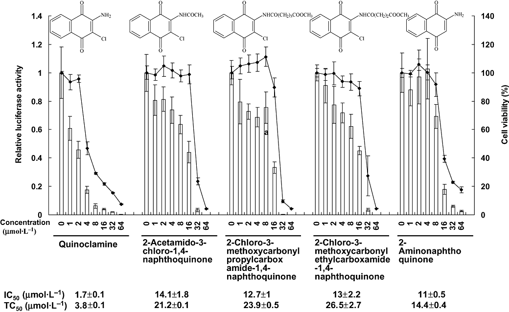
Background and purpose: The transcription factor nuclear factor-kappaB (NF-kappaB) has been linked to the cell growth, apoptosis and cell cycle progression. NF-kappaB blockade induces apoptosis of cancer cells. Therefore, NF-kappaB is suggested as a potential therapeutic target for cancer. Here, we have evaluated the anti-cancer potential of a novel NF-kappaB inhibitor, quinoclamine (2-amino-3-chloro-1,4-naphthoquinone).

Experimental approach: In a large-scale screening test, we found that quinoclamine was a novel NF-kappaB inhibitor. The global transcriptional profiling of quinoclamine in HepG2 cells was therefore analysed by transcriptomic tools in this study.

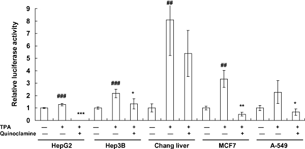
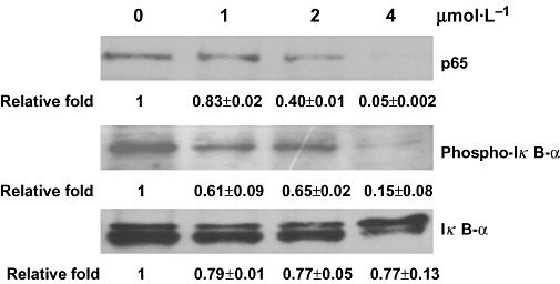
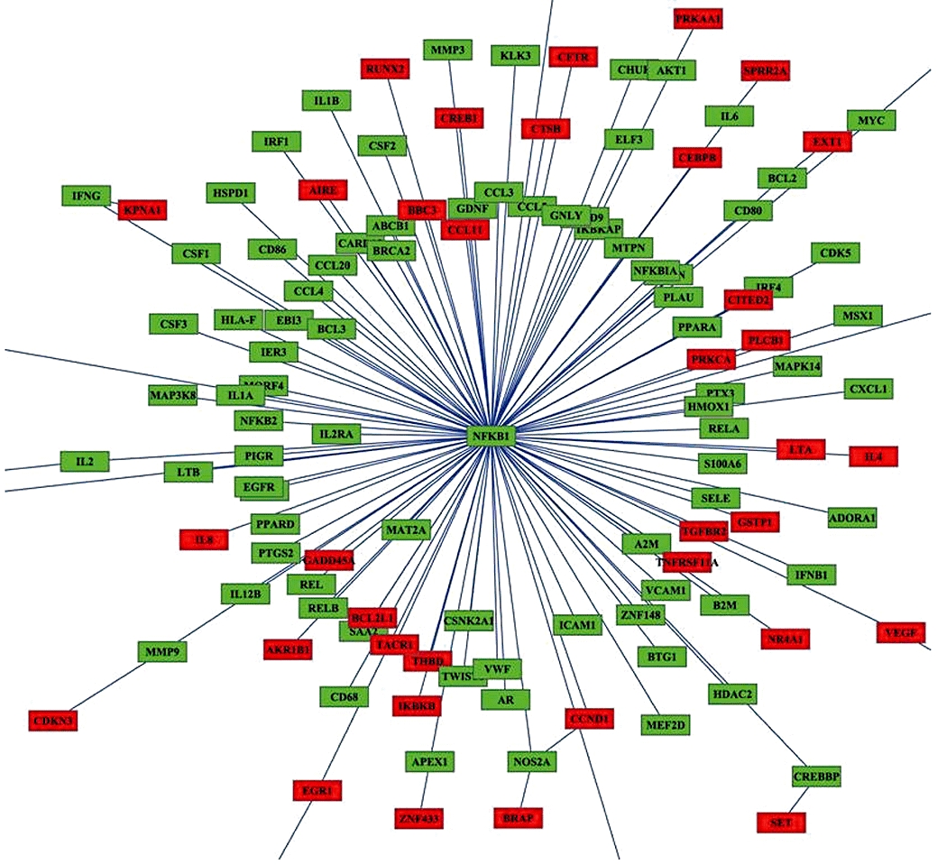
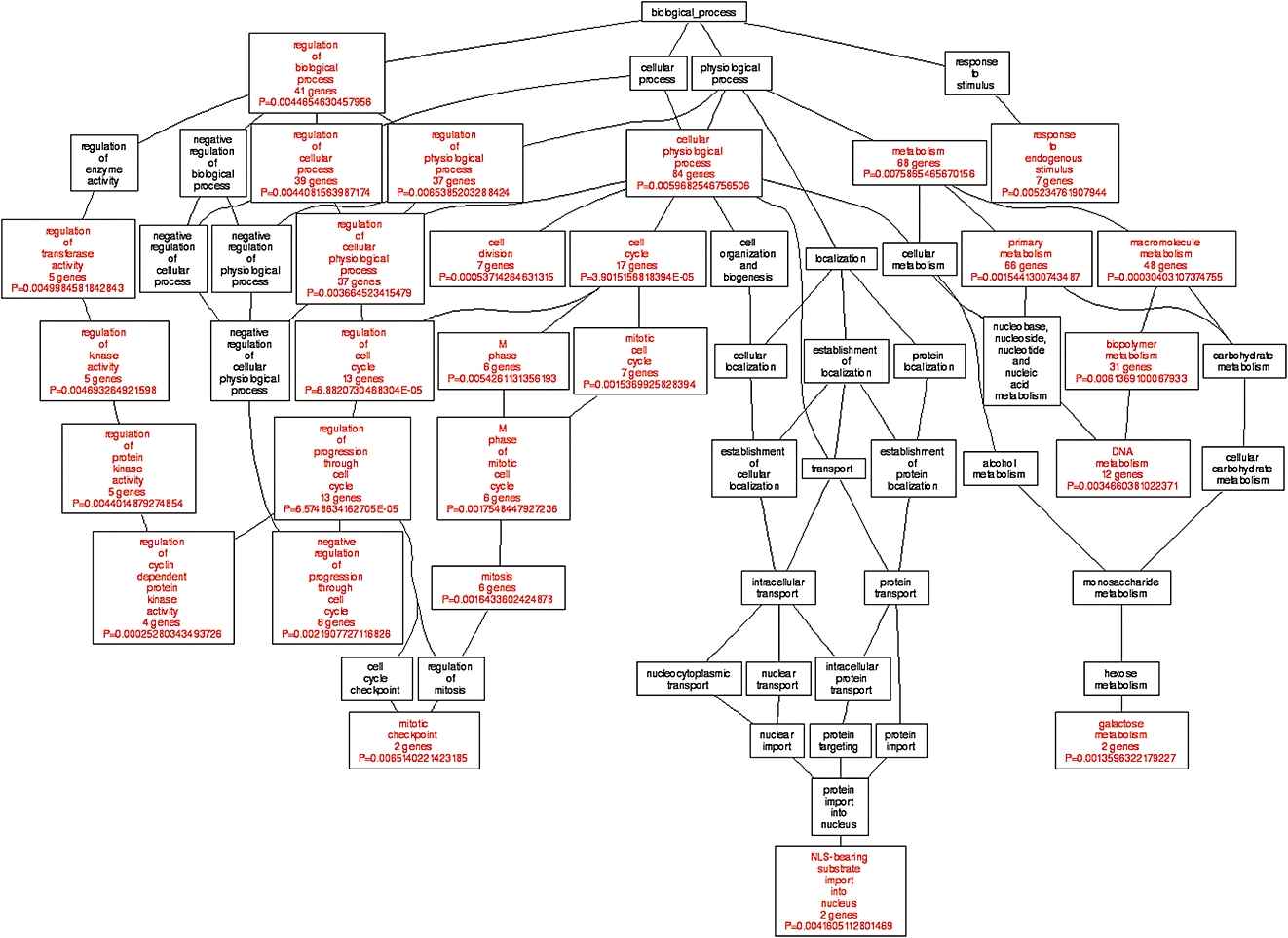
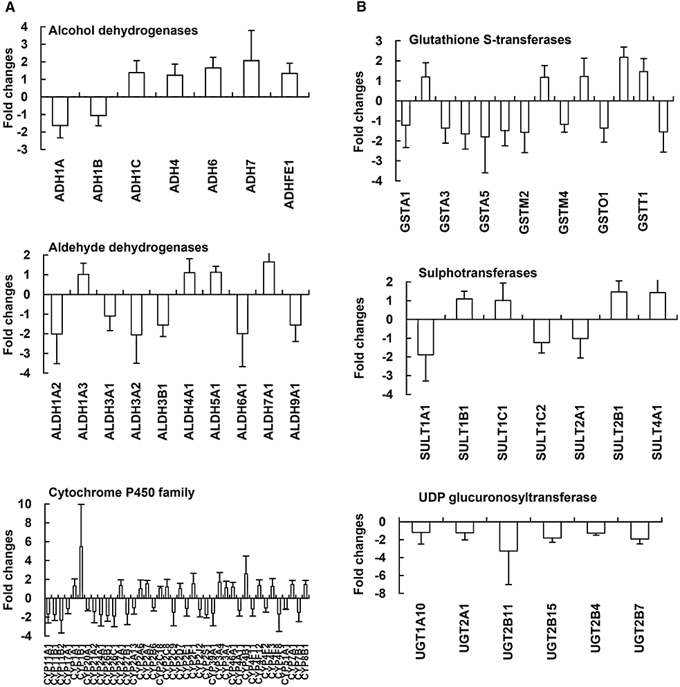
Key results: Quinoclamine suppressed endogenous NF-kappaB activity in HepG2 cells through the inhibition of IkappaB-alpha phosphorylation and p65 translocation. Quinoclamine also inhibited induced NF-kappaB activities in lung and breast cancer cell lines. Quinoclamine-regulated genes interacted with NF-kappaB or its downstream genes by network analysis. Quinoclamine affected the expression levels of genes involved in cell cycle or apoptosis, suggesting that quinoclamine exhibited anti-cancer potential. Furthermore, quinoclamine down-regulated the expressions of UDP glucuronosyltransferase genes involved in phase II drug metabolism, suggesting that quinoclamine might interfere with drug metabolism by slowing down the excretion of drugs.

Conclusion and implications: This study provides a comprehensive evaluation of quinoclamine by transcriptomic analysis. Our findings suggest that quinoclamine is a novel NF-kappaB inhibitor with anti-cancer potential.


Figures

Figure 1
Figure 1
Effects of five naphthoquinone derivatives on nuclear factor-κB (NF-κB) activities and cell viabilities in HepG2 cells. Recombinant HepG2/NF-κB cells were treated with various amounts of naphthoquinone derivatives for 24 h. The luciferase activity and cell viability were then evaluated by luciferase assay and MTT [3-(4,5-dimethylthiazol-2-yl)-2,5-diphenyltetrazolium bromide] assay, respectively. Bars represent the relative luciferase activity, and lines represent the cell viability. Structures of naphthoquinone derivatives are shown on the top. IC50 value and TC50 value of each compound are shown on the bottom. Values are mean ± standard error of four independent assays.
Figure 2
Figure 2
Effects of quinoclamine on 12-O-tetradecanoylphorbol-13-acetate (TPA)-induced nuclear factor-κB (NF-κB) activities in HepG2, Hep3B, Chang liver, MCF7 and A-549 cells. Cells were cultured in 24-well plates for 24 h, washed with Dulbecco's modified Eagle's medium and treated with 4 µmol·L−1 of quinoclamine. Thirty minutes later, TPA (100 ng·mL−1) was added to the media. After a 24 h-incubation, luciferase activities were determined. Results are expressed as relative luciferase activity, which is presented as comparison with the relative luciferase unit relative to untreated cells. Values are mean ± standard error of four independent assays. ##P < 0.01, ###P < 0.001, compared with untreated cells. *P < 0.05, **P < 0.01, ***P < 0.001, compared with TPA-treated cells.
Figure 3
Figure 3
Signal transduction pathways contributing to the inhibition of nuclear factor-κB activity by quinoclamine in HepG2 cells. HepG2 cells were treated with 0, 1, 2 and 4 µmol·L−1 of quinoclamine. The phosphorylated IκB-α and non-phosphorylated IκB-α in cellular extracts were detected by Western blot. The p65 protein in nucleus was also determined by Western blot. Relative fold, which is presented as the comparison of the band intensity relative to untreated cells, is shown on the bottom. Values are mean ± standard error of two independent assays.
Figure 4
Figure 4
Network analysis of quinoclamine-regulated genes in HepG2 cells. The relationship between nuclear factor-κB (NF-κB) target genes and differentially expressed genes responsive to 4 µmol·L−1 quinoclamine was analysed by BiblioSphere Pathway Edition software. The connection between NF-κB and quinoclamine-regulated genes was visualized by cytoscape software. Nodes for over-expressed genes and NFKB1 are colour-coded according to their log2 expression values.
Figure 5
Figure 5
Gene ontology (GO) analysis of quinoclamine-regulated genes in HepG2 cells. Differentially expressed genes responsive to 4 µmol·L−1 quinoclamine in single and dual channel hybridization experiments were organized by using Gene Ontology Tree Machine. The tree indicates the GO structure. The enriched GO categories (P < 0.01 and at least two genes) are coloured red, and non-enriched ones are coloured black.
Figure 6
Figure 6
Expression levels of genes involved in drug metabolism in quinoclamine-treated HepG2 cells. (A) Phase I drug metabolism genes. (B) Phase II drug metabolism genes. Values are mean ± standard error of three independent assays.

References

    1. Aggarwal BB, Shishodia S. Molecular targets of dietary agents for prevention and therapy of cancer. Biochem Pharmacol. 2006;71:1397–1421. - PubMed
    1. Anazawa Y, Arakawa H, Nakagawa H, Nakamura Y. Identification of STAG1 as a key mediator of a p53-dependent apoptotic pathway. Oncogene. 2004;23:7621–7627. - PubMed
    1. Arsura M, Mercurio F, Oliver AL, Thorgeirsson SS, Sonenshein GE. Role of the IκB kinase complex in oncogenic Ras- and Raf-mediated transformation of rat liver epithelial cells. Mol Cell Biol. 2000;20:5381–5391. - PMC - PubMed
    1. Bae KA, Choung SY. Differentiation inducing effects of 2-chloro-3-amino-1,4-naphthoquinone on human leukemia HL-60. Biol Pharm Bull. 1996;19:824–827. - PubMed
    1. Barnes PJ, Karin M. Nuclear factor-κB: a pivotal transcription factor in chronic inflammatory diseases. N Engl J Med. 1997;336:1066–1071. - PubMed

Publication types

MeSH terms