. 2009 Jun;14(6):669-82.
doi: 10.1111/j.1365-2443.2009.01300.x. Epub 2009 Apr 30.

Interactions between Swi1-Swi3, Mrc1 and S phase kinase, Hsk1 may regulate cellular responses to stalled replication forks in fission yeast

Affiliations

Interactions between Swi1-Swi3, Mrc1 and S phase kinase, Hsk1 may regulate cellular responses to stalled replication forks in fission yeast

Michie Shimmoto et al. Genes Cells. 2009 Jun.

Erratum in

  • Genes Cells. 2010 Mar;15(3):313

Abstract

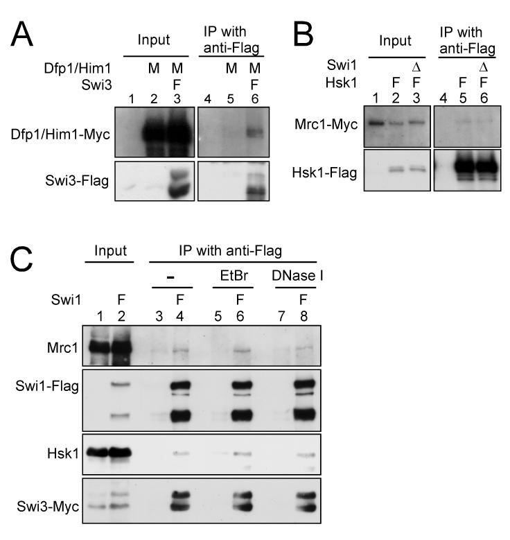
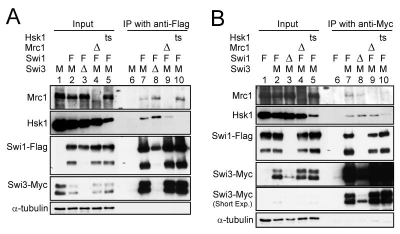
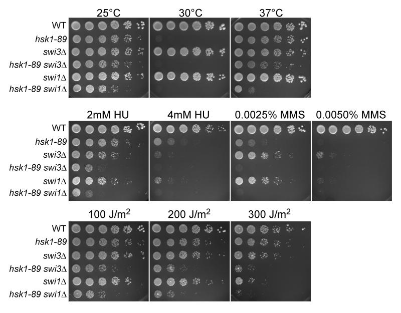
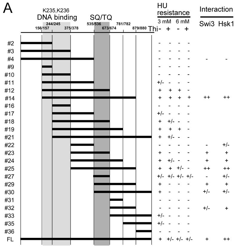
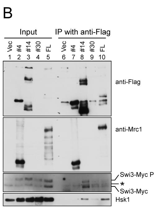
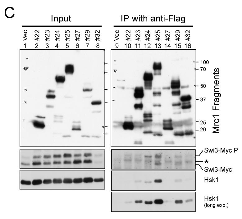
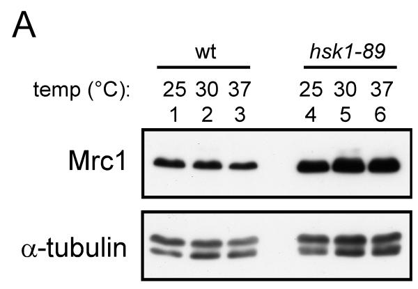
The Swi1-Swi3 replication fork protection complex and Mrc1 protein are required for stabilization of stalled replication forks in fission yeast. Hsk1 kinase also plays roles in checkpoint responses elicited by arrested replication forks. We show that both Swi1 and Swi3, the abundance of which are interdependent, are required for chromatin association of Mrc1. Co-immunoprecipitation experiments show the interactions of Swi1-Swi3, Mrc1 and Hsk1. Mrc1 interacts with Swi3 and Hsk1 proteins through its central segment (378-879) containing a SQ/TQ cluster, and this segment is sufficient for checkpoint reaction. The SQ/TQ cluster segment (536-673) is essential but not sufficient for the interactions and for resistance to replication inhibitor hydroxyurea. Mrc1 protein level is increased in hsk1-89 cells due to apparent stabilization, and we have identified a potential phosphodegron sequence. These results suggest that interactions of the Swi1-Swi3 complex and Hsk1 kinase with Mrc1 may play a role in cellular responses to stalled replication forks in fission yeast.


Figures

Figure 1
Figure 1. Interaction of Hsk1-Dfp1/Him1 with Swi3 and Mrc1
(A) Co-immunoprecipitation of Dfp1/Him1 with Swi3. (B) Co-immunoprecipitation of Mrc1 with Hsk1. The extracts were prepared from the strains indicated, and immunoprecipitation with anti-Flag antibody was performed as described in “Experimental procedures” (lanes 4-6 in (A) and lanes 4-6 in (B)). Input (lanes 1-3 in (A) and lanes 1-3 in (B)) represents 2.5 % of the extracts used for the immunoprecipitation. Western blotting analyses were conducted using the antibodies against the tag as indicated. (A) lanes 1 and 4, YM71; lanes 2 and 5, EN3404; lanes 3 and 6, SH1007. (B) lanes 1 and 4, KT2791; lanes 2 and 5, MS404; lanes 3 and 6, MS405. (C) Co-immunoprecipitation of Hsk1 with Swi1 from EtBr or DNaseI treated extracts. The extracts from the strains indicated were treated with either EtBr (50 μg/ml) or DNase I (0.35 U/μl) for 15 min at 0°C, followed by immunoprecipitation with anti-Flag antibody (lanes 3-8). Input (lanes 1 and 2) represents 1.7 % of the extracts used for the immunoprecipitation. Western blotting analyses were conducted using the antibodies indicated. Lanes 1, 3, 5 and 7, SH0987; lanes 2, 4, 6 and 8, SH1232. Genotypes are indicated above each lane with the abbreviation as follows. M, Myc-tagged; F, Flag-tagged; Δ, deletion.
Figure 2
Figure 2. Interactions among fork stabilization factors and Hsk1
(A) Co-immunoprecipitation with Swi1-Flag protein. (B) Co-immunoprecipitation with Swi3-Myc protein. The extracts were prepared from the strains indicated and immunoprecipitated with anti-Flag (A) or anti-Myc (B) antibody as described in “Experimental procedures” (lanes 6-10 in (A) and (B)). Inputs (lanes 1-5 in (A) and (B)) represent 2 % of the extracts used for the immunoprecipitation. Western blotting analyses were conducted using the antibodies indicated. (A) lanes 1 and 6, SH0987; lanes 2 and 7, SH1232; lanes 3 and 8, SH1914; lanes 4 and 9, SH2303; lanes 5 and 10, SH1302. (B) lanes 1 and 6, EN3381; lanes 2 and 7, SH1232; lanes 3 and 8, SH3309; lanes 4 and 9, SH2303; lanes 5 and 10, SH1302. The degradation products of Swi1 (lower bands) are missing in swi3Δ, since they are further degraded under this background. Abbreviations of the genotypes are the same as those in Figure 1. ts, hsk1-89.
Figure 3
Figure 3. swi3Δ causes hypersensitivity to HU, UV and MMS in hsk1-89 cells
Five-fold serial dilutions of exponentially growing S. pombe cells of the indicated genotypes were plated on YES agar medium and incubated at 25, 30, and 37°C for 5 days (upper panels). Identical five-fold serial dilutions of the indicated cultures were plated on YES plates supplemented with the indicated amounts of HU or MMS (middle panels) or on YES plates followed by UV irradiation as shown (lower panels), and incubated at 25°C for 5 days.
Figure 4
Figure 4. Chromatin association of Mrc1 is stimulated by HU treatment, and is reduced in swi1Δ or swi3Δ cells
The total cell extracts (TCE), chromatin-free (Triton-soluble) and chromatin-enriched (Triton-insoluble) fractions were prepared from the yeast strains indicated and were analyzed by western blotting using the antibodies as indicated. (A) lanes 1-6, SH1232; lanes 7-12, SH1302. (B) lanes 1-6, SH1914; lanes 7-12, SH3309. HU: treated with 12 mM HU for three hrs. The lower bands in Swi1-Flag blots are major degradation products of Swi1 protein. In B, the blots with Swi1-Flag and Swi3-Myc were exposed for a longer time in order to visualize the proteins expressed at a lower level. Note that Mrc1 in the soluble fraction is highly unstable and is degraded during the extract preparation. Chromatin-bound Mrc1 is stable in wild-type or swi1Δ cells (see Figure S1). This is why the sum of soluble and insoluble Mrc1 is less than the TCE level in some lanes. Similarly, Swi3 in the absence of Swi1 is unstable in the soluble fraction and is degraded (B. lanes 8 and 11). (C) In situ chromatin binding assay of Mrc1-GFP in cells arrested at early S-phase by treatment with 15 mM HU for 3 h at 30°C. Spheroplasts of KT2884 (wild-type) (a), MS369 (swi1Δ) (b) and MS384 (swi3Δ) (c) were untreated (-Triton X-100) or preextracted with Triton X-100 to remove soluble nuclear proteins (+Triton X-100) and then fixed for microscopic analysis, as described in “Experimental procedures”. Fluorescence of Mrc1-GFP and DAPI are shown. (d) Quantification of Mrc1-GFP localization in nuclei. The GFP-positive cells were counted and the fraction (%) of GFP-positive cells in DAPI-positive nuclei are presented.
Figure 5
Figure 5. Mrc1 segments required for resistance to HU and protein interactions
(A) A series of deletion derivatives of Mrc1 indicated were cloned into a pREP41X-3Flag which permits addition of 3xFlag-tag at the C terminus of the cloned fragment, and were expressed in mrc1Δ cells (MS252). HU resistance of each transformants were examined on EMM plates containing 3 mM or 6 mM HU. The extent of growth is indicated in the right. +. +/− and – indicate full growth, partially impaired growth and no growth, respectively. The interaction of selected deletion derivatives with Swi3 and Hsk1 is also shown. ++, +, +/− and – represents relative strength of interaction. Light gray and gray segments indicate those involved in DNA binding and containing SQ/TQ clusters, respectively. (B) Mrc1 segments 1-535 (#4), 157-879 (#14), 536-1019 (#30) and a full length of Mrc1 (FL) expressed in mrc1Δ cells (SH2309) were immunoprecipitated with anti-Flag antibody. Western blotting analyses were conducted using the antibodies indicated. (C) Mrc1 segments 376-535 (#22), 376-673 (#23), 376-781 (#24), 376-879 (#25), 536-673 (#27), 536-879 (#29) and 674-879 (#32) expressed in mrc1Δ cells (SH2309) were immunoprecipitated with anti-Flag antibody. Western blotting analyses were conducted using the antibodies indicated. * indicates a non-specific band detected in anti-Flag immunoprecipitates. Swi3-MycP indicates the mobility-shifted form of Swi3-Myc due to phosphorylation.
Figure 6
Figure 6. Stability of Swi1 and Swi3 proteins is mutually dependent
(A) The whole cell extracts were analyzed by western blotting using the antibodies against the proteins indicated. * indicates a non-specific band reacting with the anti-Flag antibody. The arrow indicates the Flag-tagged Swi3 protein. Abbreviations of the genotypes are the same as those in Figures 1 and 2. (B) Cycloheximide was added to the asynchronously growing cultures, and cells were harvested at the times indicated. The whole cell extracts were analyzed by western blotting using the antibodies against the proteins indicated. (A) lane 1,YM71; lane 2, SH2219; lane 3, SH2504; lane 4, YM71; lane 5, SH1401; lane 6, SH2611; lane 7, SH1505. (B) lanes 1-7, SH1232; lanes 8-14,SH1302; lanes 15-21, SH3309; lanes 22-28, SH1914.
Figure 7
Figure 7. Mrc1 is stabilized in hsk1-89
(A) Whole cell extracts were prepared from hsk1+ (YM71; lanes 1-3) or hsk1-89 (KO147; lanes 4-6) cells grown at 25°C (lanes 1 and 4), 30°C (lanes 2 and 5) and 37°C (lanes 3 and 6) for 6 hrs, and were analyzed by western blotting. (B) Cycloheximide (0.1 mg/ml) was added to the asynchronously growing cells (at 25°C) and the cells were harvested at the times indicated. The whole cell extracts were prepared and analyzed by western blotting. Lanes 1-5, KT2791; lanes 6-10, MS346; lanes 11-15, SH0603. (C) The band intensities of (B) were scanned, and the intensities of the Mrc1 bands were normalized to those of tubulin bands, and the normalized values are presented as relative Mrc1 protein level. For each strain, the values at time 0 were taken as 100. In (A) and (B), anti-Mrc1, anti-Myc and anti-tubulin antibodies were used for western blotting. (D) A putative phosphodegron sequence in Mrc1 protein. The mutated residues are indicated. (E) The whole cell extracts from cells carrying the wild-type or a phosphodegron mutant (SSAA) grown at 30°C were analyzed by western blotting using anti-Flag antibody. Lanes 1-4 wild-type; lanes 5-8, mutant; lanes 3-8; tagged with 3-Flag at the C-terminus. Lanes 1-2, YM71, lanes 3-4, MS424, lanes 5-6, MS164, lanes 7-8, MS165.

Similar articles

Cited by

References

    1. Alcasabas AA, Osborn AJ, Bachant J, Hu F, Werler PJ, Bousset K, Furuya K, Diffley JF, Carr AM, Elledge SJ. Mrc1 transduces signals of DNA replication stress to activate Rad53. Nat. Cell Biol. 2001;3:958–965. - PubMed
    1. Alfa C, Fantes P, Hyams J, McLeod M, Warbrick E. Experiments with fission yeast: a laboratory course manual. Cold Spring Harbor Laboratory Press; Plainview, NY: 1993.
    1. Bailis JM, Bernard P, Antonelli R, Allshire RC, Forsburg SL. Hsk1-Dfp1 is required for heterochromatin-mediated cohesion at centromeres. Nat. Cell Biol. 2003;5:1111–1116. - PubMed
    1. Boddy MN, Russell P. DNA replication checkpoint. Curr. Biol. 2001;11:R953–R956. - PubMed
    1. Brown GW, Kelly TJ. Cell cycle regulation of Dfp1, an activator of the Hsk1 protein kinase. Proc. Natl. Acad. Sci. USA. 1999;96:8443–8448. - PMC - PubMed

Publication types

MeSH terms