Review
. 2009 Apr;214(4):409-40.
doi: 10.1111/j.1469-7580.2009.01046.x.

Origin and evolution of the integumentary skeleton in non-tetrapod vertebrates

Affiliations
Review

Origin and evolution of the integumentary skeleton in non-tetrapod vertebrates

Jean-Yves Sire et al. J Anat. 2009 Apr.

Abstract

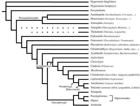
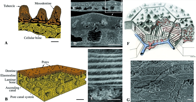
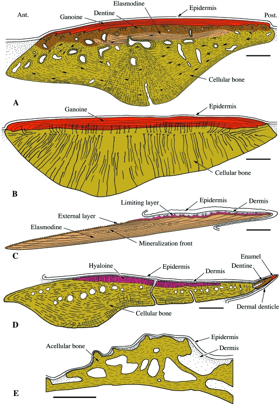
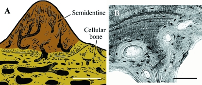
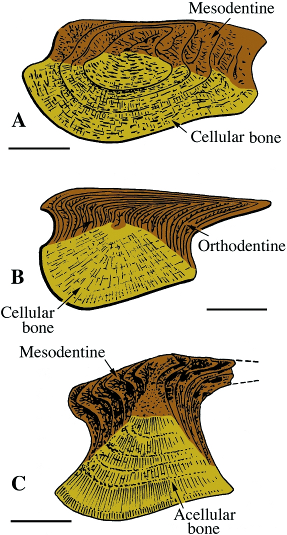
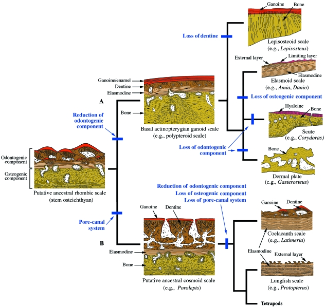
Most non-tetrapod vertebrates develop mineralized extra-oral elements within the integument. Known collectively as the integumentary skeleton, these elements represent the structurally diverse skin-bound contribution to the dermal skeleton. In this review we begin by summarizing what is known about the histological diversity of the four main groups of integumentary skeletal tissues: hypermineralized (capping) tissues; dentine; plywood-like tissues; and bone. For most modern taxa, the integumentary skeleton has undergone widespread reduction and modification often rendering the homology and relationships of these elements confused and uncertain. Fundamentally, however, all integumentary skeletal elements are derived (alone or in combination) from only two types of cell condensations: odontogenic and osteogenic condensations. We review the origin and diversification of the integumentary skeleton in aquatic non-tetrapods (including stem gnathostomes), focusing on tissues derived from odontogenic (hypermineralized tissues, dentines and elasmodine) and osteogenic (bone tissues) cell condensations. The novelty of our new scenario of integumentary skeletal evolution resides in the demonstration that elasmodine, the main component of elasmoid scales, is odontogenic in origin. Based on available data we propose that elasmodine is a form of lamellar dentine. Given its widespread distribution in non-tetrapod lineages we further propose that elasmodine is a very ancient tissue in vertebrates and predict that it will be found in ancestral rhombic scales and cosmoid scales.


Figures

Fig. 1
Fig. 1
Simplified phylogenetic tree of the vertebrates illustrating the interrelationships of the lineages discussed in the text (after Janvier, 1996, 2007; Donoghue & Smith, 2001; Hill, 2005; Donoghue et al. 2006). The relationships of Euconodonta to or within the vertebrates are still debated; the position of Anaspida and Thelodonti with respect to other stem gnathostomes remains uncertain.
Fig. 3
Fig. 3
Pteraspidomorphi (Ordovician). Odontode-like tubercles of various pteraspidomorphs presented as schematics (A–C) and original (D–F) sections. (A,B,D) Astraspis, (C,E,F) Eriptychius, demonstrating the various combinations of enameloid, orthodentine and acellular bone. Scale bars: D = 100 µm; E = 150 µm; F = 50 µm.
None
Heterostraci. (A) Schematic illustration of the integumentary skeleton of a generalized early Devonian heterostracan (e.g. Poraspis or Pteraspis). The lamellar, acellular bony plate (isopedin) is surmounted by a layer of cancellous bone and ornamented by odontode-like tubercles composed of dentine capped with enameloid. (B) SEM section of the superficial region of a portion of the integumentary skeleton of Tesseraspis demonstrating the enameloid-dentine junction (arrow). (C,D) SEM sections of the integumentary skeleton of Anglaspis. Scale bars: B = 40 µm; C = 210 µm; D = 80 µm.
Figs 2–16
Figs 2–16
The structure of various integumentary skeletal elements in extinct and extant non-tetrapod vertebrates illustrated using pictures and interpretative drawings. Vertical sections. The various tissues are identified using colours: yellow = bone (various types) and aspidin; brown = dentine (various types); beige = elasmodine; orange = enameloid; red = enamel and ganoine. Most drawings are from Janvier (1996) (with the author's permission), except Figs 9D, 12B, 12C, 13, 14A, 14E and 15B. Scanning electron micrographs (SEM) of etched sections from Donoghue & Sansom (2002) (Figs 4C, 4D, 5B, 5D, 8D), Wang et al. (2005) (Fig. 7B) and Donoghue et al. (2006) (Figs 2B, 2C, 4B, 5C, 6E), and Fig. 8G is new. Fig. 2 Pteraspidomorphi. (A) Section through the dorsal shield of Sacabambaspis (Arandaspida from the Ordovician). The main tissue identified is acellular bone, distributed into three layers. The superficial tubercles are odontode-like structures composed of dentine. (B) SEM of a section through the dorsal shield of Corvaspis (Heterostraci, Silurian). (C) SEM of a section of the integumentary skeleton of Loricopteraspis (Arandaspida, Silurian) showing the basal lamellar bone. Scale bars: B = 500 µm; C = 50 µm.
Fig. 5
Fig. 5
Anaspida. (A) Schematic illustration of a generalized early Silurian anapsid scale demonstrating the lamellar organization of the acellular (fibrous osteogenic tissue) basal plate. There is no evidence of either dentine or enameloid. (B,C,D) SEM sections of scale from Birkenia (early Silurian) demonstrating the structural organization of the bony tissue. Scale bars: A = 150 µm; B = 200 µm; C = 40 µm; D = 50 µm.
Fig. 10
Fig. 10
Chondrichthyes. Schematic illustrations (A–B,D) and section (C) of chondrichthyan integumentary elements. (A) Integumentary skeleton from the earliest known chondrichthyan, Mongolepis (early Silurian), composed of multiple superimposed layers of odontodes. (B) Elegestolepis (early Silurian) odontode comparable to those of living chondrichthyans. (C) Horizontal section of an integumentary element from an unnamed possible stem-chondrichthyan from the Late Ordovician Harding Sandstone (Sansom et al. 1996). (D) Generalized extant chondrichthyan odontode demonstrating the relationships with the underlying soft tissues of the integument. Scale bars: A = 250 µm; B = 400 µm; C = 100 µm; D = 500 µm.
Fig. 6
Fig. 6
Thelodonti. (A–D) Schematic illustrations of four of the five recognized structural forms of thelodont odontode-like scales (Janvier, 1996). (A) Kawalepis (achanolepid type, early Silurian). (B) Thelodus (thelodontid type, Silurian) (note that this form of thelodont scale is very similar to the odontodes of modern chondrichthyans). (C) Loganellia (loganiid type, late Silurian). (D) Phlebolepis (katoporid type, late Silurian). All thelodont scales are composed of a non-growing dentine-rich crown with a variably defined pulp cavity, and an attachment process of acellular bone which may be the only vestige of an osteogenic skeletal derivative. (E) SEM section of a Thelodus scale demonstrating the presence of a thin superficial layer of enameloid (arrowhead) covering the dentine. Scale bars: B = 100 µm; E = 250 µm.
Fig. 7
Fig. 7
Galeaspida (early Devonian). (A) Schematic illustration of the posterior part of the head-shield of Bannhuanaspis. The shield is thought to have formed by the fusion of multiple mineralized units. Each unit is composed of lamellar, acellular bone with many large, perpendicular fiber-like bundles (Sharpey's fibers). This lamellar bone is sometimes ornamented with small tubercles. The dermoskeleton is underlain by mineralized cartilage associated with the neurocranium; there is no evidence of a perichondrium. (B) SEM section of a polybranchiaspid scale-like element demonstrating the unique organization of the matrix. Note the perpendicular Sharpey's fibers. Scale bar: B = 20 µm.
Fig. 8
Fig. 8
Osteostraci (Silurian). Schematic illustrations (A,B,F) and SEM sections (C–E,G) of osteostracan integumentary elements. (A) Procephalaspis. The basal plate of cellular bone is covered by a layer of tubercles composed of mesodentine, and a thin layer of enameloid. (B) Diagram illustrating the structure of cosmine-like tissue (including the pore-canal system) of the integumentary skeleton of Tremataspis. (C,D) Scale-like element from an unidentified thyestiid. (E) Detail demonstrating the plywood-like tissue (putative elasmodine) of the middle layer of a Tremataspis scale. (F) Reconstruction of the inter-relationships between the polygonal plate-like tesserae of the head-shield and the underlying, richly vascularized region from the osteostracan Alaspis rosamundae (from an unpublished manuscript by Tor Ørvig). Two series of canals are figured in blue and red. (G) Close-up of putative elasmodine in the integumentary skeleton of Hemicyclaspis. Scale bars: A–D: 150 µm; E: 60 µm; G = 10 µm.
Fig. 13
Fig. 13
Actinopterygii. Schematic illustrations of integumentary elements from various extant actinopteryians. (A) Polypterus senegalus (polypteroid-type ganoid scale). (B) Lepisosteus ocellatus (lepisosteoid-type ganoid scale). (C) Danio rerio (elasmoid scale). (D) Corydoras aeneus (scute). (E) Gasterosteus aculeatus (dermal plate). Scale bars: A–D: 250 µm; E = 50 µm.
Fig. 9
Fig. 9
Placodermi. (A) Schematic illustration of a section of the integumentary skeleton from a generalized arthrodiran. This tissue composition includes a basal layer of cellular bone ornamented by tubercles made of semidentine (odontocytes embedded in the matrix). To date enameloid has not been reported in placoderms. (B) Bothriolepis (Late Devonian). Section demonstrating bone remodeling. Scale bars: A = 300 µm; B = 160 µm.
Fig. 11
Fig. 11
Acanthodii. Schematic illustrations of acanthodian scales. (A) Nostolepis (Silurian). (B) Gomphonctus (Silurian). (C) Machairacanthus (early Devonian). Each scale grows by accretion, with successive layers of bone (either cellular or acellular) covered by dentine (either mesodentine or orthodentine). It is generally accepted that the combination of bone and dentine is comparable with odontodes. A ganoine-like tissue has been reported but most taxa appear to lack hypermineralized tissues such as enameloid or enamel. Scale bars: A = 150 µm; B = 200 µm; C = 300 µm.
Fig. 12
Fig. 12
Actinopterygii. Schematic illustrations of palaeoniscoid-type ganoid scales. (A) Andreolepis (late Silurian). (B) Cheirolepis (middle Devonian). (C) Moythomasia (late Devonian). (D) Scanilepis (Triassic). Palaeoniscoid-type ganoid scales grow by deposition of successive layers of dentine and ganoine (= cf. odontocomplexes) (A,C) or apposition of successive odontodes (B,D) onto a deep basal plate made of cellular bone. Scale bars: A,C,D: 100 µm; B: 125 µm.
Fig. 14
Fig. 14
Sarcopterygii. Schematic illustrations of integumentary elements from various non-tetrapodan aquatic sarcopterygians. (A) Glyptolepis (Dipnomorpha, middle to late Devonian). Scale demonstrating the superposition of multiple odontodes embedded in cellular bone. (B–D) Upper surface of the scales of three species of basal dipnomorphans. (B) Dipterus (late Devonian), (C) Osteolepis (middle Devonian), and (D) Porolepis (early Devonian). Cosmine, consisting of a pore-canal system covered by juxtaposed odontodes composed of dentine and ganoine, is common to many basal dipnomorphans (dipnoans and porolepiforms). (E) Latimeria chalumnae (Actinistia, Extant). As demonstrated by this section through the posterior field, in the coelacanth the scale is of elasmoid type; the upper region is composed of numerous overlapping odontodes, with dentine and ganoine, covering a thick, unmineralized basal plate of elasmodine. Scale bars: A = 250 µm; B = 70 µm; C = 100 µm; D = 140 µm; E = 200 µm.
Fig. 16
Fig. 16
A revised scenario depicting the evolution of the integumentary skeleton in non-tetrapods. Although at present uncertain, we hypothesize that elasmodine is present in the ancestral rhombic scales and in cosmoid scales. Furthermore, we propose that similar to modern polypteroid-type ganoid scales, the ancestral rhombic scale was composed of tissues derived from two discrete skeletogenic cell populations: odontogenic and osteogenic. (A) Among actinopterygians, the diversity of scale-type structure can be explained due to the loss and/or modification of these skeletogenic cell components. Loss of the odontogenic component results in the absence of dentine from the lepisosteoid-type ganoid scale. In contrast, loss of the osteogenic component (along with reduction and modification of the odontogenic component) gives rise to the elasmoid scale. Independent losses of the odontogenic component gives rise to the development of dermal plates and to the scutes. Similar to lepisosteoid-type ganoid scales, scutes are characterized by the presence of a well-mineralized layer (hyaloine), similar if not equivalent to ancestral ganoine. (B) Among sarcopterygians, the pore-canal system of the cosmoid scale is lost, followed by reduced and modified expression of the odontogenic-derived components. In actinistians and dipnomorphans these modifications, combined with the loss of the osteogenic component, occur independently (in parallel), giving rise to the sarcopterygian elasmoid scale. Unlike modern dipnoans, extant coelacanths continue to develop superficial odontodes.
Fig. 15
Fig. 15
Schematic illustrations demonstrating structural similarities in the developing scales of (A) Tremataspis (Osteostraci, Silurian), determined from a rare growth series (Denison, 1947), and (B) Polypterus senegalus (Actinopterygii, Extant); polypteroid-type ganoid scale (after Sire, 1989). Scale bars: A = 250 µm; B = 200 µm.

Comment in

References

    1. Alibardi L, Dalla Valle L, Nardi A, Toni M. Evolution of hard proteins in sauropsid integument in relation to cornification of skin derivatives in amniotes. J Anat. 2009;214:560–586. - PMC - PubMed
    1. Arsenault M, Janvier P. The anaspid-like craniates of the Escuminac Formation (Upper Devonian) from Miguasha (Québec, Canada), with remarks on anaspid-petromyzontid relationships. In: Chang MM, Liu YH, Zhang GR, editors. Early Vertebrates and Related Problems of Evolutionary Biology. Beijing: Science Press; 1991. pp. 19–40.
    1. Atchley WR, Hall BK. A model for development and evolution of complex morphological structures. Biol Rev. 1991;66:101–157. - PubMed
    1. Belamie E, Mosser G, Gobeaux F, Giraud-Guille MM. Possible transient liquid crystal phase during the laying out of connective tissues, a chitin and collagen as models. J Phys Condens Matter. 2006;18:115–129.
    1. Bemis WE, Northcutt RG. Skin and blood vessels of the snout of the Australian lungfish, Neoceratodus forsteri, and their significance for interpreting the cosmine of Devonian lungfishes. Acta Zool (Stockh) 1992;73:115–139.

LinkOut - more resources