Review
. 2009 Apr;214(4):441-64.
doi: 10.1111/j.1469-7580.2008.01043.x.

The integumentary skeleton of tetrapods: origin, evolution, and development

Affiliations
Review

The integumentary skeleton of tetrapods: origin, evolution, and development

Matthew K Vickaryous et al. J Anat. 2009 Apr.

Abstract

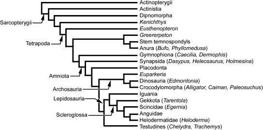
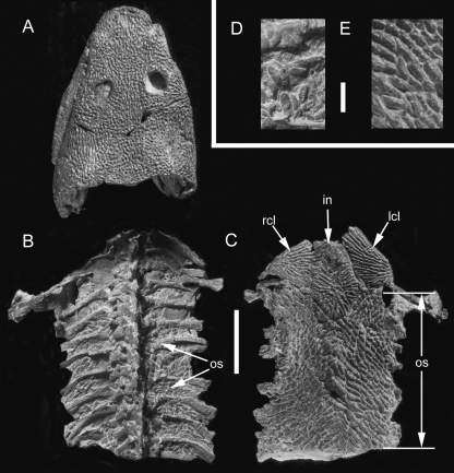
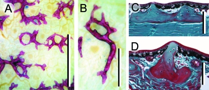
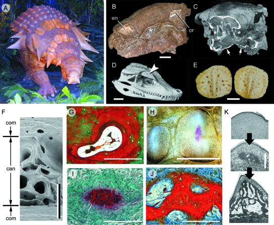
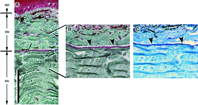
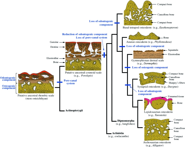
Although often overlooked, the integument of many tetrapods is reinforced by a morphologically and structurally diverse assemblage of skeletal elements. These elements are widely understood to be derivatives of the once all-encompassing dermal skeleton of stem-gnathostomes but most details of their evolution and development remain confused and uncertain. Herein we re-evaluate the tetrapod integumentary skeleton by integrating comparative developmental and tissue structure data. Three types of tetrapod integumentary elements are recognized: (1) osteoderms, common to representatives of most major taxonomic lineages; (2) dermal scales, unique to gymnophionans; and (3) the lamina calcarea, an enigmatic tissue found only in some anurans. As presently understood, all are derivatives of the ancestral cosmoid scale and all originate from scleroblastic neural crest cells. Osteoderms are plesiomorphic for tetrapods but demonstrate considerable lineage-specific variability in size, shape, and tissue structure and composition. While metaplastic ossification often plays a role in osteoderm development, it is not the exclusive mode of skeletogenesis. All osteoderms share a common origin within the dermis (at or adjacent to the stratum superficiale) and are composed primarily (but not exclusively) of osseous tissue. These data support the notion that all osteoderms are derivatives of a neural crest-derived osteogenic cell population (with possible matrix contributions from the overlying epidermis) and share a deep homology associated with the skeletogenic competence of the dermis. Gymnophionan dermal scales are structurally similar to the elasmoid scales of most teleosts and are not comparable with osteoderms. Whereas details of development are lacking, it is hypothesized that dermal scales are derivatives of an odontogenic neural crest cell population and that skeletogenesis is comparable with the formation of elasmoid scales. Little is known about the lamina calcarea. It is proposed that this tissue layer is also odontogenic in origin, but clearly further study is necessary. Although not homologous as organs, all elements of the integumentary skeleton share a basic and essential relationship with the integument, connecting them with the ancestral rhombic scale.


Figures

Fig. 1
Fig. 1
Simplified phylogeny of Tetrapoda demonstrating the interrelationships of the taxa discussed in the text. The phylogenetic arrangement follows the hypotheses of a diphyletic origin of modern amphibians, turtles as the sister group to lepidosaurs, and iguanians as the sister group to scleroglossans based on the work of Janvier (1996, 2007), Hill (2005), Anderson (2007), Anderson et al. (2008) and Conrad (2008). See text for details.
Fig. 2
Fig. 2
Basal tetrapod osteoderms. Greererpeton burkemorani (Stem temnospondyl, Early Carboniferous: Cleveland Museum of Natural History 11090) in (A,B,D) dorsal and (C,E) ventral views. Osteoderms are absent from across the skull (A), modestly developed across the dorsal body surface (B), but are abundant and highly organized along the ventral body surface (C), beginning immediately caudal to the pectoral apparatus. Note the prominent ornamentation embossing the skull (A), and the interclavicle and clavicles (C), coinciding with a lack of osteoderms across these regions. Close-up views of osteoderms from the dorsal (D) and ventral body surfaces (E). in (interclavicle), lcl (left clavicle), os (osteoderms), rcl (right clavicle). Scale bars: A–C = 50 mm, D–E = 3 mm. Photographs courtesy of L. Russell and Dr. M. Ryan, Cleveland Museum of Natural History, Cleveland, OH, USA.
Fig. 3
Fig. 3
Anuran osteoderms. Phyllomedusa bicolor (Phyllomedusinae, Extant). (A,B) Adult osteoderms from the dorsal body surface, prepared as whole-mounts using Alizarin red (single-stained). (C,D) Transverse sections (dorsal towards the top) of dorsal body surface osteoderms, Masson's trichrome staining. Osteoderms reside entirely within the stratum superficiale. Note the development of large spines displacing the epidermis dorsally. Scale bars: A,B = 0.25 mm; C,D = 100 µm. Specimens courtesy of Dr. J. Bogart and M.-T. Rush, University of Guelph, Guelph, Canada.
Fig. 4
Fig. 4
Archosaur osteoderms. (A–C) Edmontonia rugosidens (Ankylosauria, Late Cretaceous). (A) Reconstruction on display at the Royal Tyrrell Museum of Palaeontology, Drumheller, Alberta, demonstrating various plate-like and spine-shaped osteoderms. (B) Skull (American Museum of Natural History, New York, 5381) in right rostrodorsal view. (C) Computed tomography reconstruction of the skull in (B) with the rostrum truncated to indicate the in situ position of the cheek region osteoderms, and a series of small, granular osteoderms across the throat (small arrows). The asterisks (*) in both (A) and (B) identify the presence of an osteoderm embedded in the cheek region, lateral to the tooth rows. (D) Paleosuchus palpebrosus skull (Crocodylia, Extant: Royal Ontario Museum, Toronto, Paleobiology collection R6692) demonstrating a well-developed osteoderm (= palpebral) within the eyelid (large arrowhead). (E) Alligator mississippiensis (Crocodylia, Extant). Cervical osteoderms demonstrating a common pattern of ornamentation among archosaurs: superficial pitting. (F) Caiman c. crocodilus (Crocodylia, Extant: Royal Ontario Museum, Toronto, Paleobiology collection R7719). Transverse section through a cervical osteoderm. The structural organization includes an outer and an inner cortex of compact bone (com) surrounding a cancellous core (can). (G,I–K) Longitudinal sections (dorsal towards the top). (G) A. mississippiensis adult, cervical osteoderm, Masson's trichrome staining. Note the resorption of woven-fibred bone and newly deposited lamellar bone matrix. (H) A. mississippiensis subadult, cervical osteoderms prepared as whole-mounts using Alizarin red (single-stained). The initial site of mineralization (red staining) is within the keel of the largest presumptive element. (I) Same specimen as (H), sectioned and stained with a modified Masson's trichrome (Cole & Hall, 2004). Mineralization (red staining) is initiated without the formation of a cell condensation. This mode of ossification is consistent with bone metaplasia. (J) A. mississippiensis subadult [slightly older than (I)], cervical osteoderm, Mallory's trichrome. Numerous extrinsic collagen fibres are becoming incorporated into the osteoderm matrix. Note the absence of a clear osteoblastic front. (K) Sequence of osteoderm development beginning as a weakly defined primordium of dense irregular connective tissue (top panel), followed by mineralization within the centre of the keel (middle panel), and finally expansion of the osteoderm into the majority of the keel and the deposition of bone (bottom panel). Scale bars: B–C = 50 mm, D = 100 mm, E = 30 mm, F,K = 1 mm, G,I,J = 40 µm, H = 5 mm. en (external naris), or (orbit). Micrograph (F) courtesy of M. Burns, University of Alberta, Edmonton, Canada.
Fig. 5
Fig. 5
Lepidosaur osteoderms. (A–C) Alizarin red single-staining. (D,F–H) Longitudinal sections (dorsal towards the top). (A,B) Egernia sp. (Scincidae, Extant). Among scincids, most postcranial osteoderms overlap one another and demonstrate a compound or fractured morphology. (C) Tarentola mauritanica (Gekkota, Extant) postcranial osteoderms with a granular morphology. (D) Postcranial osteoderms from T. annularis (Gekkota, Extant) stained with Masson's trichrome. Each osteoderm has two distinct tissue regions. The superficial region resides entirely within the stratum superficiale, and is collagen-poor with virtually no incorporated cells. The basal region resides within the stratum compactum, and consists of compact (cellular) bone. Sharpey's fibres anchor both regions within the surrounding dermis. (E–H) Heloderma horridum (Helodermatidae, Extant). (E) Adult skull demonstrating the presence of osteoderms. Osteoderms from the left lateral surface have been removed to reveal the underlying cranial elements. Heloderma horridum osteoderms stained with Masson's trichrome (F,H) and toluidine blue (G). Similar to Tarentolaspp., H. horridum skeletally mature osteoderms (F,G) have a superficial collagen-poor region and a basal region composed of compact bone. (H) Skeletally immature osteoderm (white asterisk) demonstrating the earliest stages of mineralization. Note the absence of an osteoblast-rich condensation. This mode of ossification is consistent with bone metaplasia. The superficial region develops later during ontogeny. bh (basal region of bone-rich tissue), sf (Sharpey's fibres), sh (superficial region of unidentified skeletal tissue), stc (stratum compactum of the dermis), sts (stratum superficiale of the dermis). Scale bars: A = 1 mm, C = 0.5 mm, D = 50 µm, E = 20 mm, F–H = 100 µm.
Fig. 6
Fig. 6
Synapsid osteoderms. (A,B) Heleosaurus scholtzi (Varanopidae, Permian: Iziko South African Museum of Cape Town SAM-PK-K8305), a rare example of a non-xenarthran osteoderm-bearing synapsid. (A) A fossil specimen consisting of five individuals, with the largest (B) demonstrating in situ osteoderms (black arrowheads) across the cervical region (see Botha-Brink & Modesto, 2007). (C–H) Morphology and development of osteoderms in xenarthrans. (C) Holmesina occidentalis (Pampatheriidae, Late Pleistocene: Royal Ontario Museum 39257, 40046, 40047). Note the distinctive cranial (overlapped) and caudal (overlapping) margins on each osteoderm. (D–H) Dasypus novemcinctus (structural-grade armadillo, Extant). (D) Scanning electron micrograph of an adult osteoderm with a polygonal morphology. (E,F,G) Osteoderm sections stained with Masson's trichrome. (E) Frontal section demonstrating the development of cancellous bone within the centre of the osteoderm, and Sharpey-fibred bone along the lateral margins. (F) Longitudinal section (dorsal towards the top of the panel) demonstrating the presence of cancellous bone sandwiched between layers of compact bone, and Sharpey-fibred bone at the lateral margins. (G) Late term embryo with Alizarin red single-stained whole-mounted skin superimposed. Development of osteoderms is asynchronous, with the elements first developing over the pectoral apparatus and mid-trunk area before those above the pelvic apparatus. (H) Early mineralization of a presumptive osteoderm, characterized by many large osteoblasts and a thick seam of osteoid. This mode of skeletogenesis is consistent with intramembranous ossification. cab (cancellous bone), cam (caudal margin), cob (compact bone) crm (cranial margin), sfb (Sharpey-fibred bone). Scale bars: A = 1 mm, C = 40 mm, D = 50 µm, E = 20 mm, F,H = 40 µm, G = 30 mm. Photographs (A,B) courtesy of Dr. Jennifer Botha-Brink, University of the Free State, Bloemfontein, Republic of South Africa.
Fig. 7
Fig. 7
Turtle carapace. (A) Trachemys scripta (Cryptodira, Extant). Dorsal view of the cranial portion of a subadult carapace. As for most turtles, the carapace of T. scripta is composed of a complex of dermal (thecal) elements, ribs, and vertebrae. (B,C) Chelydra serpentina embryos (Cryptodira, Extant), serially sectioned. (B) Yntema stage 16, stained with Celestine blue and Direct red (the Hall-Brunt Quadruple stain). Cells of the carapacial ridge synthesize fibroblast growth factors, attracting the developing rib and drawing it into the future dermis (indicated by black arrow heads). (C) Yntema stage 22, stained with Mallory's trichrome. The developing rib is now firmly invested within the dermis. Note the development of an intramembranously derived bony spicule (black arrows). The adjacent rib is undergoing perichondral ossification. co (costal bone), cr (carapacial ridge), ne (neural bone), nu (nuchal bone), pe (peripheral bone), ri (rib). Scale bars: A = 10 mm, B = 500 µm, C = 400 µm.
Fig. 8
Fig. 8
Gymnophionan dermal scale. (A) Schematic dermal scale in dorsal view (modified from Zylberberg & Wake, 1990) demonstrating the presence of numerous irregularly shaped squamulae across the dorsal surface. (B) Dermophis mexicanus (Caeciliidae, Extant) computed tomographic rendering of a segment of the trunk demonstrating the presence of dermal scales within the dorsal integument. Note the correspondence between annuli (body rings) and concentrations of dermal scales. (C) Caecilia thompsoni (Caeciliidae, Extant), longitudinal section of the integument (dorsal towards the top), Mallory's azan staining. Each dermal scale resides within a separate connective tissue pocket, with multiple scale-bearing pockets nested in a connective tissue pouch. Adjacent to the pocket are various glands. an (annulus), bp (basal plate), gd (gland), sq (squamula), stc (stratum compactum of the dermis). Scale bar: A = 200 µm, B = 2 mm, C = 100 µm. Specimens (B,C) courtesy of Dr. M. Wake, University of California, Berkeley, CA, USA.
Fig. 9
Fig. 9
Anuran lamina calcarea. (A–C) Bufo borealis (Bufonidae, Extant: University of Calgary Museum of Zoology/Amphibia 1975.30). Longitudinal sections (dorsal towards the top), stained with (A,B) Masson's trichrome and (C) toluidine blue. The lamina calcarea is situated at the interface between the stratum superficiale and the stratum compactum (black arrowheads). It lacks intrinsic cells and collagen fibres, and stains positive for glycosaminoglycans. Scale bar: 100 µm. Specimens courtesy of W. Fitch, University of Calgary, Calgary, Canada.
Fig. 10
Fig. 10
A revised scenario depicting the evolution of the integumentary skeleton in tetrapods. The plesiomorphic integumentary skeletal element of osteichthyans is the rhombic scale. Following the work of Sire et al. (2009, this volume), we hypothesize that this ancestral scale is comparable to modern polypteroid-type ganoid scales with a stratified sequence of tissues derived from two separate populations of scleroblasts. Odontogenic cells give rise to layers of ganoine, dentine and elasmodine (a tissue as of yet unidentified in early fossil forms but predicted to exist). Osteogenic cells give rise to the bony basal plate. Among basal sarcopterygians, the superficial ganoine and dentine are penetrated by a ramifying pore-canal system. For most tetrapods, the pore-canal system and odontogenic tissue are lost (or no longer expressed), resulting in a bone-rich integumentary element known as an osteoderm. In gymnophionans, the odontogenic potential is retained (or reexpressed) while the osteogenic tissues are lost (or no longer expressed). In some lepidosaurs the osteoderm is capped by an unnamed tissue that is highly mineralized and collagen- and cell-poor. Although outwardly similar to ganoine, it is hypothesized that this tissue is an osteogenic derivative resulting from an inductive interaction with the overlying epidermis. See text for details. Schematic illustrations depicting longitudinal sections. Bone-rich tissue yellow, dentine brown, elasmodine beige, ganoine and hypermineralized tissues red. Not to scale.

Comment in

References

    1. Ahlberg PE. A re-examination of sarcopterygian interrelationships, with special reference to the Porolepiformes. Zool J Linn Soc. 1991;103:241–287.
    1. Alexander RMN, Fariña R, Vizcaíno SF. Tail blow energy and carapace fractures in a large glyptodont (Mammalia, Xenarthra) Zool J Linn Soc. 1999;126:41–46.
    1. Anderson JS. Incorporating ontogeny into the matrix: a phylogenetic evaluation of developmental evidence for the origin of modern amphibians. In: Anderson JS, Sues H-D, editors. Major Transitions in Vertebrate Evolution. Bloomington, IN: Indiana University Press; 2007. pp. 182–227.
    1. Anderson JS, Reisz RR, Scott D, Frobisch NB, Sumida SS. A stem batrachian from the Early Permian of Texas and the origin of frogs and salamanders. Nature. 2008;453:515–518. - PubMed
    1. Barrett PM, Clarck J, Brinkman DB, Chapman SD, Ensom PC. Morphology, histology and identification of the ‘granicones’ from the Purbeck Limestone Formation (Lower Cretaceous: Berriasian) of Dorset, southern England. Cretac Res. 2002;23:279–295.