

Evolutionary and developmental origins of the vertebrate dentition
- PMID: 19422425
- PMCID: PMC2736119
- DOI: 10.1111/j.1469-7580.2009.01053.x
Evolutionary and developmental origins of the vertebrate dentition
Abstract
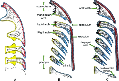
According to the classical theory, teeth derive from odontodes that invaded the oral cavity in conjunction with the origin of jaws (the 'outside in' theory). A recent alternative hypothesis suggests that teeth evolved prior to the origin of jaws as endodermal derivatives (the 'inside out' hypothesis). We compare the two theories in the light of current data and propose a third scenario, a revised 'outside in' hypothesis. We suggest that teeth may have arisen before the origin of jaws, as a result of competent, odontode-forming ectoderm invading the oropharyngeal cavity through the mouth as well as through the gill slits, interacting with neural crest-derived mesenchyme. This hypothesis revives the homology between skin denticles (odontodes) and teeth. Our hypothesis is based on (1) the assumption that endoderm alone, together with neural crest, cannot form teeth; (2) the observation that pharyngeal teeth are present only in species known to possess gill slits, and disappear from the pharyngeal region in early tetrapods concomitant with the closure of gill slits, and (3) the observation that the dental lamina (sensu Reif, 1982) is not a prerequisite for teeth to form. We next discuss the progress that has been made to understand the spatially restricted loss of teeth from certain arches, and the many questions that remain regarding the ontogenetic loss of teeth in specific taxa. The recent advances that have been made in our knowledge on the molecular control of tooth formation in non-mammalians (mostly in some teleost model species) will undoubtedly contribute to answering these questions in the coming years.
Figures
Comment in
-
'The integument story: origins, evolution and current knowledge'.J Anat. 2009 Apr;214(4):407-8. doi: 10.1111/j.1469-7580.2009.01051.x. J Anat. 2009. PMID: 19422422 Free PMC article. No abstract available.
References
-
- Adams AE. An experimental study of the development of the mouth in the amphibian embryo. J Exp Zool. 1924;40:311–379.
-
- Ahlberg PE, Smith MM, Johanson Z. Developmental plasticity and disparity in early dipnoan (lungfish) dentitions. Evol Dev. 2006;8:331–349. - PubMed
-
- Barlow LA, Northcutt RG. Embryonic origin of amphibian taste buds. Dev Biol. 1995;169:273–285. - PubMed
-
- Berman DS. A trimerorhachid amphibian from the upper Pennsylvanian of New Mexico. J Paleontol. 1973;47:932–945.
-
- Bjerring HC. A contribution to structural analysis of the head of craniate animals. Zool Scr. 1977;6:127–183.
Publication types
MeSH terms
LinkOut - more resources
Full Text Sources
