Review
. 2009 Apr;214(4):560-86.
doi: 10.1111/j.1469-7580.2009.01045.x.

Evolution of hard proteins in the sauropsid integument in relation to the cornification of skin derivatives in amniotes

Affiliations
Review

Evolution of hard proteins in the sauropsid integument in relation to the cornification of skin derivatives in amniotes

Lorenzo Alibardi et al. J Anat. 2009 Apr.

Abstract

Hard skin appendages in amniotes comprise scales, feathers and hairs. The cell organization of these appendages probably derived from the localization of specialized areas of dermal-epidermal interaction in the integument. The horny scales and the other derivatives were formed from large areas of dermal-epidermal interaction. The evolution of these skin appendages was characterized by the production of specific coiled-coil keratins and associated proteins in the inter-filament matrix. Unlike mammalian keratin-associated proteins, those of sauropsids contain a double beta-folded sequence of about 20 amino acids, known as the core-box. The core-box shows 60%-95% sequence identity with known reptilian and avian proteins. The core-box determines the polymerization of these proteins into filaments indicated as beta-keratin filaments. The nucleotide and derived amino acid sequences for these sauropsid keratin-associated proteins are presented in conjunction with a hypothesis about their evolution in reptiles-birds compared to mammalian keratin-associated proteins. It is suggested that genes coding for ancestral glycine-serine-rich sequences of alpha-keratins produced a new class of small matrix proteins. In sauropsids, matrix proteins may have originated after mutation and enrichment in proline, probably in a central region of the ancestral protein. This mutation gave rise to the core-box, and other regions of the original protein evolved differently in the various reptilians orders. In lepidosaurians, two main groups, the high glycine proline and the high cysteine proline proteins, were formed. In archosaurians and chelonians two main groups later diversified into the high glycine proline tyrosine, non-feather proteins, and into the glycine-tyrosine-poor group of feather proteins, which evolved in birds. The latter proteins were particularly suited for making the elongated barb/barbule cells of feathers. In therapsids-mammals, mutations of the ancestral proteins formed the high glycine-tyrosine or the high cysteine proteins but no core-box was produced in the matrix proteins of the hard corneous material of mammalian derivatives.


Figures
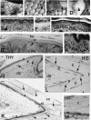
Fig. 1
Fig. 1
Macroscopic aspect of reptilian scales (A–D) and histology of the epidermis in scales of different reptiles (E–M). (A) Overlapped trunk scale of snake (Natrix natrix). Bar, 0.5 mm. (B) Little overlapping scale of ventral region of midtrunk region of the tuatara (Sphenodon punctatus). Bar, 0.5 mm. (C) Large dorsal scales (arrow) and lateral small scales (arrowhead) of the alligator (Alligator mississippiensis). Bar, 1 mm. (D) Plastron scale (gular, arrowhead) and the darker neck epidermis (arrow) of the turtle Emydura macquarii. The latter shows a rough surface with a scale-like pattern, represented by a rough skin. Bar, 1 mm. (E) Epidermis of the snake N. natrix showing differentiating fusiform beta-cells beneath the shedding line (arrow). Bar, 15 µm. (F) Normal epidermis (resting) of the lizard Podarcis sicula showing the outer beta-layer and the complete alpha-layer. Bar, 15 µm. (G) Epidermis of S. punctatus in resting phase showing the outer beta-layer and inner alpha layer (arrows). Bar, 15 µm. (H) Stratified wound epidermis of S. punctatus showing a thick corneous alpha-layer. Bar, 10 µm. (I) Epidermis of ventral scale in saltwater crocodile (Crocodylus porosus) showing flat cells in the transitional layer before the pale, lower part of the corneous layer. Bar, 15 µm. (J) Tail scale of the turtle Chrysemys picta showing thymidine-labeled nuclei (arrows) 1 day after injection and autoradiography. The arrowhead indicates the transitional layer of the epidermis in the outer scale surface. Bar, 20 µm. (K) Tip of a plastron scute of C. picta showing the shedding line (arrows) along which the outer part of the corneous layer will detach. Bar, 20 µm. (L) Tip of plastron scute of C. picta 3 days post-injection of tritiated histidine. Most autoradiographic labeling (arrows) is present in cells of the intermediate and transitional layer beneath the thick corneous layer. Bar, 20 µm. (M) Autoradiographic detail of the intense silver grains localized in the transitional layer of the carapace 1 day after injection of tritiated histidine in C. picta. Bar, 10 µm. a, alpha-layer; ba, basal layer; be, beta-layer; c, corneous layer; ci, inner corneous layer; co, outer corneous layer; de, dermis; e, epidermis; h, hinge region; in, intermediate layer beneath the wound epidermis; pc, pre-corneous or transitional layer; sb, suprabasal layer; t, tip of scute; tr, transitional layer; w, wound (regenerating) epidermis. Dashes underline the basal layer of the epidermis.
Fig. 2
Fig. 2
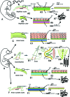
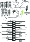
Drawing illustrating the embryonic layers formed on the scale of embryonic crocodilians (A) in comparison with those of bird embryos (B). The arrowed square illustrates the sequence of layers in crocodilian scale (A1) vs. avian beak (C), downfeather (D), claw (E), and scutate scales (F). The same colors indicate homologous layers, including those of downfeathers after the cell displacement within the barb ridge (see details in the text). The arrows pointing down in downfeathers indicate the direction of formation of barb ridges. Whereas feather-keratin form oriented and parallel bundles (arrow to D6), the other beta-keratins form an irregular orientation (arrows to A7, E3, F3. a, alpha (intermediate) layer; ADEI, areas of dermal–epidermal interaction; (B) Definitive (post-hatching and adult) beta-keratin layer containing different beta-keratins in scales, claws, beak and feather (colored with different colors); BA, barb (ramus); BL, barbule cells (equivalent to subperiderm cells); BR, barb ridges; BBK, adult beak beta-keratin with specific epitopes in black; CBK, adult claw beta-keratin with specific epitopes in black; CCBK, crocodilian claw beta-keratin; CSBK, crocodilian scale beta-keratin; CY, cylindrical cells; FBK, feather beta-keratin; FF, forming follicle; G, germinal layer; GFG, growing feather germ; HIS, tritiated histidine autoradiography; IP, inner periderm; OP, outer periderm; RB/PG, reticulate or periderm granules of the periderm; S, sheath (supportive cells corresponding to external layers derived from inner periderm cells); SP, subperiderm (containing the feather-keratin epitope, FBK); SBK, adult scale beta-keratin with specific epitopes in black; T, transitional layer along which the embryonic epidermis is shed around hatching (A3). THY, tritiated thymidine autoradiography; V, barb vane ridge cells of the axial plate of barb ridges (supportive cells equivalent to inner layers derived from inner periderm cells).
Fig. 3
Fig. 3
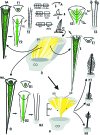
Hypothetical evolution of feathers and their keratins from reptilian scales (A), through coniform scales (B) and a long coniform proto-feather (C). The latter contained a specific feather-keratin box (FK-box) together with the ancestral core-box. The glycine-rich box (GG-box) was lost from B to C. The formation of barb ridges (D) shows the inside view of the base of a feather filament and the different cell displacement (E–G) gives rise to three different branching phenotypes (see text for explanation). Finally, the fusion of barb ridges with the rachidial ridge gives rise to pennaceous feathers (H1, contour feathers; H2, bristles; H3, filoplume). AX, axial plate; BA, barbula; BL, barbule; BR, barb ridge; CO, collar; NA, no axial plate; RA, ramus; RM, ramogenic zone; RR, rachidial ridge; SA, short axial plate.
Fig. 4
Fig. 4
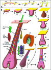
A) Schematic representation of the hypothesis of hair evolution following the reduction of areas of dermal–epidermal interactions (ADEI) (A 1–3). (A1) In a large synapsid scale the area of contact (red layer) between the active dermis (green beads) and epidermis was extended over most of the outer scale surface, forming expanded ADEIs. (A2) ADEIs become smaller and smaller in later mammal-like reptiles (therapsids), producing dermal condensation near the hinge region while scales became reduced or disappeared. The concentration of active dermal cells, a precursor of the dermal papilla, induced the proliferation of a rod of corneous material out of the hinge region, a proto-hair. (A3) In advanced therapsids or true mammals the formation of a (condensed) dermal papilla stimulated the production of longer rods of corneous cells, the hairs. (B) Schematic representation of hair morphogenesis (1–6) to the mature hair (7). From the linear epidermis (1) a hair placode is formed (2) that grows downward into a hair germ (3). The latter forms a hair peg (4 and 4′) that grows into a bulbous peg in which the active dermis starts to penetrate into its cup-like basal epithelium to form a dermal papilla (5–6). From the matrix cell precursor of the hair initially only trichocytic keratins synthesize, and later KAPs are formed at different levels of the differentiating cortical and cuticle cells. Also cells of the inner root sheath express specific keratins and later trichohyalin (7). AD, active dermis (competent to send inductive signals to the epidermis); ADEIs, areas of dermal–epidermal interactions; BU, bulge (stem cell repository); CL, companion layer; CO, cortical cells (containing long bundles of corneous proteins); CU, hair cuticle; DC, dermal condensation; DP, dermal papilla; EP, epidermis; EIRS, elongating inner root sheath; FDP, forming dermal papilla; FIRS, forming inner root sheath; GE, germinal epidermis (matrix); HCU, cuticle of the hair; HE, Henle's layer; HGT, high glycine tyrosine proteins; HL, hair-like primordia; HR, hair; HS, high sulfur proteins; S, high-sulfur proteins; HUX, Huxley layer; ICU, cuticle of the IRS (serrations correspond to those of the IRS); IK, intermediate filament keratins; IRS, inner root sheath; KAPs, keratin-associated proteins; MD, medulla cells; P, periderm; SG, sebaceous gland; SZ, sloughing zone; TH, trichohyalin; TK, trichocyte keratins; UD, undifferentiated dermis (does not produce signaling molecules); UHS, ultra-high sulfur proteins; VE, vesicles.
Fig. 5
Fig. 5
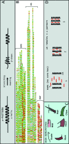
Mammalian proteins involved in cornification. (A) Alpha keratins that normally produce an alpha-pattern can be deformed under some conditions (stretching or steaming the corneous material above 70–80 °C). These conditions induce a change of secondary conformation, from alpha-helix to random coil and even beta-sheets, which produce a beta-keratin. (B) Some examples of mammalian KAPs are shown. The key amino acids are colored with different colors to illustrate the three main groups – HGT, HS and UHS matrix proteins. (C) Examples of the main mammalian skin derivatives containing trichocytic keratins and KAPs to form very hard corneous layers. (D) Schematic representation of the progressive phases (1–3) of association between coiled alpha-keratins (trichocytic) and KAPs. In phase 3, KAPs are probably regularly ordered around alpha-keratins and the resulting pattern at X-ray is indicated as alpha pattern.
Fig. 6
Fig. 6
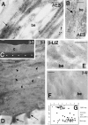
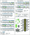
Localization of alpha-keratins (A–B) and beta-keratins (C–F) in reptilian corneous layers. (A) AE3-immunogold labeling (arrows) in lizard (Podarcis sicula) beta cells among pale beta-keratin bundles. Bar, 150 nm. (B) Detail of AE3-labeling over filaments (arrow) surrounding the pale beta-keratin bundle in a beta-cell of lizard (P. sicula). Bar, 100 nm. (C) Beta-1 immunolabeled, thick corneous layer of carapace scute in turtle (Chrysemys picta). Dashes underline the basal epidermis. Bar 10 µm. (D) Beta-1 gold immunolabeled corneous layer of scute of C. picta that disappears along the border with the underlying living keratinocyte (arrows). Bar, 250 nm. (E) Beta-lizard (A68B-antibody) immunolabeled keratin bundles in a beta-cell of P. sicula. Bar, 250 nm. (F) Beta-universal (β-U) labeling over keratin bundles of a differentiating beta-cell in snake (Natrix natrix). Bar, 250 nm. (G) Schematic general pattern of epidermal proteins from the epidermis of different species of reptiles (lizard, gecko lizard, turtle, alligator, crocodile and snake) showing the relative amount of alpha- vs. beta-keratins. AE3, immunolabeling positive for alpha-keratin AE3; be, beta-keratin bundles; β1, immunolabeling for beta-keratins using the chick scale β1 antibody; c, corneous layer.
Fig. 7
Fig. 7
Genomic organization (A–F) and examples of nucleotide structure (G–I) of avian and reptilian beta-keratin genes. A cluster of genes coding for related proteins is localized in a chromosome locus (A) and they are linearly arranged in multiple copies (B). In the genes of chick (chicken feather-keratin gene, AC J00847, and snake (Elaphe guttata,AC AM404188), a variably-long intron is present in the 5′-non coding region (C,F,G,H). In contrast, in the gecko lizard species studied so far (here exemplified by one sequence of Tarentola mauritanica, ACAM162665), no intron has been found (E,I). ATG (boxed) initiation codon; TAA or TAG (boxed), termination codon; AATAAA (boxed), polyadenylation signal. Uppercase letters, coding region; lowercase letters, 5′ and 3′ untranslated terminal regions; bold lowercase letters, intron sequence. The splice signals of introns are boxed and shaded.
Fig. 8
Fig. 8
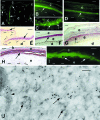
Examples of in situ hybridization localization using antisense probes for specific messengers coding for beta-keratins, and visualized by immunofluorescence (A–D,I), alkaline-phosphatase (E–H), or ultrastructural localization (M). (A) Regenerating scale of gecko (Tarentola mauritanica) with expression (arrows) in cells of the forming beta-layer (arrows). Bar, 10 µm. (B) Beta layer (arrow) of snake (Elaphe guttata). Bar, 10 µm. (C) transitional (arrow) and lower corneous layer of turtle (Pseudemys nelsonii). Bar, 10 µm. (D) transitional layer (arrow) of crocodile (Crocodylus niloticus). Bar, 10 µm. (E) beta-cell layers (arrows) of regenerating scales in lizard (Podarcis sicula). Bar, 20 µm. (F) beta-layer cells (arrow) in scales in renewal stage of snake (E. guttata). Bar, 10 µm. G, fusiform cells of the transitional layer (arrow) of scutes in Chrysemys picta. Bar, 10 µm. H, forming beta-layer (arrow) of scale in the tuatara(Sphenodon punctatus). Bar, 10 µm. I, detail on the spindle-shaped cells of the transitional layer of crocodile (C. niloticus). Bar, 10 µm. J, ultrastructural detail of the cytoplasm of differentiating beta-cell in lizard (P. sicula). Clusters of 5-nm diameter gold particles indicate the position of mRNAs among small keratin material (arrows). The arrowhead points to gold particles associated with a larger keratin bundle. Bar, 50 nm. a, alpha-layer; b, beta-layer; c, corneous layer; d, dermis; e, epidermis.
Fig. 9
Fig. 9
Examples of protein prediction using the PSIPRED Protein Structure Prediction Server at http://bioinf.cs.ucl.ac.uk/psipred/ for representative proteins of lizard, snake, crocodile, and turtle. The molecular weight and isoelectric point (pI) of these proteins are indicated. They all present inner amino acid regions with two close strands (beta-folded sheats) indicated as core-box (boxed). Note the variable extension of the alpha-helical region (green rods) in relation to the random-coiled regions (indicated with black lines).
Fig. 11
Fig. 11
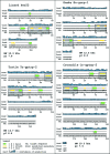
Examples of the specific amino acid sequences that characterize each group of reptiles. (Top panel) Comparative representation of some examples of proteins in which the position of three key amino acids such as tyrosine, valine and isoleucine is indicated. Tyrosine is mainly lateral (hydrophilic interactions?), whereas valine and isoleucine are more concentrated in the core-box and nearby regions, where they contribute to the formation of strand regions. (Other panels) Examples of group-specific (lepidosaurians vs. chelonians/arcosaurians) amino acid sequences found so far in pre- and post-core-boxes of sauropsids (see text).
Fig. 10
Fig. 10
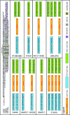
Comparative representation of some sKAPs from all different reptilian groups. Some key amino acids are specifically colored to reveal their preferred location within these proteins. Note in particular the conservation in the core-box region and the presence of cysteines mainly toward the N- and C-regions in all groups of reptiles and birds, where they probably intervene in cross-linking. Glycine-rich regions in HGPS proteins of lepidosaurians are present toward the N- and C-regions. Cysteine residues in lepidosaurian HCPS proteins are also present toward the N- and C-terminals. Serines are mainly present outside the core-box where they may be involved in phosphorylation or in other post-translational processes. In the chimeric-like chelonian/archosaurian proteins a cysteine-rich region is particularly evident, while the glycine-rich region is mainly present toward the C-terminus. Finally, the glycine-rich region disappears in the specialized, smallest feather proteins (FK). Goanna-claw (Inglis et al. 1987); Anolis Ker 20, 22, 23, 24, 25; Ge-gprp-1 (CAJ44302); Ge-gprp-3 (CAK19321); Ge-gprp-4 (CAJ90467); Ge-gprp-5 (CAK19322); Li-gprp-1 (CAJ67601); Li-gprp-3 (CAJ90483); Li-gprp-5 (CAJ90485); Sn-gprp-1 (CAL49457); Sn-gprp-5 (CA CAL51276); Sn-gcrp-1 (work in progress); Tu-gptrp-1 (CAO78677); Tu-gptrp-2 (CAO78678); Cr- gptrp-1 (CAO78748); Cr-gptrp-2 (CAO78749); Chick-scale (P04459), Chick-beak (AAO85139), Chick-claw (AAA62730), Chick-feather I–IV (P02450, P04458, P20307, P20308); Chick-keratinocyte (NP-001001310); Turkey-vulture-feather (Q98U06); Wood-stork-feather (Q98U05); Pigeon-feather (BAA33471); Duck-feather (PO8335).
None
Schematic drawing illustrating the synthesis of corneous material in beta-keratin cells of various skin derivatives in sauropsids. Beta-cells (B) of claw (A), scale (A1) and beak (A2) accumulate proteins with glycine-rich regions which produce irregularly organized filaments (B2) for high mechanical resistance. Only feather-keratins (A3) do not possess glycine-rich sequences. Beta-keratin filaments (in black in B3 and B4) increase among the decreasing alpha-keratin filaments. The molecular organization of keratin-associated proteins (KAPs) and histidine rich proteins (HRPs) among fibrous proteins is not known. The detail in B5 schematically shows the antiparallel overlapping of keratin monomers (see orientation of arrows inside the core-box) to form the framework of the protein polymer. Numerous inter- and intra-molecular disulfide bonds strengthen the stability of the filament/s.
Fig. 13
Fig. 13
Protein predictions using the PSIPRED Protein Structure Prediction Server at http://bioinf.cs.ucl.ac.uk/psipred/ of avian scale and claw keratins (both HTPS proteins) in comparison with the shorter feather protein (HCPS protein). The molecular weight, isoelectric point and type of proteins are indicated. The core-box is present in all these proteins despite the specific amino acid length and composition in other regions. The lack of a glycine-rich region in the feather protein allows its polymerization into long filaments/bundles tending to be parallel, as indicated in the lower right panel (compare Figs 13 and 12). The accumulation of corneous material mainly with a parallel orientation in feather cells during progressive stages of morphogenesis is indicated by the drawing in the bottom right panel. The small amount of alpha-keratin (represented by short coils) is rapidly replaced by the small feather protein (BFK). The detail in the drawing schematically shows the antiparallel overlapping of keratin monomers (see orientation of arrows in the core-box) to form the framework of feather-keratin polymer. Numerous inter- and intra-molecular disulfide bonds strengthen the stability of the filament(s). BL, barbule cells; BCC, barb cortical cells; BMC, barb medullary cells (become vacuolated); HCPS, high cysteine proline serine proteins; HRP, histidine-rich proteins; HTPS, high tyrosine proline serine proteins; HCPS, high cysteine proline serine protein. KAP, keratin-associated proteins; KB, keratin bundles; PF, parallel filaments.
Fig. 15
Fig. 15
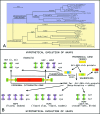
A) Summarizing cladogram showing the affinities of some representative of sKAPs in all groups (see text for further details). Note in particular the initial separation between lepidosaurian and chelonian/archosaurian proteins, and the following separation of HGPS from HCPS in lepidosaurian. In chelonian/archosaurians note the early separation between feather vs. non-feather proteins. The scale bar indicates the percentage of distance. (B) Hypothetical scheme of the point mutations in ancestral genes responsible for the transformation of a small glycine-serine-rich protein into mammalian or sauropsid KAPs, or histidine-rich proteins in arcosaurians (see text for a more detailed explanation). Whereas in sauropsids similar mutations invested a region that originated the core-box (green) in the lineage of synapsid-mammals, the mutations invested another region (blue) from which no core-box was (generally) formed. Also note that the mutated gene might or not belong to that coding for the variable regions of cytokeratins, assumed to be phylogenetically more ancient than KAPs. Finally, in the mammalian line, the enrichment of cysteine in the V1 and V2 regions of cytokeratins may also have contributed to the formation of the E-regions of trichocytic keratins.
Fig. 14
Fig. 14
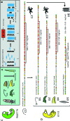
Skin derivatives of sauropsids (A), and schematic mechanism of formation of their hard corneous material (B). This occurs as in mammals (compare with panel D in Fig. 5) but, because of the core-box, sauropsid KAPs form themselves into filaments that replace most intermediate filaments. This process determines the prevalence of a beta-keratin pattern over the initial alpha-pattern. The embryo of a crocodilian (C) and its scale (C1) synthesize a protein with a long glycine-rich region that forms irregularly oriented corneous bundles (C2). (D) A bird embryo, with scale keratin (D1) and claw keratin (D2) with a shorter glycine-rich sequence. These keratins form irregular corneous bundles (D3) as for beak keratin (D4). (D5) The short feather-keratin where the glycine-rich regions have disappeared but corneous bundles parallel (D6). One possibility is that an ancestral, crocodilian-like protein gave rise (short arrow on the left) to proteins present in avian scales, claws and beak and, eventually, feathers. The other possibility (long arrow on the right) is that feather-keratin was directly derived from a large deletion of the glycine-rich region from an ancestral protein, and that a specific evolution gave rise to the feather-specific sequence. According to immunological studies (see Fig. 2) the feather-epitope may be very ancient in arcosaurian (embryonic) epidermis but is later eliminated in scale, claw or beak proteins. Color codes: Glycine (red), Serine (yellow), Cysteine (green), Proline (blue).

Comment in

References

    1. Alibardi L. Keratinization and lipogenesis in epidermal derivatives of the zebrafinch, Taeniopygia guttata castanotis (Aves, Passeriformes, Ploecidae) during embryonic development. J Morphol. 2002;251:294–308. - PubMed
    1. Alibardi L. Adaptation to the land: The skin of reptiles in comparison to that of amphibians and endotherm amniotes. J Exp Zoolog B Mol Dev Evol. 2003;298:12–41. - PubMed
    1. Alibardi L. Comparative aspects of the inner root sheath in adult and developing hairs of mammals in relation to the evolution of hairs. J Anat. 2004a;205:179–200. - PMC - PubMed
    1. Alibardi L. Dermo-epidermal interactions in reptilian scales: speculations on the evolution of scales, feathers, and hairs. J Exp Zoolog B Mol Dev Evol. 2004b;302:365–383. - PubMed
    1. Alibardi L. Fine structure and immunocytochemistry of monotreme hairs, with emphasis on the inner root sheath and trichohyalin-based cornification during hair evolution. J Morphol. 2004c;261:345–363. - PubMed

Publication types

MeSH terms

LinkOut - more resources