. 2009 Apr;214(4):607-19.
doi: 10.1111/j.1469-7580.2009.01052.x.

The anatomy and development of the claws of Xenopus laevis (Lissamphibia: Anura) reveal alternate pathways of structural evolution in the integument of tetrapods

Affiliations

The anatomy and development of the claws of Xenopus laevis (Lissamphibia: Anura) reveal alternate pathways of structural evolution in the integument of tetrapods

Hillary C Maddin et al. J Anat. 2009 Apr.

Abstract

Digital end organs composed of hard, modified epidermis, generally referred to as claws, are present in mammals and reptiles as well as in several non-amniote taxa such as clawed salamanders and frogs, including Xenopus laevis. So far, only the claws and nails of mammals have been characterized extensively and the question of whether claws were present in the common ancestor of all extant tetrapods is as yet unresolved. To provide a basis for comparisons between amniote and non-amniote claws, we investigated the development, growth and ultrastructure of the epidermal component of the claws of X. laevis. Histological examination of developing claws of X. laevis shows that claw formation is initiated at the tip of the toe by the appearance of superficial cornified cells that are dark brown. Subsequent accumulation of new, proximally extended claw sheath corneocyte layers increases the length of the claw. Histological studies of adult claws show that proliferation of cornifying claw sheath cells occurs along the entire length of the claw-forming epidermis. Living epidermal cells that are converting into the cornified claw sheath corneocytes undergo a form of programmed cell death that is accompanied by degradation of nuclear DNA. Subsequently, the cytoplasm and the nuclear remnants acquire a brown colour by an as-yet unknown mechanism that is likely homologous to the colouration mechanism that occurs in other hard, cornified structures of amphibians such as nuptial pads and tadpole beaks. Transmission electron microscopy revealed that the cornified claw sheath consists of parallel layers of corneocytes with interdigitations being confined to intra-layer contacts and a cementing substance filling the intercorneocyte spaces. Together with recent reports that showed the main molecular components of amniote claws are absent in Xenopus, our data support the hypothesis that claws of amphibians likely represent clade-specific innovations, non-homologous to amniote claws.


Figures

Fig. 1
Fig. 1
Distribution of claws among tetrapod taxa. The presence (+) or absence (−) of claws is mapped onto a phylogenetic tree that is based on San Mauro et al. (2005), with relationships among Sirenoidea being shown according to Frost et al. (2006).
Fig. 2
Fig. 2
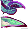
Comparison of the claws of Xenopus and Mus. (A) Longitudinal section of an adult mouse claw, and (B) longitudinal section of the claw of Xenopus laevis (used with permission from Maddin et al. 2007). The cornified layers of the epidermis form the claw sheaths in the mouse and in Xenopus. Scale bar 1 mm. Abbreviations for all figures: bg, basal germinative layer; d, dermis; ds, desmosome; g, gland; gm, germinative matrix region; is, intermediate spinous layers; le, living epidermal layers; lm, lateral margin; rl, replacement layer; mp, melanophore; msc, modified corneocytes of the corneous layer; tp, bony terminal phalanx; tp*, cartilaginous terminal phalanx; usc, unmodified corneocytes of the corneous layer.
Fig. 4
Fig. 4
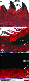
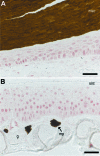
Close-up views of epidermis from forelimb and hindlimb digits showing details of the modified epidermis forming hard keratinous structures in X. laevis. (A) Epidermis from the ventral surface of a forelimb digit showing the dark hook-like nuptial structures comprising modified corneocytes (msc) with a similar distinctive, dark appearance to those of the cornified claw sheath in comparison with the adjacent unmodified corneocytes (usc). (B) Close-up view of a section through the epidermis of the dorsal surface at the base of the claw of X. laevis showing the abrupt change at the border between the modified claw sheath corneocytes and the adjacent unmodified corneocytes. (C) Close-up view of the claw region epidermis of X. laevis midway along the length of the claw, revealing the four epidermal layers typical of unmodified epidermis and the dramatically modified cornified layer. Masson's trichrome. Scale bars 50 µm.
Fig. 3
Fig. 3
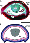
Cross-sectional view of the claw of Xenopus laevis. (A) Histological image of a slightly oblique cross-section showing the roughly ovoid shape of the cornified claw sheath. This section is near the proximal base of the cornified claw sheath, therefore remnants of the older unmodified corneocyte layers are present atop of the younger modified corneocyte layers of the claw sheath. (B) Schematic reconstruction of an ideal cross-section of the claw of X. laevis. The three corneocyte layers depicted (light, medium and dark blue) wrap around the lateral and medial surfaces of the digit tip as continuous layers. A pair of ventral ridges (asterisks) formed by the epidermis of the cornified claw sheath extend proximodistally along the length of the claw. Scale bar 1 mm.
Fig. 6
Fig. 6
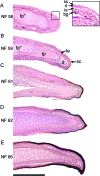
Longitudinal sections of the left pedal digit I showing the pattern of claw sheath growth in X. laevis. (A) There is no sign of a claw in NF stage 58 but the epidermis is locally stratified (inset of digit tip). (B) Modified claw sheath corneocytes appear at NF stage 59. (C–E) Sheath growth proceeds in the proximal direction by the addition of more proximally extending modified claw sheath corneocyte layers basal to the older layer and sheath thickness increases as corneocytes are retained. Haematoxylin and eosin. Scale bar 200 µm. Abbreviations in Fig. 2.
Fig. 5
Fig. 5
Developmental series of X. laevis from NF 58 stage (no claws present) to NF 66 stage (recently transformed froglet) shows the first appearance of cornified claw sheath and their subsequent growth. Upper row (A–G): preserved whole specimens of the stages documenting claw development. Scale bar 1 cm. Lower row: right pes in dorsal view of the stages illustrated in the upper row. Scale bar 3 mm.
Fig. 7
Fig. 7
Schematic illustration of the pattern of claw sheath development in X. laevis. (A–C) Successively older stages in the sheath development sequence. Initial differentiation of modified sheath-type corneocytes occurs at the distal tip of the digit (A, dark blue). Subsequent differentiation takes place underneath this layer and extends further proximally (B, medium blue), with successive additions extending even further proximally (C, light blue). Grey, terminal phalanx; pink, living epidermal layers; yellow, unmodified corneocytes of adjacent epidermis.
Fig. 8
Fig. 8
Immunostaining of proliferating cell nuclear antigen (PCNA) of the modified claw epidermis of X. laevis shows the proliferation of epidermal cells contributing to the cornified claw sheath is uniformly distributed underneath the entire length of the claw (A), a pattern like that observed in the unmodified epidermis of the digit (B). PCNA-positive nuclei are stained red. Note that haematoxylin counterstaining has been omitted. Scale bar 40 µm.
Fig. 9
Fig. 9
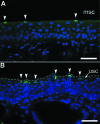
Detection of DNA fragmentation in the epidermis of X. laevis. Thin-sections of digits were subjected to transferase-mediated fluorescein-dUTP nick end labelling (TUNEL) to visualize DNA fragments. (A) TUNEL-positive DNA fragments (green nuclei, arrowheads) were seen in the replacement layer underneath the modified claw sheath epidermis, (B) as well as in replacement layer of the unmodified epidermis, sampled from a different animal. Absence of concomitant TUNEL-positivity in modified claw and unmodified epidermis of the same sample (not shown) may indicate that DNA breakdown occurs in a non-simultaneous manner at both sites. The thick corneous layer of the claw sheath occupies the entire black space at the top of panel (A). In (B), the superficial border of the epidermis is marked by a discontinuous line. Scale bar 40 µm.
Fig. 10
Fig. 10
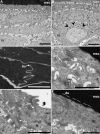
TEM micrographs of the modified claw and unmodified epidermis of X. laevis. Longitudinal section through the claw (A) and the tip of a non-clawed digit (B) comparing the overall epidermal organization. Large mucous granules (arrowheads) are more prevalent in unmodified epidermis than in the modified claw region epidermis (scale bars 5 µm). (C) Close-up of the modified claw sheath corneocytes. Highly interdigitated cell borders are restricted to the lateral borders of the corneocytes of the cornified claw sheath and resemble those seen in the replacement layer under claw (D), and the corneous layer of unmodified digit epidermis (E). (F) The replacement layer and claw are held in close contact by numerous, elongate desmosomes. Scale bars 1 µm. Abbreviations in Fig. 2.

Comment in

Similar articles

Cited by

References

    1. Alibardi L. Keratinization in the epidermis of amphibians and the lungfish: comparison with amniote keratinization. Tissue Cell. 2001;33:439–449. - PubMed
    1. Alibardi L. Ultrastructural localization of histidine-labelled interkeratin matrix during keratinization of amphibian epidermis. Acta Histochemica. 2003;105:273–283. - PubMed
    1. Alibardi L. Microscopic analysis of lizard claw morphogenesis and hypothesis on its evolution. Acta Zool (Stockholm) 2008;89:169–178.
    1. Alibardi L, Toni M. Immuno-cross reactivity of Transglutaminase and cornification marker proteins in the epidermis of vertebrates suggests common processes of soft cornification across species. J Exp Zool (Mol Dev Evol) 2004;302B:526–549. - PubMed
    1. Alibardi L, Toni M, Valle LD. Hard cornification in reptilian epidermis in comparison to cornification in mammalian epidermis. Exp Dermatol. 2007;16:961–976. - PubMed

Publication types

MeSH terms

LinkOut - more resources