. 2009 Apr;214(4):620-43.
doi: 10.1111/j.1469-7580.2009.01068.x.

The structure of the cornified claw sheath in the domesticated cat (Felis catus): implications for the claw-shedding mechanism and the evolution of cornified digital end organs

Affiliations

The structure of the cornified claw sheath in the domesticated cat (Felis catus): implications for the claw-shedding mechanism and the evolution of cornified digital end organs

Dominique G Homberger et al. J Anat. 2009 Apr.

Abstract

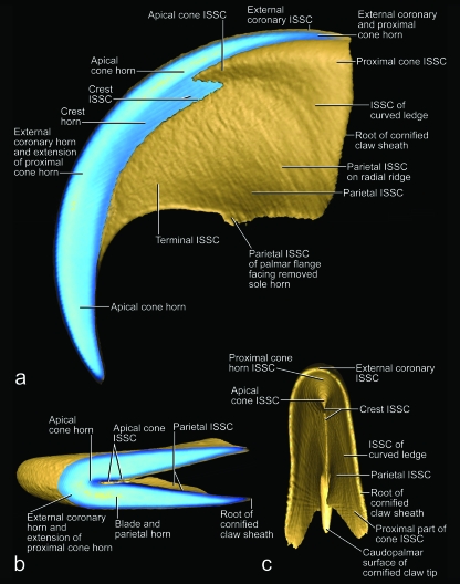
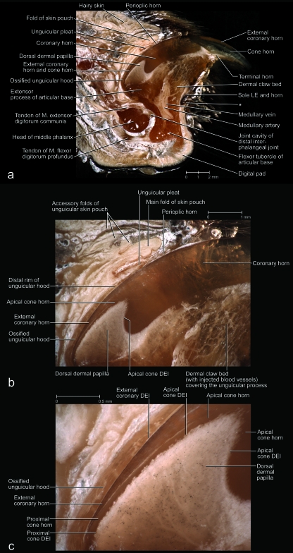
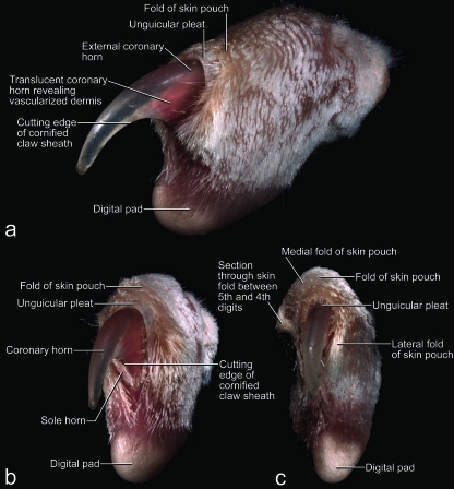
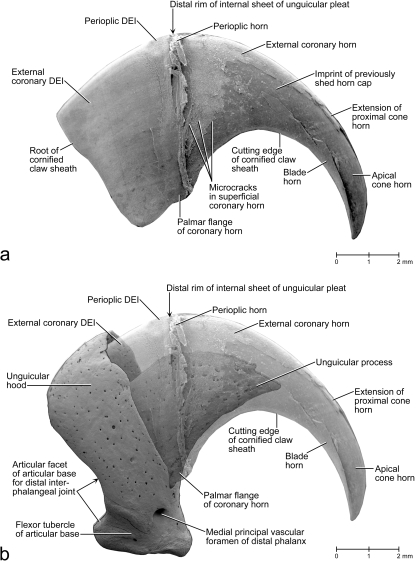
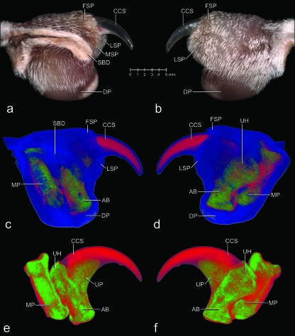
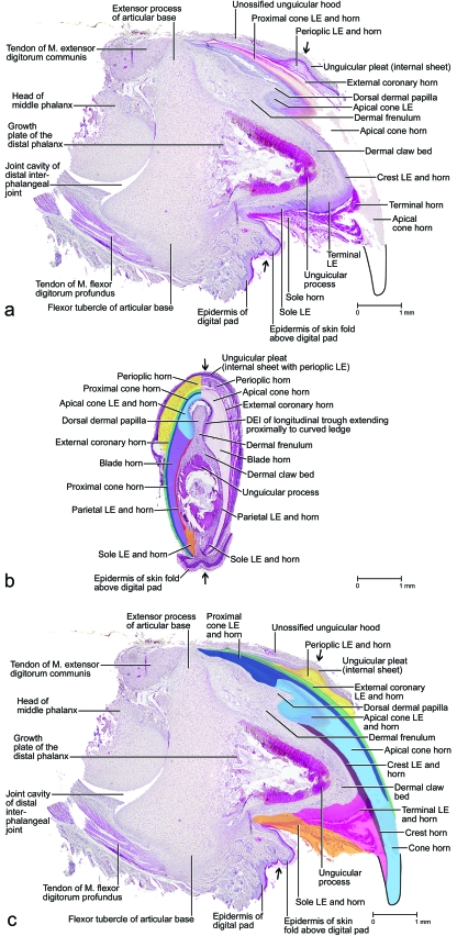
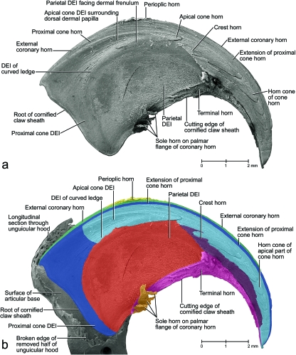
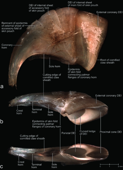
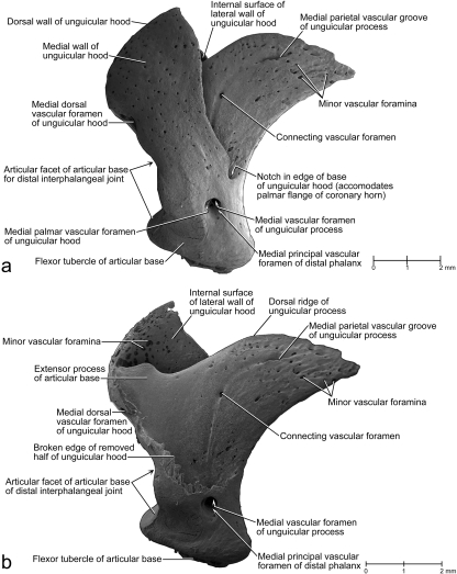
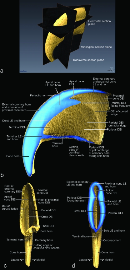
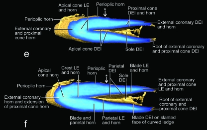
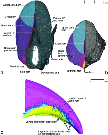
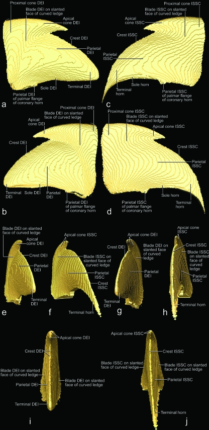
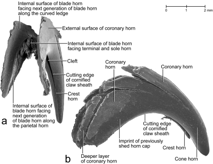
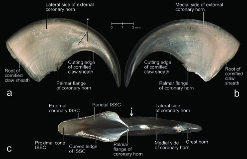
The morphology of cornified structures is notoriously difficult to analyse because of the extreme range of hardness of their component tissues. Hence, a correlative approach using light microscopy, scanning electron microscopy, three-dimensional reconstructions based on x-ray computed tomography data, and graphic modeling was applied to study the morphology of the cornified claw sheath of the domesticated cat as a model for cornified digital end organs. The highly complex architecture of the cornified claw sheath is generated by the living epidermis that is supported by the dermis and distal phalanx. The latter is characterized by an ossified unguicular hood, which overhangs the bony articular base and unguicular process of the distal phalanx and creates an unguicular recess. The dermis covers the complex surface of the bony distal phalanx but also creates special structures, such as a dorsal dermal papilla that points distally and a curved ledge on the medial and lateral sides of the unguicular process. The hard-cornified external coronary horn and proximal cone horn form the root of the cornified claw sheath within the unguicular recess, which is deeper on the dorsal side than on the medial and lateral sides. As a consequence, their rate of horn production is greater dorsally, which contributes to the overall palmo-apical curvature of the cornified claw sheath. The external coronary and proximal cone horn is worn down through normal use as it is pushed apically. The hard-cornified apical cone horn is generated by the living epidermis enveloping the base and free part of the dorsal dermal papilla. It forms nested horn cones that eventually form the core of the hardened tip of the cornified claw. The sides of the cornified claw sheath are formed by the newly described hard-cornified blade horn, which originates from the living epidermis located on the slanted face of the curved ledge. As the blade horn is moved apically, it entrains and integrates the hard-cornified parietal horn on its internal side. It is covered by the external coronary and proximal cone horn on its external side. The soft-cornified terminal horn extends distally from the parietal horn and covers the dermal claw bed at the tip of the uniguicular process, thereby filling the space created by the converging apical cone and blade horn. The soft-cornified sole horn fills the space between the cutting edges of blade horn on the palmar side of the cornified claw sheath. The superficial soft-cornified perioplic horn is produced on the internal side of the unguicular pleat, which surrounds the root of the cornified claw sheath. The shedding of apical horn caps is made possible by the appearance of microcracks in the superficial layers of the external coronary and proximal cone horn in the course of deformations of the cornified claw sheath, which is subjected to tensile forces during climbing or prey catching. These microcracks propagate tangentially through the coronary horn and do not injure the underlying living epidermal and dermal tissues. This built-in shedding mechanism maintains sharp claw tips and ensures the freeing of the claws from the substrate.


Figures

Fig. 11
Fig. 11
Images of an isolated unpreserved hard-cornified claw sheath without living and soft-cornified epidermis of a cat (cat no. 45R, spayed adult female, fifth digit of right forepaw) reconstructed and visualized from x-ray computed tomography data (40 kVp polychromatic x-ray beam; field of view of 28 mm in diameter and 19 mm in height; voxel side length of 37 µm). Sectioned planes are shown in blue (although lighter shades are indicative of greater density, this relation is not absolute in this rendering because of distortions along the edges); surfaces are shown in gold. The actual three-dimensional profile of the internal surface of the stratum corneum (i.e. the interface between the living and cornified epidermal layers) (ISSC) may be easier to visualize if the figure is held upside-down because of an optical illusion. (a) Medial view of median section. (b) Oblique horizontal section at the level of the maximal extent of the dorsal dermal papilla. (c) Proximal view.
Fig. 3
Fig. 3
Mesoscopic digital photographs of lateral views of a slightly oblique paramedian section through the third digit with a retracted claw of a cat (cat no. 1, adult male, right forepaw). The vascular system was injected with white 100% BaSO4 to distinguish the vascularized dermis and bone from the avascular epidermis (some depressions and cavities were contaminated with BaSO4). The specimen was embedded in resin prior to sectioning. (a) Section through the entire digit to show the complex relationships among the various structural elements supporting the cornified claw sheath. Part of the coronary horn at the tip of the claw was broken off during the sectioning process, thereby revealing the underlying core of the tip of the cornified claw sheath. (b) Magnified section to show the relationships among the bony unguicular hood, unguicular pleat, unguicular skin fold, and cornified claw sheath. (c) Magnified section to show the relationships between the external coronary horn and proximal cone horn. Note the small dermal papillae of the cone dermo–epidermal interface (DEI) and the injected blood vessels within the ossified unguicular hood and dorsal dermal papilla. The white longitudinal lines within the coronary horn are microcracks that have become contaminated with BaSO4during the sectioning process. LE, living epidermis; *, skin fold connecting the palmar flanges of coronary horn.
Fig. 1
Fig. 1
Mesoscopic digital photographs of the unpreserved fifth digit in resting position with a maximally retracted (i.e. retroflexed or hyperextended) claw of a cat (cat no. 50, tricolored adult female, left forepaw) to show the configuration of the skin pouch and unguicular pleat in situ. The fold of the skin pouch was slightly pulled back to reveal the underlying unguicular pleat. The long hairs of the furry skin fold originally covered most of the claw; they were clipped with microscissors. (a) Dorsolateral view. (b) Distolateral view. (c) Distal view.
Fig. 6
Fig. 6
Scanning electron microscopy images of a lateral view of the cornified claw sheath (including soft-cornified horn and living epidermis) of a cat (cat no. 18, fifth digit of right forepaw). (a) Surface view of the extracted and isolated cornified claw sheath (including soft-cornified horn and living epidermis) to show the superficial microcracks leading eventually to the shedding of horn caps off the tip of the cornified claw sheath, as well as the claw portion hidden from view in vivo. (b) Lateral view of the translucent cornified claw sheath (including soft-cornified horn and living epidermis) and medial view of the isolated distal phalanx of a cat (cat no. 21, male, older than 22–24 months; fifth digit of right forepaw; see also Fig. 5) to show the topographical relationship between the cornified claw sheath and distal phalanx. The mixing of the views and specimens was necessitated by the scarcity of intact specimens. DEI, dermo–epidermal interface.
Fig. 2
Fig. 2
Images of the unpreserved fifth digit in resting position with a maximally retracted (i.e. retroflexed or hyperextended) claw of a cat (cat no. 50, tricolored adult female, left forepaw) to show the surface structures as well as the internal configuration of the cornified claw sheath and skeletal elements in situ. (a and b) Mesoscopic digital photographs. (a) Medial view. (b) Lateral view. (c–f) Images based on three-dimensional visualization of x-ray computed tomography data (40 kVp x-ray beam; field of view of 28 mm in diameter and 19 mm in height; voxel side length 37 µm). Red: Density of hard-cornified epidermis, as well as transitional tissue density between blue and green. Green: Density of bone. Blue: Density of soft tissue (living and soft-cornified epidermis, and dermis). (c and d) Soft tissues shown slightly transparent. (c) Medial view. (d) Lateral view. (e and f) Only bone and hard-cornified epidermis shown. (e) Medial view. (f) Lateral view. AB, articular base of distal phalanx; CCS, cornified claw sheath; DP, digital pad; FSP, fold of skin pouch; LSP, lateral fold of skin pouch; MP, middle phalanx; MSP, medial fold of skin pouch; SBD, section through the skin fold between the fifth and fourth digits; UH, unguicular hood of distal phalanx; UP, unguicular process of distal phalanx.
Fig. 4
Fig. 4
Microscopy images of stained histological sections of claws of a kitten (cat G, 10 days old, third or fourth digit of a forepaw) to show the tissue composition in situ. At this stage of development, only the unguicular process of the distal phalanx is ossifying, while the articular base is still cartilaginous and the unguicular hood is still fibrous. (a) Paramedian section at the level indicated by a pair of opposite arrows in (b); much of the apical part of the cone horn was torn away during the sectioning process due to its extreme hardness. (b) Transverse section at the level indicated by the pair of opposite arrows in (a); the epidermal elements of the cornified claw sheath are color-coded on the left half of the section. (c) Paramedian section at the level indicated by a pair of opposite arrows in (b); the epidermal elements of the cornified claw sheath are color-coded. DEI, dermo–epidermal interface; LE, living epidermis; M., Musculus.
Fig. 7
Fig. 7
Scanning electron microscopy images of the internal view of the medial half of the isolated cornified claw sheath (including the soft-cornified horn and living epidermis) of a cat (cat no. 18, fifth digit of right forepaw) to show the topography and architecture of the horn elements of the cornified claw sheath. The perioplic, external coronary, crest, terminal and sole horn, as well as the superficial parts of the proximal cone horn and the horn cones of the apical cone horn, are shown in section. The dermo–epidermal interface (DEI) of the cone, blade and parietal horn is shown in a surface view. The three-dimensional profile of the dermis can be inferred from the three-dimensional profile of the DEI. The microcracks between layers of sectioned horn segments develop during dehydration along stress lines that are dictated by the architecture of the horn material. (a) Isolated cornified claw sheath. The artifactual crack that is visible in the parietal DEI developed in the horn external to the parietal horn; it parallels the radial stress lines visible in the superficial coronary horn in Figs 8a, 11a,b. (b) Color-coded isolated claw sheath reinserted within the unguicular recess of the distal phalanx (after the removal of the medial half of the unguicular hood) of a cat (cat no. 21, male, older than 22–24 months; fifth digit of left forepaw; see Fig. 5b) to show the topographical relationships of the internal architecture of the cornified claw sheath and distal phalanx. The articular base of the distal phalanx was graphically cut along the root of the cornified claw sheath.
Fig. 8
Fig. 8
Mesoscopic digital photographs of the unpreserved isolated cornified claw sheath (including the living, horn-generating epidermis and parts of the epidermis of the unguicular pleat, skin pouch and skin fold connecting the palmar flanges of the coronary horn) of a cat (cat no. 24, Persian adult male, fifth digit of left forepaw). The frayed tip of the cornified claw sheath may be the result of the maceration procedure. The faint radial ridges of opaque tissue [visible in (a) and (b)], which diverge from the proximo-palmar end of the cornified claw sheath, are formed by the proximal cone epidermis underneath the external coronary horn. (a) Lateral view. (b) Latero-proximo-palmar view. (c) Proximo-palmar view. DEI, dermo–epidermal interface.
Fig. 5
Fig. 5
Scanning electron microscopy images of a medial view of the isolated distal phalanx of a cat (cat no. 21, male, older than 22–24 months; fifth digit of left forepaw) to show the complex structure of the distal phalanx and the dimension of the unguicular recess. (a) Intact distal phalanx. (b) Distal phalanx after the removal of the medial half of the unguicular hood.
Fig. 9
Fig. 9
Images of an unpreserved isolated cornified claw sheath (including the underlying living, horn-generating epidermis) of a cat (cat no. 24, Persian adult male, fifth digit of left forepaw) reconstructed and visualized from x-ray computed tomography data (40 kVp polychromatic x-ray beam; field of view of 28 mm in diameter and 19 mm in height; voxel side length of 37 µm). Section planes are shown in blue (although lighter shades are indicative of greater density, this relationship is not absolute in this rendering because of distortions along the edges); surfaces are shown in gold. The striations on the gold surfaces are artifacts. The epidermis of the unguicular pleat, skin pouch and skin fold connecting the palmar flanges of coronary horn was apparently not dense enough to absorb x-rays and be visualized. (a) Oblique rostro-dorso-lateral view with section planes that were applied in (b–f). (b) Medial view of median section. (c) Proximal view of the isolated cornified claw sheath. (d) Proximal view after a virtual transverse section at the level of the dermal frenulum. (e) Horizontal section at the level of the maximal extent of the dorsal dermal papilla. The space framed by the cone dermo–epithelial interface (DEI) would be filled by the dermal frenulum in the intact claw. (f) Horizontal section at the level just below the palmar edge of the base of the dorsal dermal papilla. LE, living epidermis; *, distal rim of the unguicular pleat.
Fig. 9
Fig. 9
Images of an unpreserved isolated cornified claw sheath (including the underlying living, horn-generating epidermis) of a cat (cat no. 24, Persian adult male, fifth digit of left forepaw) reconstructed and visualized from x-ray computed tomography data (40 kVp polychromatic x-ray beam; field of view of 28 mm in diameter and 19 mm in height; voxel side length of 37 µm). Section planes are shown in blue (although lighter shades are indicative of greater density, this relationship is not absolute in this rendering because of distortions along the edges); surfaces are shown in gold. The striations on the gold surfaces are artifacts. The epidermis of the unguicular pleat, skin pouch and skin fold connecting the palmar flanges of coronary horn was apparently not dense enough to absorb x-rays and be visualized. (a) Oblique rostro-dorso-lateral view with section planes that were applied in (b–f). (b) Medial view of median section. (c) Proximal view of the isolated cornified claw sheath. (d) Proximal view after a virtual transverse section at the level of the dermal frenulum. (e) Horizontal section at the level of the maximal extent of the dorsal dermal papilla. The space framed by the cone dermo–epithelial interface (DEI) would be filled by the dermal frenulum in the intact claw. (f) Horizontal section at the level just below the palmar edge of the base of the dorsal dermal papilla. LE, living epidermis; *, distal rim of the unguicular pleat.
Fig. 13
Fig. 13
Images of the tip of a claw of a cat (cat no. 12, adult, fifth digit of left forepaw) based on phase-contrast enhanced x-ray computed tomography data (20 keV monochromatic synchrotron x-ray beam; field of view of 7.19 mm in diameter and 5.7 mm in height; scintillator distance to specimen 330 mm; voxel side length 11.06 µm) and manual segmentation. (a and b) Transverse sections to show the development of microcracks through the coronary horn (including the external and internal coronary horn and blade horn). (a) Section at the level near the distal tip of the unguicular process. (b) Section at the level slightly distal to the distal tip of the unguicular process. (c) Visualization of a three-dimensional reconstruction of the distal one-third of the cornified claw sheath to show the surface configuration of horn blades that are separated by microcracks. The color scheme used in (c) does not correspond to the color scheme in (a), (b) and the other color-coded figures. LE, living epidermis.
Fig. 12
Fig. 12
Images of virtual casts of the internal volumes enclosed by isolated cornified claw sheaths of cats and reconstructed from x-ray computed tomography data (40 kVp polychromatic x-ray beam; field of view of 28 mm in diameter and 19 mm in height; voxel side length 37 µm) and processed with visualization software that was supplemented by external computational processing. The palmar surface of the virtual casts are artificial and do not represent the actual surfaces. (a, b, e, g and i) Three-dimensional profiles of the dermis reconstructed from virtual casts of the internal volume enclosed by the complete epidermis, i.e. the isolated cornified claw sheath with the underlying living, horn-generating epidermis (cat no. 24, Persian adult male, fifth digit of left forepaw). (c, d, f, h and j) Three-dimensional profiles of the living and soft-cornified epidermis reconstructed from virtual casts of the internal volume enclosed by the hard-cornified epidermis, i.e. the isolated hard-cornified claw sheath without living and soft-cornified epidermis (cat no. 45R, spayed adult female, fifth digit of right forepaw). (a and c) Medial views. (b and d) Lateral views. (e and h) Disto-medial views. (f and g) Disto-lateral views. (i and j) Distal views. DEI, dermo–epidermal interface; ISSC, internal surface of the stratum corneum (i.e. the interface between the living and cornified epidermal layers).
Fig. 14
Fig. 14
Scanning electron microscopy picture of shed horn caps off the tips of cornified claw sheaths of an adult female cat. (a) Proximal view (specimen SC no. 3). (b) Side view (specimen SC no. 5).
Fig. 10
Fig. 10
Mesoscopic digital photographs of an unpreserved isolated hard-cornified claw sheath without soft-cornified horn or living epidermis (cat no. 45R, spayed adult female, fifth digit of right forepaw). The radial ridges of opaque tissue [visible in (a) and (b)], which diverge from the proximo-palmar end of the cornified claw sheath, are formed by the proximal cone epidermis underneath the external coronary horn. (a) Lateral view. (b) Medial view. The curved ledge, as well as the blade horn growing distally from it, are visible through the translucent external coronary horn. (c) Palmar view. Notice the several blades of blade horn, especially on the lateral side of the cornified claw surface. ISSC, internal surface of the stratum corneum (i.e. the interface between the living and cornified epidermal layers); *, superficial layer of coronary horn to be shed eventually as part of a horn cap (see Fig. 14b).

Comment in

References

    1. Al-Bagdadi F. The integument. In: Evans H, editor. Miller's Anatomy of the Dog. 3rd edn. Philadelphia, Pennsylvania: W. B. Saunders Company; 1993. pp. 98–121.
    1. Banks WJ. Applied Veterinary Histology. Baltimore, Maryland: Williams & Wilkins; 1986.
    1. Boas JEV. Krallen (inkl. Nägel, Hufe, Klauen) In: Bolk L, Göppert E, Kallius E, Lubosch W, editors. Handbuch der vergleichenden Anatomie der Wirbeltiere. Vol. 1. Berlin: Urban & Schwarzenberg; 1931. pp. 521–544. Reprinted: Amsterdam: A. Asher & Co.
    1. Bragulla H, Ernsberger S, Budras K-D. On the development of the papillary body of the feline claw. Anat Histol Embryol. 2001;30:211–217. - PubMed
    1. Bragulla HH, Homberger DG. The role of the specific, profilaggrin-containing keratohyalin granules in the developing epidermis of the fetal horse hoof. Pferdeheilkunde. 2007;23:5–20.

Publication types