

Choroideremia: new findings from ocular pathology and review of recent literature
- PMID: 19422966
- PMCID: PMC2679958
- DOI: 10.1016/j.survophthal.2009.02.008
Choroideremia: new findings from ocular pathology and review of recent literature
Abstract
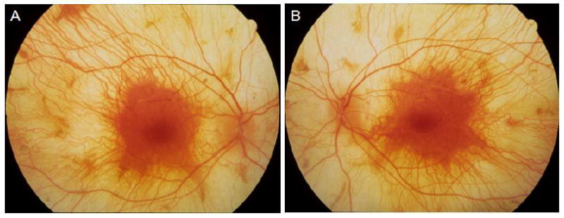
Histopathology of young individuals affected by choroideremia is rarely available to allow correlation with the clinical presentation. A 30-year-old man with choroideremia died in a motor vehicle accident and one eye was subjected to histopathological examination. Immunoblot analysis of protein derived from white blood cells of a living brother, also affected with choroideremia, confirmed the absence of Rab escort protein-1, the normal CHM gene product. Direct sequencing of the coding region and adjacent splice sites of the CHM gene was undertaken on genomic DNA from the living brother and revealed a transition mutation, C to T, in exon 6 (R253X) which resulted in a stop codon and was predicted to truncate the protein product. Histopathological examination of the eye of the deceased brother showed relative independent degeneration of choriocapillaris, retinal pigment epithelium, and retina, similar to observations in the mouse model of choroideremia. In addition, mild T-lymphocytic infiltration was found within the choroid. The ophthalmic features and the pathology of choroideremia are discussed in light of new findings in the current case.
Figures


References
-
- Bringmann A, Pannicke T, Grosche J, et al. Muller cells in the health and diseased retina. Prog Retin Eye Res. 2006;25:397–424. - PubMed
-
- Cameron JD, Fine BS, Shapiro I. Histopathologic Observations in choroideremia with emphasis on vascular changes of the uveal tract. Ophthalmology. 1987;94:187–96. - PubMed
-
- Chamberlain M, Baird P, Dirani M, Guymer R. Unraveling a complex genetic disease : age-related macular degeneration. Surv Ophthalmol. 2006;51:576–86. - PubMed
-
- Chan CC, Shen DF. Newer methodologies in immunohistochemistry and diagnosis. Dev Ophthalmol. 1999;31:1–13. - PubMed
Publication types
MeSH terms
Substances
Grants and funding
LinkOut - more resources
Full Text Sources
Research Materials
