. 2009 Aug;8(8):1972-87.
doi: 10.1074/mcp.M800495-MCP200. Epub 2009 May 7.

A protein interaction network for the large conductance Ca(2+)-activated K(+) channel in the mouse cochlea

Affiliations

A protein interaction network for the large conductance Ca(2+)-activated K(+) channel in the mouse cochlea

Thandavarayan Kathiresan et al. Mol Cell Proteomics. 2009 Aug.

Abstract

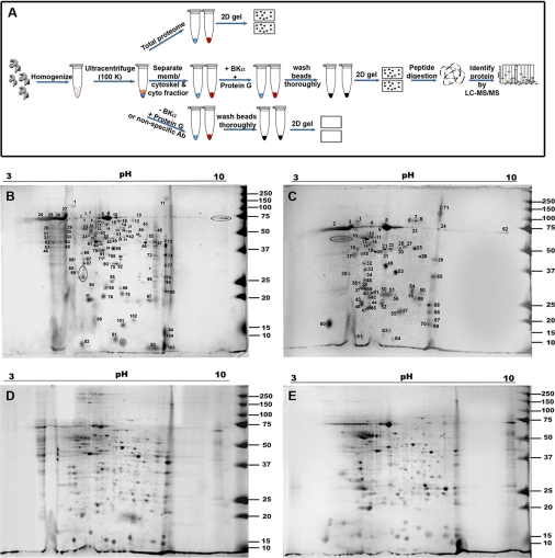
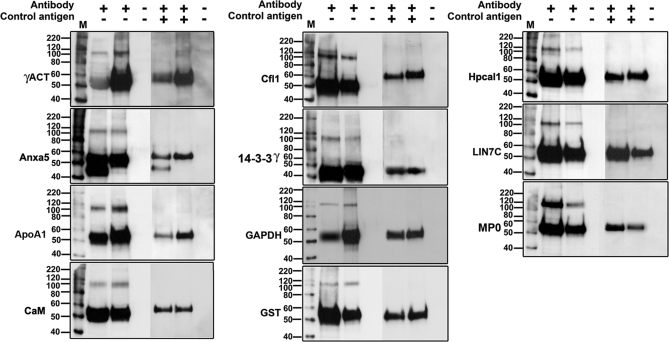
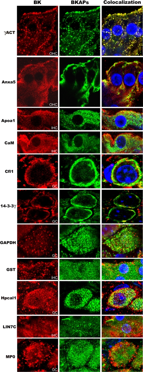
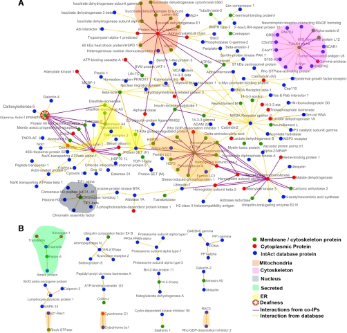
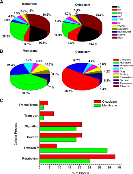
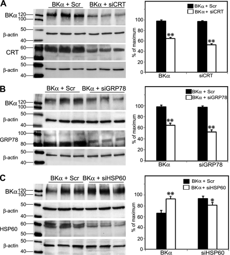
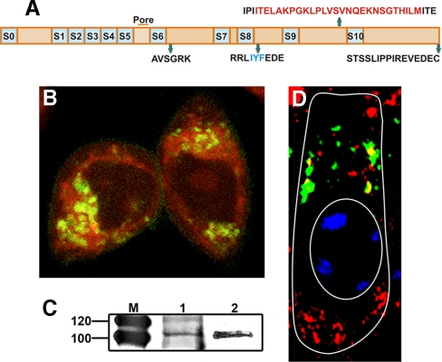
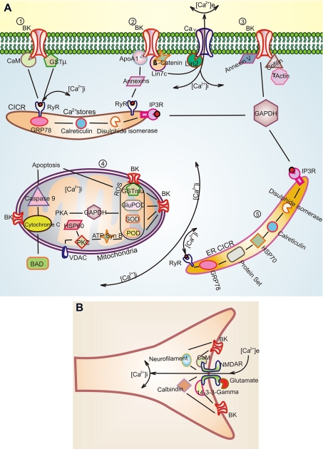
The large conductance Ca(2+)-activated K(+) or BK channel has a role in sensory/neuronal excitation, intracellular signaling, and metabolism. In the non-mammalian cochlea, the onset of BK during development correlates with increased hearing sensitivity and underlies frequency tuning in non-mammals, whereas its role is less clear in mammalian hearing. To gain insights into BK function in mammals, coimmunoprecipitation and two-dimensional PAGE, combined with mass spectrometry, were used to reveal 174 putative BKAPs from cytoplasmic and membrane/cytoskeletal fractions of mouse cochlea. Eleven BKAPs were verified using reciprocal coimmunoprecipitation, including annexin, apolipoprotein, calmodulin, hippocalcin, and myelin P0, among others. These proteins were immunocolocalized with BK in sensory and neuronal cells. A bioinformatics approach was used to mine databases to reveal binary partners and the resultant protein network, as well as to determine previous ion channel affiliations, subcellular localization, and cellular processes. The search for binary partners using the IntAct molecular interaction database produced a putative global network of 160 nodes connected with 188 edges that contained 12 major hubs. Additional mining of databases revealed that more than 50% of primary BKAPs had prior affiliations with K(+) and Ca(2+) channels. Although a majority of BKAPs are found in either the cytoplasm or membrane and contribute to cellular processes that primarily involve metabolism (30.5%) and trafficking/scaffolding (23.6%), at least 20% are mitochondrial-related. Among the BKAPs are chaperonins such as calreticulin, GRP78, and HSP60 that, when reduced with siRNAs, alter BKalpha expression in CHO cells. Studies of BKalpha in mitochondria revealed compartmentalization in sensory cells, whereas heterologous expression of a BK-DEC splice variant cloned from cochlea revealed a BK mitochondrial candidate. The studies described herein provide insights into BK-related functions that include not only cell excitation, but also cell signaling and apoptosis, and involve proteins concerned with Ca(2+) regulation, structure, and hearing loss.


Figures

Fig. 1.
Fig. 1.
Schema and results of BKα coIP assay. A, anti-BKα antibody bound to protein G beads was used to coimmunoprecipitate putative BKAPs from cochlear fractions. Resultant complexes were fractionated on a two-dimensional gel and analyzed using LC-MS/MS. Controls consisted of using membrane/cytoskeletal and cytoplasmic lysates in the absence of beads and antibody (total proteome), beads alone, or a nonspecific antibody (supplemental Fig. 1). Two-demensional gel electrophoresis of BKAPs for the membrane/cytoskeletal (B) and cytoplasmic fractions (C) shows 112 and 74 features resolved on these gels, respectively. All the numbered spots from the immunoprecipitated gels were subjected to LC-MS/MS analysis. Regions delimited by ovals represent proteins that are common to both fractions. Two-dimensional gel electrophoresis of the total proteome for the membrane/cytoskeletal (D) and cytoplasmic fractions (E) shows 322 and 282 visible features resolved on the gels, respectively.
Fig. 2.
Fig. 2.
Verification of BKAPs by reciprocal coIP. Eleven representative examples of BKAP reciprocal coIPs (lane 2; +,−) and BKα IPs (lane 3; +,−) reveal prominent immunoreactive peptide species of ∼110 kDa, the expected weight of the BK α-subunit. The negative control, in which anti-BKα antibody was pre-adsorbed with peptide, did not produce immunoreactive bands for either the BKAP reciprocal coIPs (lane 5; +,+) or the BKα IPs (lane 6; +,+). The ∼55 kDa bands correspond to heavy immunoglobulin (IgG), resulting from the use of polyclonal antibodies to both precipitate the proteins and probe the blots. Bead controls consisted of lysate mixed with protein G beads without antibody (lanes 4 and 7; −, −). The lane marked “M” is the molecular weight marker. BKAPs represented include γ-actin (γACT), annexin V (Anxa5), apolipoprotein A1 (ApoA1), calmodulin (CaM), cofilin (Cfl1), 14-3-3γ, GAPDH, GST, hippocalcin 1 (Hpcal1), Lin7 homolog c (LIN7C), and myelin P0 (MP0).
Fig. 3.
Fig. 3.
Coimmunolocalization of BKα and 11 BKAPs in various tissues from mouse cochlea. Cochlear tissues were triple-stained for BKα (red), BKAPs (green), DAPI (blue), with colocalization shown in orange or yellow. Immunoreactivity for BKAPs and BKα is shown in IHC, OHC, and ganglion cells (GC). BKAPs colocalized with BK include γ-actin (γACT), annexin V (Anxa5), apolipoprotein A1 (ApoA1), CaM, cofilin (Cfl1), 14-3-3γ, GAPDH, GST, hippocalcin 1 (Hpcal1), Lin7 homolog c (LIN7C), and myelin P0 (MP0).
Fig. 4.
Fig. 4.
The BKAP interactome of mouse cochlea. Visualization of primary and secondary BKAPs using Cytoscape revealed 13 networks involving 199 proteins and 254 interactions. A, of these proteins, 160 are nodes that are linked with 188 edges to form a single global network. Within this network are 12 major hubs consisting of a single node connected to six or more nodes that may or may not be linked to the larger network. The central nodes in these hubs include protein kinase C ε, α-tubulin, calmodulin, cytoplasmic actin, NMDA receptor, calreticulin, γ-actin, protein SET, ATP synthase β, ubiquitin, and chromobox homolog. B, the remaining BKAPs consist of 39 nodes and 28 edges that form 12 smaller distinct modules, each with five or fewer nodes. Different-colored nodes represent contributions from either membrane/cytoskeletal or cytoplasmic fractions or from the IntAct database source. Different-colored edges indicate interactions derived from the BKα coIP assays or from IntAct. Colored fields represent portions of the network that are located in different subcellular locations. BKAPs involved in deafness/NIHL are indicated by an additional symbol.
Fig. 5.
Fig. 5.
Primary BKAPs and their relation with ion channels, subcellular localization, and cellular process. A, charts show previous ion channel associations of BKAPs as reported in PubMed for membrane/cytoskeleton and cytoplasmic fractions of mouse cochlea. The two major ion channel groups that show previous associations with BKAPs are K+ and Ca2+ channels. However, ∼37% of the putative isolated BKAPs have a new association with BK. The remaining BKAPs interact with TRP, Na+, Cl, VDAC, aquaporin, nucleic acid, and cation channels. B, subcellular localization of BKAPs isolated from membrane/cytoskeletal and cytoplasmic fractions localized to various organelles and cellular matrices as determined by UniProtKB. Although a majority of BKAPs was found in the membrane and cytoplasm, a third major group was localized to the mitochondrion. Other proteins were dispersed among various organelles including ER, Golgi, and nucleus. C, BKAPs are classified according to the cellular process with which they are involved, as determined by mining Gene Ontology, GO Slim, and PubMed literature databases. A majority of BKAPS was involved in cellular processes related to metabolism and trafficking/scaffolding followed by development/differentiation, signaling, transport, and transcription/translation.
Fig. 6.
Fig. 6.
Regulation of BKα expression by chaperonins in CHO cells. A, mouse siRNAs were used to reduce the expression of endogenous calreticulin (CRT) in cells transfected with BK-DEC (BKα + SiCRT). Controls consisted of CHO cells treated with BK-DEC and scrambled RNAs (BKα + Scr). Plots derived from densitometry measurements made for BKα and CRT show that a >30% reduction in BKα coincided with an ∼50% reduction in CRT following 48 h of incubation. B, siRNAs reduced the expression of endogenous GRP78/BiP by ∼40%, which resulted in a >30% decrease in BKα. Measurements for BKα and GRP78 were made relative to controls consisting of cells transfected with BK-DEC and ScrRNA. C, in contrast to the previous chaperonins, cells treated with HSP60 siRNA showed an ∼13% reduction in HSP60 that resulted in a 26% increase in the expression of BKα. All lanes were loaded with equivalent amounts of protein that were calibrated as described under “Experimental Procedures.” Experiments were done in triplicate as independent samples for both si- and ScrRNA groups and β-actin served as a loading control for all experiments. Densitometry measurements for each band in a lane were normalized to the highest densitometric value (normalized to 100%) within a given set of six lanes, consisting of triplicates for Scr- and siRNA-treated cells. Statistical significance was determined using an unpaired, two-tailed t test to obtain *, p ≤ 0.05, **, p ≤ 0.001. Error bars represent the standard error of the mean.
Fig. 7.
Fig. 7.
Expression of mitochondrial BKα in vitro and in vivo. A, splice sequences shown in relation to the different regions of a BK-DEC variant cloned from mouse cochlea and used in CHO expression studies. B, live CHO cells transfected with pcDNA3.1 containing Cerulean in fusion with the C terminus of the BK-DEC variant and treated with MitoTracker (red) to identify mitochondria. Cerulean was pseudo-colored (dark green) to visualize overlap with red as yellow. Dark green immunostaining of BKα alone is observed at the plasmalemma. C, immunoprecipitation of BKα, using a pure mitochondrial preparation from mouse cochlea (left lane) and brain (right lane), shows bands at the expected weight of BK. D, tangential section of an outer hair cell, as outlined in white, showing the supranuclear region where BKα (red) is colocalized (yellow) in mitochondria with VDAC channels (green). The nucleus (oval) is partially stained with blue DAPI.
Fig. 8.
Fig. 8.
Putative BK-BKAP interactions derived from the BK interactome in mouse cochlea. A, 1), putative primary BKAPs at the membrane include calmodulin, GSTμ, 14-3-3, ApoA1, γ-actin, Lin7c, annexin V, cofilin. BK channels are known to have a functional link to RyR (30) and, thus, possibly to intracellular stores. This link may occur via CaM or GSTμ. Another putative link is through annexin, which is known to regulate intracellular Ca2+ stores (–35) and also binds to ApoA1 (39). ApoA1 alters the functional characteristics of BK. A, 2) Lin7, a primary BKAP, potentially interacts with BK as part of the Lin7·β-catenin complex. A BK link to Ca2+ channels in presynaptic sites may occur via the Lin7·Lin2 complex because Lin2 is known to interact with Ca2+ channels (46). A, 3) The cytoskeletal protein γ-actin and cofilin, a protein involved in disassembly of actin, are BKAPs that may link BK to the cytoskeleton. Thus, the link between BK and γ-actin may be disrupted in DFNA20/26, a mutation of γ-actin that causes deafness in humans. Protein 40 kDa and calreticulin are part of this matrix as determined from the interactome. A, 4) BKAPs associated with BKmito may play a role in regulating intracellular Ca2+ in mitochondria, thus influencing apoptosis and phosphorylation. This premise is based on the interaction with cytochrome C, ATP synthase, and GAPDH, which regulate mitochondrial Ca2+. BKα interactions with antioxidants such as SOD, GSTμ, and glutathione peroxidase (GluPOD) may be involved in mediating hair cell apoptosis initiated by the activation of ROS that can cause NIHL. The BKAP, cytochrome C, is shown in relation to BAD and Caspase-9, which are part of the hair cell apoptosis pathway (60). In addition, our data suggest HSP60 as a part of this pathway because it regulates BK expression inversely. A, 5) While the ER is another source for Ca2+, here BKAPs such as GRP78 and calreticulin regulate the folding and assembly of BK. B, NMDAR is a binary partner of BK, forming a major hub in the BK interactome. There is a functional relationship to BK as NMDARs are known to provide an extracellular source for Ca2+ that activates BK. BKAPs common to both BKα and NMDAR include the structural and signaling proteins, neurofilament, and 14-3-3, respectively, and the Ca2+-binding proteins, calbindin, and calmodulin.

Similar articles

Cited by

References

    1. Marty A. ( 1981) Ca-dependent K channels with large unitary conductance in chromaffin cell membranes. Nature 291, 497– 500 - PubMed
    1. Brainard A. M., Miller A. J., Martens J. R., England S. K. ( 2005) Maxi-K channels localize to caveolae in human myometrium: a role for an actin-channel-caveolin complex in the regulation of myometrial smooth muscle K+ current. Am. J. Physiol. Cell Physiol. 289, C49– C57 - PubMed
    1. Issa N. P., Hudspeth A. J. ( 1994) Clustering of Ca2+ channels and Ca(2+)-activated K+ channels at fluorescently labeled presynaptic active zones of hair cells. Proc. Natl. Acad. Sci. 91, 7578– 7582 - PMC - PubMed
    1. Robitaille R., Adler E. M., Charlton M. P. ( 1993) Calcium channels and calcium-gated potassium channels at the frog neuromuscular junction. J. Physiol. Paris 87, 15– 24 - PubMed
    1. Pattillo J. M., Yazejian B., DiGregorio D. A., Vergara J. L., Grinnell A. D., Meriney S. D. ( 2001) Contribution of presynaptic calcium-activated potassium currents to transmitter release regulation in cultured Xenopus nerve-muscle synapses. Neuroscience 102, 229– 240 - PubMed

Publication types

MeSH terms

Substances

Associated data