. 2009 May 26;106(21):8483-8.
doi: 10.1073/pnas.0812930106. Epub 2009 May 7.

The tail binds to the head-neck domain, inhibiting ATPase activity of myosin VIIA

Affiliations

The tail binds to the head-neck domain, inhibiting ATPase activity of myosin VIIA

Nobuhisa Umeki et al. Proc Natl Acad Sci U S A. .

Abstract

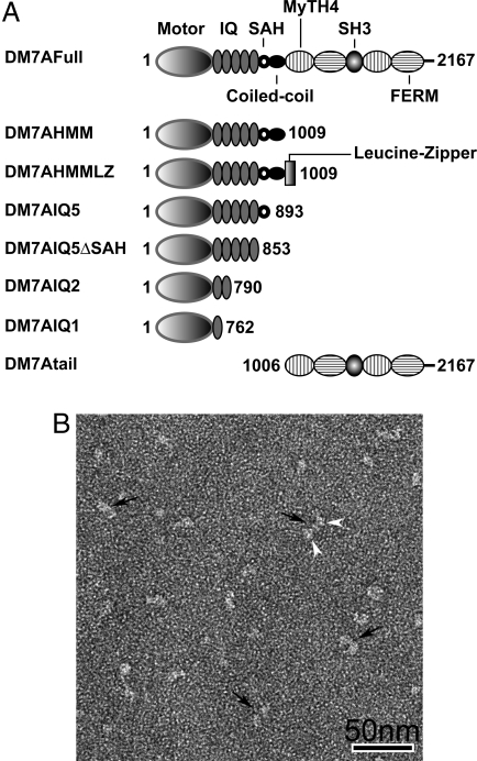
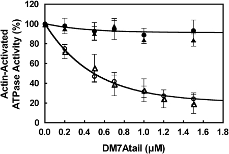
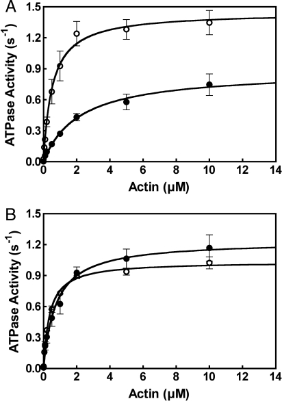
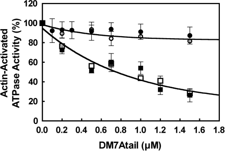
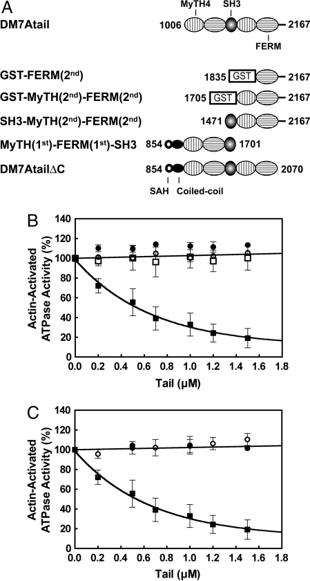
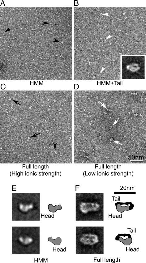
Myosin VIIA is an unconventional myosin, responsible for human Usher syndrome type 1B, which causes hearing and visual loss. Here, we studied the molecular mechanism of regulation of myosin VIIA, which is currently unknown. Although it was originally thought that myosin VIIA is a dimeric myosin, our electron microscopic (EM) observations revealed that full-length Drosophila myosin VIIA (DM7A) is a monomer. Interestingly, the tail domain markedly inhibits the actin-activated ATPase activity of tailless DM7A at low Ca(2+) but not high Ca(2+). By examining various deletion constructs, we found that deletion of the distal IQ domain, the C-terminal region of the tail, and the N-terminal region of the tail abolishes the tail-induced inhibition of ATPase activity. Single-particle EM analysis of full-length DM7A at low Ca(2+) suggests that the tail folds back on to the head, where it contacts both the motor core domain and the neck domain, forming an inhibited conformation. We concluded that unconventional myosin that may be present a monomer in the cell can be regulated by intramolecular interaction of the tail with the head.


Conflict of interest statement

The authors declare no conflict of interest.

Figures

Fig. 1.
Fig. 1.
DM7A constructs and subunit structure of myosin VIIA. (A) Schematic diagram of the DM7A constructs. The amino acid numbers of the constructs are indicated. (B) General appearance of DM7AHMMLZ, showing dimeric conformation. White arrowheads point to 2-pear shaped heads of individual molecules (black arrows). (Scale bar: 50 nm.)
Fig. 2.
Fig. 2.
Inhibition of the actin-activated ATPase activity of DM7AHMM by exogenous tail domain. Effect of exogenous tail domain (DM7Atail) on the actin-activated ATPase activity of DM7AHMMLZ and DM7AHMM is shown. ○, DM7AHMMLZ (1 mM EGTA); ●, DM7AHMMLZ (pCa4); △, DM7AHMM (1 mM EGTA); ▲, DM7AHMM (pCa4). Assay conditions are as described in Materials and Methods. Values are mean with SE from 4 independent experiments. One hundred percent activities in EGTA and pCa4 conditions were 1.4 and 1.1 s−1, respectively.
Fig. 3.
Fig. 3.
Decreased actin-activated ATPase activity of DM7AFull in EGTA but not in pCa4. The actin-activated ATPase activities of DM7AFull and DM7AHMM were measured as a function of actin concentration in EGTA (A) and pCa4 (B), respectively. ○, DM7AHMM; ●, DM7AFull. Values are mean with SE from 4 independent experiments. Assay conditions are as described in Materials and Methods.
Fig. 4.
Fig. 4.
Determination of the domain in DM7AHMM critical for the tail-induced inhibition of the actin-activated ATPase activity. Effect of exogenous tail domain on the actin-activated ATPase activity of various truncated DM7A constructs under EGTA conditions is shown. ○, DM7AIQ2; ●, DM7AIQ1; □, DM7AIQ5ΔSAH; ■, DM7AIQ5. Values are mean with SE from 4 independent experiments. One hundred percent represents as follows: DM7AIQ2, 1.8 s−1; DM7AIQ1, 1.7 s−1; DM7AIQ5ΔSAH, 1.3 s−1; DM7AIQ5, 1.5 s−1.
Fig. 5.
Fig. 5.
Determination of the regions in the tail domain critical for the inhibition of the actin-activated ATPase activity of DM7AHMM. (A) Schematic representation of the various tail domain constructs. (B) Effect of tail domain constructs, in which the N-terminal domains are truncated to various extent on the actin-activated ATPase activity of DM7AHMM under EGTA conditions is shown. ○, GST-MyTH(2nd)-FERM(2nd); ●, SH3-MyTH(2nd)-FERM(2nd); □, GST-FERM(2nd); ■, DM7Atail. (C) Effect of tail domain constructs, in which the C-terminal domains are truncated to various extent on the actin-activated ATPase activity of DM7AHMM under EGTA conditions is shown. ○, MyTH(1st)-FERM(1st)-SH3; ●, DM7AtailΔC; ■, DM7Atail. One hundred percent represents 1.4 s−1.
Fig. 6.
Fig. 6.
EM of DM7AHMM and DM7AFull in EGTA. (A and B) Negatively-stained fields of DM7AHMM in the absence and presence of the tail domain. Black and white arrowheads indicate individual HMM molecules and molecules interacting with the tail domain, respectively. (B Inset) Averaged image of head–tail interacting molecules (containing 37 individual images). (C and D) Fields of DM7AFull in 300 mM NaCl and 50 mM NaAcetate solution. Black and white arrows indicate the appearance of full-length molecules in the high and low ionic strength conditions, appearing narrower and wider, respectively. (E and F) Averaged images of HMM and full-length molecules (compare A and D, respectively). Averaged images in E show 2 different views (thought to represent molecules face-up and face-down on the carbon substrate), containing 104 images (Upper) and 80 images (Lower). Averaged images of full-length molecules in F, containing 21 images in each, were selected based on separation between the head and the tail domains in the closely-packed structures. Diagrams in E and F suggest possible interpretation of densities of the head (gray) and the tail domain (black), identified from the averages.

References

    1. Brown ME, Bridgman PC. Myosin function in nervous and sensory systems. J Neurobiol. 2004;58:118–130. - PubMed
    1. Mallik R, Gross SP. Molecular motors: Strategies to get along. Curr Biol. 2004;14:R971–R982. - PubMed
    1. Weil D, et al. Defective myosin VIIA gene responsible for Usher syndrome type 1B. Nature. 1995;374:60–61. - PubMed
    1. Hasson T, et al. Unconventional myosins in inner-ear sensory epithelia. J Cell Biol. 1997;137:1287–1307. - PMC - PubMed
    1. Liu X, Udovichenko IP, Brown SD, Steel KP, Williams DS. Myosin VIIa participates in opsin transport through the photoreceptor cilium. J Neurosci. 1999;19:6267–6274. - PMC - PubMed

LinkOut - more resources