. 2009 Jul 3;284(27):18302-10.
doi: 10.1074/jbc.M109.016717. Epub 2009 May 7.

PALB2 regulates recombinational repair through chromatin association and oligomerization

Affiliations

PALB2 regulates recombinational repair through chromatin association and oligomerization

Shirley M-H Sy et al. J Biol Chem. .

Abstract

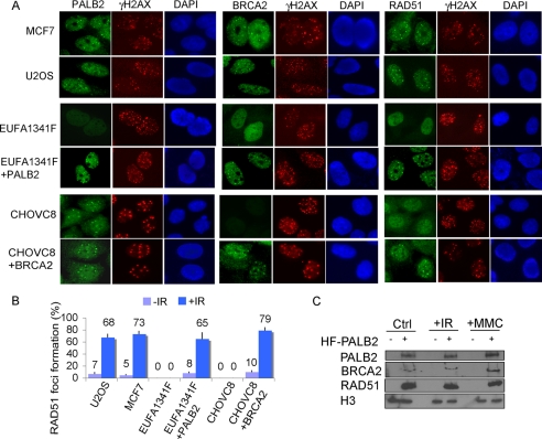
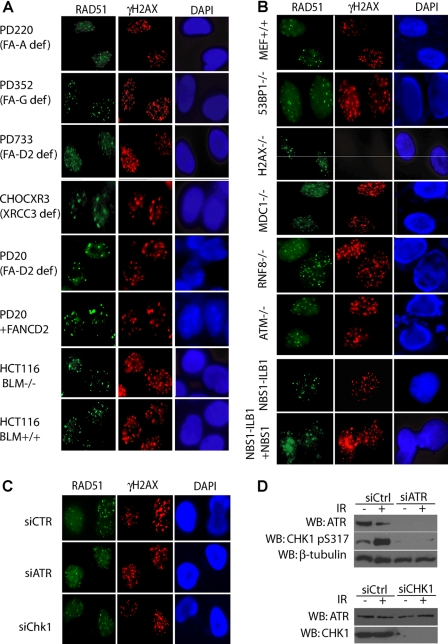
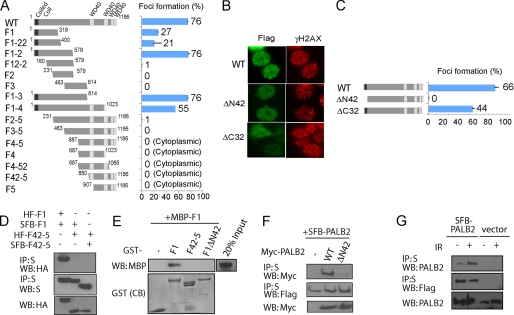
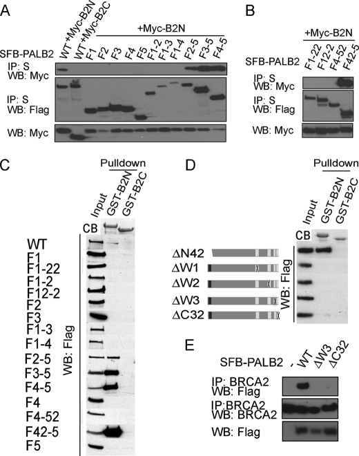
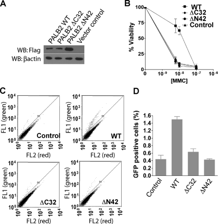
Maintenance of genomic stability ensures faithful transmission of genetic information and helps suppress neoplastic transformation and tumorigenesis. Although recent progress has advanced our understanding of DNA damage checkpoint regulations, little is known as to how DNA repair, especially the RAD51-dependent homologous recombination repair pathway, is executed in vivo. Here, we reveal novel properties of the BRCA2-associated protein PALB2 in the assembly of the recombinational DNA repair machinery at DNA damage sites. Although the chromatin association of PALB2 is a prerequisite for subsequent BRCA2 and RAD51 loading, the focal accumulation of the PALB2 x BRCA2 x RAD51 complex at DSBs occurs independently of known DNA damage checkpoint and repair proteins. We provide evidence to support that PALB2 exists as homo-oligomers and that PALB2 oligomerization is essential for its focal accumulation at DNA breaks in vivo. We propose that both PALB2 chromatin association and its oligomerization serve to secure the BRCA2 x RAD51 repair machinery at the sites of DNA damage. These attributes of PALB2 are likely instrumental for proficient homologous recombination DNA repair in the cell.


Figures

FIGURE 1.
FIGURE 1.
PALB2 controls focal accumulation of BRCA2 and RAD51 at DNA damage site. A, cells were irradiated with 10 Gy of ionizing radiation and recovered for 5 h. Immunofluorescence staining was performed using antibodies as indicated. C, PALB2-deficient EUFA1341 cells and its derivative (EUFA1341+PALB2) were treated with 10 Gy of ionizing radiation and recovered for 5 h or 1 mm MMC for 24 h. Cell lysates were separated by fractionation to retrieve both nuclear and chromatin-associated proteins. The presence of PALB2, BRCA2, and RAD51 was examined by Western blot analyses as indicated. B, RAD51 foci formation was examined in indicated cell lines using anti-RAD51 antibody. DAPI, 4′,6-diamidino-2-phenylindole.
FIGURE 2.
FIGURE 2.
RAD51 focus formation is not dependent on major DNA repair and DNA damage checkpoint pathway components. Cells deficient in various components in DNA repair (A) or DNA damage signaling and checkpoint pathways (B and C) were examined for the formation of ionizing radiation induced RAD51 foci. Cells were all collected 5 h after radiation (10 Gy), and immunostaining experiments were performed with anti-RAD51 and anti-γH2AX antibodies. D, expression levels of ATR and CHK1 after siRNA knockdown were monitored by Western blot (WB) analyses. DAPI, 4′,6-diamidino-2-phenylindole. siCTR, control siRNA.
FIGURE 3.
FIGURE 3.
PALB2 focus formation at DSBs requires its N terminus. A, HEK293T cells transfected with plasmids encoding SFB-tagged wild type (WT) or deletion mutants of PALB2 were exposed to 10 Gy of ionizing radiation. Cells were fixed, and immunostaining was performed with anti-FLAG and anti-pH2AX antibodies. The percentage of cells showing foci overlapping with γH2AX was plotted. B, foci accumulation of wild type and PALB2 internal deletion mutants. The percentage of foci positive cells was plotted in C. D, co-immunoprecipitation (IP) experiments using hemagglutinin (HA)-FLAG-tagged and SFB-tagged PALB2 F1 and F42–5 fragments were performed (5% input was showed). WB, Western blot. E, recombinant GST and MBP-tagged PALB2 fragments were subjected to pulldown assays. CB, Coomassie Blue staining. F, Myc-tagged wild-type and ΔN42 PALB2 mutant were subjected to co-immunoprecipitation experiments along with wild-type SFB-tagged PALB2 (5% input was showed). G, 293T cells stably expressing SFB-PALB2 or vector alone were mock-treated or irradiated. Cell lysates were subjected to S beads pulldown (5% input was showed). The amounts of SFB-tagged and endogenous PALB2 presented in the precipitates were determined by Western blotting analyses using anti-FLAG or anti-PALB2 antibodies.
FIGURE 4.
FIGURE 4.
PALB2 interacts with BRCA2 via its C-terminal WD40 repeats. A and B, SFB-tagged wild type and deletion mutants of PALB2 were expressed in HEK293T and subjected to co-immunoprecipitation (IP) with Myc-B2N or Myc-B2C. C, pulldown assays using GST-B2N or control GST-B2C purified from E. coli. Western blotting (WB) using anti-FLAG antibody was performed to verify the interaction between wild type or mutants of PALB2 with BRCA2. D, internal deletion mutants lacking each of the four WD40 domains at the C terminus of PALB2 and the N-terminal mutant of PALB2 with deletion of the coiled-coil domain were subjected to pulldown assays similar to that described in C. E, extracts prepared from 293T cells expressing SFB-tagged wild-type, ΔW3, or ΔC32 mutants of PALB2 were subjected to immunoprecipitation using anti-BRCA2 antibody. Western blotting experiments were conducted with anti-BRCA2 or anti-FLAG antibodies. B2N, BRCA2 N terminus; B2C, BRCA2 C terminus; CB, Coomassie Blue stain.
FIGURE 5.
FIGURE 5.
PALB2 elevates cell survival following DNA damage by restoring homologous recombination repair. A, EUFA1341 cells were transduced with vector control or HA-FLAG-tagged wild-type PALB2 (WT), the C-terminal deletion mutant (ΔC32), and the N-terminal deletion mutant (ΔN42). PALB2 expression was confirmed by Western blot (WB) as indicated. B, clonogenic assay after MMC treatment indicated that both the N-terminal and C-terminal regions of PALB2 are required for cell survival after MMC treatment. Results are the averages of three independent experiments. C and D, gene conversion in EUFA1341 reconstituted with wild-type or mutant PALB2. GFP-positive cells indicative of gene conversion (R1) were quantified by flow cytometry analysis. Results are the average of three independent experiments and were presented as mean ± S.E.

References

    1. Bartek J., Lukas C., Lukas J. ( 2004) Nat. Rev. Mol. Cell Biol. 5, 792– 804 - PubMed
    1. Kennedy R. D., D'Andrea A. D. ( 2006) J. Clin. Oncol. 24, 3799– 3808 - PubMed
    1. Wang W. ( 2007) Nat. Rev. Genet. 8, 735– 748 - PubMed
    1. Zhang H., Tombline G., Weber B. L. ( 1998) Cell 92, 433– 436 - PubMed
    1. Gudmundsdottir K., Ashworth A. ( 2006) Oncogene 25, 5864– 5874 - PubMed

Publication types

MeSH terms