

doi: 10.1074/jbc.M109.010744.
Epub 2009 May 7.
Neuroserpin polymers activate NF-kappaB by a calcium signaling pathway that is independent of the unfolded protein response
Affiliations
- PMID: 19423713
- PMCID: PMC2709363
- DOI: 10.1074/jbc.M109.010744
Item in Clipboard
Neuroserpin polymers activate NF-kappaB by a calcium signaling pathway that is independent of the unfolded protein response
J Biol Chem.
.
Abstract
The autosomal dominant dementia familial encephalopathy with neuroserpin inclusion bodies is characterized by the accumulation of ordered polymers of mutant neuroserpin within the endoplasmic reticulum of neurones. We show here that intracellular neuroserpin polymers activate NF-kappaB by a pathway that is independent of the IRE1, ATF6, and PERK limbs of the canonical unfolded protein response but is dependent on intracellular calcium. This pathway provides a mechanism for cells to sense and react to the accumulation of folded structures of mutant serpins within the endoplasmic reticulum. Our results provide strong support for the endoplasmic reticulum overload response being independent of the unfolded protein response.
Figures
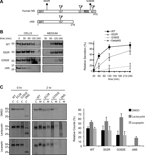
Characterization of PC12 Tet-On cell lines expressing neuroserpin variants. A, schematic representation of neuroserpin variants used in this study. Full-length human neuroserpin is 410 amino acids long, with N-linked glycans at positions 157, 321, and 401, a signal peptide (SP) and reactive center loop (RCL) as indicated. The polymerogenic mutants have amino acid substitutions at positions 52 (Ser to Arg) and 392 (Gly to Glu), respectively, and ΔNS has a premature stop codon at position 276. We generated PC12 Tet-On cell lines that stably expressed these constructs under the control of the tetracycline response element in the pTRE-Tight expression vector. B, secretion of neuroserpin variants. PC12 cells expressing WT, S52R, G392E, and ΔNS neuroserpin were metabolically labeled with 35S-Met/Cys for 15 min and chased for the times indicated. Neuroserpin was immunoprecipitated from cell lysates and culture medium, resolved by 10% w/v SDS-PAGE, and quantified by autoradiography using a PhosphorImager. The graph shows average ± S.D. for three independent repeats. C, intracellular degradation of neuroserpin variants. PC12 cells treated with DMSO (vehicle), 25 μm lactacystin (irreversible inhibitor of the proteasome), or 200 μm leupeptin (inhibitor of lysosomal proteases) were metabolically labeled and analyzed as described in B. The histograms show the amount of radiolabeled neuroserpin remaining after the chase normalized to the initial amount of neuroserpin for each condition, and values are averages ± S.E. of three independent repeats. Control experiments in PC12 cells with leupeptin and lactacystin showed the efficacy of the inhibitor treatments in blocking processing of pro-cathepsin D to mature cathepsin D and the degradation of cyclin B1 respectively (data not shown).
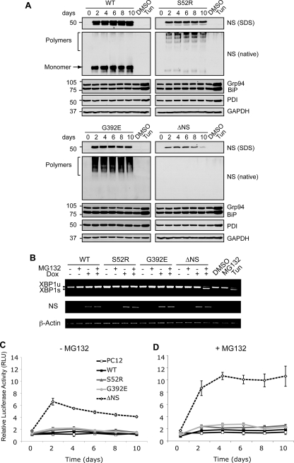
Accumulation of neuroserpin polymers within the ER does not activate the UPR. A, polymers of neuroserpin do not up-regulate ER luminal chaperones. Neuroserpin expression was induced in PC12 cells with 10 μg/ml doxycycline for 10 days, and cell lysates were resolved by 10% w/v SDS- and 7.5% w/v/non-denaturing PAGE. Western blot analysis for neuroserpin of SDS-PAGE (NS, SDS) revealed a 50-kDa band in WT, S52R, and G392E neuroserpin-expressing cells and 28 kDa in ΔNS neuroserpin cells. On non-denaturing PAGE (NS, native), WT neuroserpin migrated as a single monomer band (arrow), whereas S52R and G392E neuroserpin formed high molecular mass ladders that are characteristic of polymers (bracket). There was no detectable signal for ΔNS on non-denaturing PAGE. The expression levels of ER luminal chaperones regulated by the UPR (glucose regulated protein 94 (Grp94), immunoglobulin heavy chain-binding protein (BiP), and protein disulfide isomerase (PDI)) were determined in the same membranes by Western blotting. Treatment with 2 μg/ml tunicamycin (Tun) for 16 h compared with vehicle alone (DMSO) was used as a positive control for UPR induction, and protein loading was assessed by Western blot analysis for GAPDH. B, polymers of neuroserpin do not activate IRE1. The expression of WT, S52R, G392E, and ΔNS neuroserpin was induced for 4 days with 10 μg/ml doxycycline (Dox), and cells were either harvested or treated 16 h prior to RNA isolation with 100 nm MG132, a reversible inhibitor of the proteasome (46). XBP1 mRNA was amplified by PCR and resolved by 2% w/v TBE agarose gel electrophoresis. Unspliced XBP1 (XBP1u) and spliced XBP1 (XBP1s) products migrated at 486 and 457 bp, respectively. Neuroserpin (NS) and β-actin cDNAs were amplified to demonstrate transgene induction and as loading controls, respectively. PC12 cells treated with 2 μg/ml tunicamycin (Tun) were used as a positive control for UPR-induced XBP1 mRNA splicing. C and D, polymers of neuroserpin do not activate ATF6. Neuroserpin expression was induced in PC12 cell lines with 10 μg/ml doxycycline for 10 days. Twenty-four hours prior to lysis the cells were co-transfected with a plasmid encoding firefly luciferase under the control of a UPR response element (p5×ATF6-Luc) and with the transfection efficiency reporter pRL-TK Renilla luciferase. The graphs show firefly luciferase normalized to Renilla luciferase as averages ± S.D. of three repeats, and values are expressed in relative light units (RLU). Cells were mock-treated (C) or treated with 100 nm MG132 (D) for 16 h prior to harvesting.
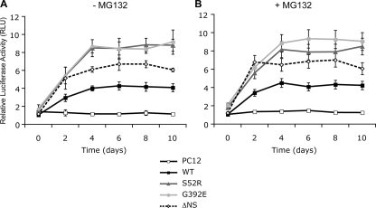
The ER accumulation of neuroserpin polymers activates NF-κB. Neuroserpin expression was induced in PC12 cell lines with 10 μg/ml doxycycline, and cells were harvested at 2-day intervals for up to 10 days. Twenty-four hours prior to collection cells were co-transfected with a plasmid encoding a NF-κB-driven luciferase reporter (pELAM1-Luc) and with the transfection efficiency reporter pRL-TK Renilla luciferase. The graphs show firefly luciferase normalized to Renilla luciferase as averages ± S.D. of three repeats, and values are expressed in relative light units (RLU). NF-κB was measured in cells mock-treated (A) or treated with 100 nm MG132 (B) for 16 h prior to harvesting.
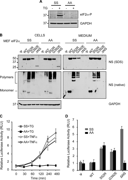
Neuroserpin polymers expressed in eIF2α S51A/A MEF cells can still activate NF-κB. Wild-type eIF2α S51S/S and PERK signaling-deficient eIF2α S51A/A MEF cells were transduced with pBABE retrovirus alone (ψ) or encoding WT, S52R, G392E, or ΔNS neuroserpin. A, only wild-type eIF2α S51S/S cells can phosphorylate eIF2α. eIF2α S51S/S and eIF2α S51A/A MEF cells were treated as indicated with 1 μm thapsigargin (TG) for 30 min and allowed to recover for 3 h in fresh medium. Cell lysates were resolved by 10% w/v SDS-PAGE and phosphorylated-eIF2α detected by Western blot analysis. GAPDH was used as a protein loading control. B, neuroserpin is similarly expressed and handled in eIF2α S51S/S and eIF2α S51A/A MEF cells. Cell lysates and culture media from puromycin-selected stable pools of MEF cells expressing neuroserpin variants were resolved by 10% w/v SDS or 7.5% w/v non-denaturing PAGE (native), followed by Western blot analysis for neuroserpin or GAPDH as a protein loading control. C, eIF2α S51A/A MEF cells fail to activate NF-κB upon UPR induction with thapsigargin. eIF2α S51S/S and eIF2α S51A/A MEF cells were co-transfected with the NF-κB (pELAM1-Luc) and pRL-TK reporter plasmids as described in Fig. 3. Cells were either treated with 1 μm thapsigargin (TG) for 30 min with recovery in fresh medium or 200 ng/ml TNF-α for the indicated times prior to lysis and luciferase assay. The graph shows firefly luciferase normalized to Renilla luciferase as averages ± S.D. of three repeats, and values are expressed in relative light units (RLU). D, eIF2α S51A/A MEF cells expressing polymer-forming mutants of neuroserpin can activate NF-κB. eIF2α S51S/S and eIF2α S51A/A cells expressing WT or mutant neuroserpin were co-transfected with the NF-κB (pELAM1-Luc) and pRL-TK reporter plasmids as described in Fig. 3, and the normalized activity of NF-κB was expressed in RLUs. The data are averages ± S.D. of six independent experiments.
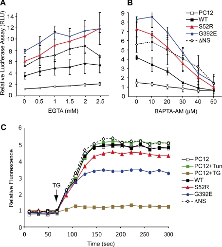
The activation of NF-κB by neuroserpin polymers depends on intracellular Ca2+. Neuroserpin expression in PC12 cells was induced for 5 days with 10 μg/ml doxycycline, cells were co-transfected with NF-κB (pELAM1-Luc) and pRL-TK reporter plasmids as described in Fig. 3, and the normalized activity of NF-κB was expressed in RLU. The culture medium was supplemented 16 h prior to harvesting with increasing concentrations of EGTA, pH 7.4 (A), a membrane-impermeable extracellular Ca2+ chelator or BAPTA-AM (B), a membrane-permeable intracellular Ca2+ chelator. The values are averages ± S.D. of three independent repeats. C, expression of neuroserpin in PC12 cell lines was induced for 5 days with 10 μg/ml doxycycline, or PC12 cells were treated with either 2 μg/ml tunicamycin (Tun) for 16 h or 1 μm thapsigargin (TG) for 30 min. All cells were subsequently loaded with Fluo-4 (Molecular Probes, Invitrogen). Intracellular Ca2+ mobilization was measured with an emission optical density of 488 nm over time with the addition of 1 μm thapsigargin (arrow), which induces Ca2+ efflux into the cytoplasm and excites the Fluo-4 chromophore. The data were collected using a FACSCalibur (BD Biosciences) flow cytometer and analyzed with FlowJo; a representative dataset of three independent repeats is shown.
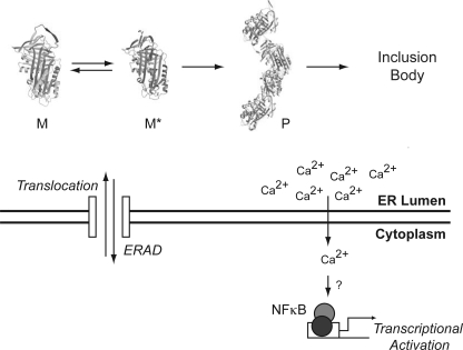
Molecular mechanism of NF-κB activation by serpin polymers. Newly synthesized serpin molecules enter the ER by co-translational translocation. Correctly folded molecules (native protein) are packaged into ER-to-Golgi transport vesicles for secretion (not shown), whereas folding intermediates and misfolded species are retained in the ER, where they can be targeted into the ERAD pathway. Some mutant serpins monomers (M) form homogenous ordered polymers (P) via partial RCL insertion intermediates (M*), which escape the ER quality control system and form inclusion bodies. These inclusion bodies are in effect “insoluble” to the cell and may no longer be seen by the canonical quality control systems. Accumulated polymers of mutant serpins can elicit the specific activation of the transcription factor NF-κB independently of the UPR by increasing cytosolic Ca2+.
References
-
- Carrell R. W., Lomas D. A. ( 1997) Lancet 350, 134– 138 - PubMed
-
- Lomas D. A., Carrell R. W. ( 2002) Nat. Rev. Genet. 3, 759– 768 - PubMed
-
- Sivasothy P., Dafforn T. R., Gettins P. G., Lomas D. A. ( 2000) J. Biol. Chem. 275, 33663– 33668 - PubMed
-
- Davis R. L., Shrimpton A. E., Holohan P. D., Bradshaw C., Feiglin D., Collins G. H., Sonderegger P., Kinter J., Becker L. M., Lacbawan F., Krasnewich D., Muenke M., Lawrence D. A., Yerby M. S., Shaw C. M., Gooptu B., Elliott P. R., Finch J. T., Carrell R. W., Lomas D. A. ( 1999) Nature 401, 376– 379 - PubMed
