. 2009 Jul 22;28(14):2042-52.
doi: 10.1038/emboj.2009.121. Epub 2009 May 7.

Site-specific binding of a PPR protein defines and stabilizes 5' and 3' mRNA termini in chloroplasts

Affiliations

Site-specific binding of a PPR protein defines and stabilizes 5' and 3' mRNA termini in chloroplasts

Jeannette Pfalz et al. EMBO J. .

Abstract

Chloroplast mRNA populations are characterized by overlapping transcripts derived by processing from polycistronic precursors. The mechanisms and functional significance of these processing events are poorly understood. We describe a pentatricopeptide repeat (PPR) protein, PPR10, whose binding defines mRNA segments derived from two transcription units in maize chloroplasts. PPR10 interacts in vivo and in vitro with two intergenic RNA regions of similar sequence. The processed 5' and 3' RNA termini in these regions overlap by approximately 25 nucleotides. The PPR10-binding sites map precisely to these overlapping sequences, and PPR10 is required specifically for the accumulation of RNAs with these termini. These findings show that PPR10 serves as a barrier to RNA decay from either the 5' or 3' direction and that a bound protein provides an alternative to an RNA hairpin as a barrier to 3' exonucleases. The results imply that protein 'caps' at both 5' and 3' ends can define the termini of chloroplast mRNA segments. These results, together with recent insights into bacterial RNA decay, suggest a unifying model for the biogenesis of chloroplast transcript populations and for the determinants of chloroplast mRNA stability.


Conflict of interest statement

The authors declare that they have no conflict of interest.

Figures
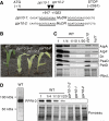
Figure 1
Figure 1
ppr10 mutants. (A) Transposon insertions in ppr10. The PPR10 coding region is indicated by a rectangle. Sequences flanking the insertions are shown below with the target-site duplications underlined. Polymorphisms in the terminal inverted repeats identify the insertions as related to the MuDR member of the Mu family. (B) Phenotypes of ppr10 mutants. Seedlings were grown for 8 days in soil. ppr10-1/-2 is the progeny of a complementation cross. (C) Immunoblot analysis of photosynthetic complex subunits. Immunoblots of leaf extracts were probed with antibodies to proteins indicated to the right; the Ponceau S-stained blot below illustrates sample loading and the abundance of RbcL, the large subunit of Rubisco. AtpA and AtpF are subunits of the CF1 and CF0 portions of the ATP synthase, respectively. D1, PsaD and PetD are subunits of photosystem II, photosystem I and the cytochrome b6f complex, respectively. hcf7 illustrates protein losses resulting from a global decrease in plastid translation (Barkan, 1993). (D) Immunoblot detection of PPR10 in leaf extract, showing antibody specificity and loss of PPR10 in ppr10 mutants. A full-colour version of this figure is available at The EMBO Journal Online.
Figure 2
Figure 2
PPR10 is localized to the chloroplast stroma. (A) Immunoblots of leaf and subcellular fractions. Chloroplast (Cp) subfractions were loaded on the basis of equal chloroplast number. Replicate blots were probed with antibodies to the proteins indicated at left. Cpn60, PetD, IM35 and PDH were used as markers for stroma (Str), thylakoid (Thy), envelope (Env) and mitochondria (Mito), respectively. (B) Size distribution of PPR10-containing particles. Stroma was incubated with ribonuclease A (RNAse) or mock-treated and fractionated by sedimentation through sucrose gradients. An equal volume of each gradient fraction was analysed by probing immunoblots with PPR10 antibody. The Ponceau S-stained blots illustrate the position of Rubisco (∼550 kDa). P, pelleted material.
Figure 3
Figure 3
Coimmunoprecipitation assays identifying RNAs associated with PPR10. (A) Summary of RIP-chip data. The median log2-transformed enrichment ratios (F635/F532) for two replicate PPR10 immunoprecipitations are plotted according to chromosomal position after subtracting the corresponding values for a control immunoprecipitation with OE16 antibody. Data points that show significant differential enrichment between the PPR10 and control assays (P-value <1E-4) are marked with diamonds and are annotated with the locus name. The underlying data for the highest-ranking fragments are summarized in Supplementary Table 1. (B) Validation of RIP-chip data. Immunoprecipitations were performed as for RIP-chip assays except that ribonuclease inhibitor was not included. One-sixth of the RNA from each immunoprecipitation pellet (P) and one-twelfth of the RNA from the corresponding supernatant (S) were applied to replicate slot blots. The two PPR10 immunoprecipitations (a and b) used sera from different immunized rabbits. Blots were probed with oligonucleotides specific for the atpH 5′ UTR, the psaJ 3′ UTR, the petA 5′ UTR and the coding regions of the other genes indicated.
Figure 4
Figure 4
Fine-mapping RNAs that coimmunoprecipitate with PPR10. (A, B) Replicate slot blots were prepared from immunoprecipitation pellet (P) and supernatant (S) RNAs, as described in Figure 3B. The positions of the 60-mer oligonucleotide probes are diagrammed, using line weights that reflect the degree to which corresponding RNAs were enriched in PPR10 immunoprecipitations. The most strongly enriched sequences are shown below, with the consensus residues shared by the atpH and psaJ sites highlighted in bold. The data were quantified with a phosphorimager, and are graphed at right. (C) Alignment of sequences near atpH and psaJ that coimmunoprecipitate with PPR10, with the consensus highlighted.
Figure 5
Figure 5
RNA gel blot analysis of atpH (A) and psaJ (B) RNAs in ppr10 mutants. Probes are diagrammed above the maps as black lines. Numbered probes correspond to the same 60-mer oligonucleotides used for slot blot hybridizations in Figure 4. Transcript maps were generated based on transcript size, the probes with which they hybridized and by the cRT–PCR data in Supplementary Table 2. RNAs missing in ppr10 mutants are in grey. Dashed lines indicate the spliced atpF intron. The band marked with an asterisk on the psaJ-6 blot derives from a prior probing with the petG probe. These transcript maps are consistent with and expand on those reported earlier (Stahl et al, 1993; Miyagi et al, 1998; Yamazaki et al, 2004); remaining ambiguities are indicated with question marks. All lanes in each panel come from the same gel.
Figure 6
Figure 6
Positions of PPR10-dependent RNA termini. (A) Primer extension mapping of the PPR10-dependent atpH-5′ end. A sequencing ladder generated with the same primer on an in vitro transcript is shown to the right. The U-tract disrupts the fidelity of reverse transcriptase, making the distal sequence ambiguous. (B) cRT–PCR procedure used to map transcript termini. Primer RP1 was used for reverse transcription. PCR was performed initially with RP1 and FP1 primers; nested PCR was then performed with RP1 and FP2. (C) Overlapping transcript termini in the atpI/atpH and psaJ/rpl33 intergenic regions. Residue numbers refer to position relative to the start codons of atpH and rpl33 or the stop codons of atpI and psaJ. Triangle size reflects relative transcript frequency, based on the data in Supplementary Table 2. Residues that are most highly conserved between the two sites are highlighted.
Figure 7
Figure 7
rPPR10 binds with specificity to the consensus sequences shared by the atpH 5′ UTR and psaJ 3′ UTR. (A) Elution of rPPR10 from a size-exclusion column. Column fractions were fractionated by SDS–PAGE and stained with Coomassie blue. The elution positions of β-amylase (200 kDa) and BSA (67 kDa) are shown. Fractions 10, 11 and 12 were pooled for binding assays. (B) RNA oligonucleotides used for binding assays. The atpH-a and psaJ-a sequences are shown below, aligned according to their consensus sequence. The positions of mapped RNA termini (see Figure 6) are marked with arrows. (C) Gel mobility shift assays. Binding reactions contained RNA at100 pM and the indicated concentration of rPPR10. The control RNAs (atpH-b and psaJ-b) were matched to the corresponding RNAs in length but migrated differently due to distinct structures.
Figure 8
Figure 8
Model linking mechanisms for intercistronic RNA processing and differential mRNA stability in chloroplasts. In this model, intercistronic processing and RNA degradation are both initiated by endonucleolytic cleavage of AU-rich sequences that are not masked by RNA structure, ribosomes, or proteins. The cleaved products are substrates for 3′ → 5′ and 5′ → 3′ exonucleases, which proceed until blocked by a strong RNA structure or bound protein. The ribonucleolytic activities are proposed to reside in homologues of the bacterial enzymes indicated in the key. Protective functions are attributed here to PPR proteins, but other protein classes could function analogously. The relative accumulation of the different transcripts is indicated by line thickness. Transcript level is proposed to be determined primarily by the accessibility of the AU-rich targets for RNAses E and J in untranslated regions, which determines both the rate of processing and the stabilities of the processed products. For example, ORF3 mRNA is most abundant because the PPR-B-binding site abuts the ribosome-binding site and the 3′ UTR is short and structured. Only a subset of potential processed RNAs is shown. A full-colour version of this figure is available at The EMBO Journal Online.

Comment in

References

    1. Asakura Y, Barkan A (2006) Arabidopsis orthologs of maize chloroplast splicing factors promote splicing of orthologous and species-specific group II introns. Plant Physiol 142: 1656–1663 - PMC - PubMed
    1. Barkan A (1993) Nuclear mutants of maize with defects in chloroplast polysome assembly have altered chloroplast RNA metabolism. Plant Cell 5: 389–402 - PMC - PubMed
    1. Barkan A (1998) Approaches to investigating nuclear genes that function in chloroplast biogenesis in land plants. Methods Enzymol 297: 38–57
    1. Barkan A, Walker M, Nolasco M, Johnson D (1994) A nuclear mutation in maize blocks the processing and translation of several chloroplast mRNAs and provides evidence for the differential translation of alternative mRNA forms. EMBO J 13: 3170–3181 - PMC - PubMed
    1. Beick S, Schmitz-Linneweber C, Williams-Carrier R, Jensen B, Barkan A (2008) The pentatricopeptide repeat protein PPR5 stabilizes a specific tRNA precursor in maize chloroplasts. Mol Cell Biol 28: 5337–5347 - PMC - PubMed

Publication types

MeSH terms

Associated data

LinkOut - more resources