

Comment
. 2009 May;15(5):483-4; author reply 484-5.
doi: 10.1038/nm0509-483.
Interleukin-17A is not expressed by CD207(+) cells in Langerhans cell histiocytosis lesions
- PMID: 19424201
- PMCID: PMC2820828
- DOI: 10.1038/nm0509-483
Item in Clipboard
Comment
Interleukin-17A is not expressed by CD207(+) cells in Langerhans cell histiocytosis lesions
Nat Med.
2009 May.
Abstract
Interleukin-17 (IL-17A) is a pro-inflammatory cytokine that has recently been implicated in pathogenesis of Langerhans Cell Histiocytosis (LCH), a potentially fatal disease characterized by lesions including CD207+ (langerin +) histiocytes. However, in this study we were unable to identify IL-17A gene expression in Langerhans cell lesions, and plasma levels of IL-17A did not correlate with disease activity. Therefore, this study does not support a central role for IL-17A in LCH pathogenesis.
Figures
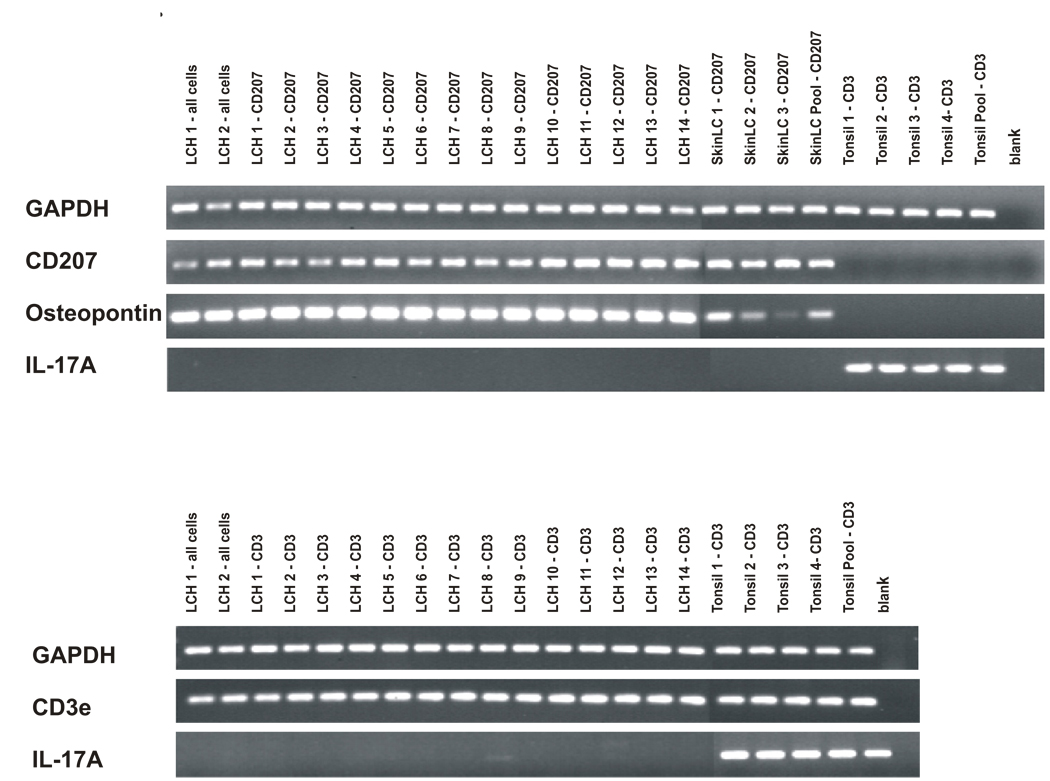
Upper Panel: Cells were isolated from LCH lesions and normal skin and tonsil control samples. After RNA extraction and cDNA amplification, PCR was performed using primers for GAPDH, CD207, osteopontin-1, and IL-17A. Samples were run on agarose gels stained with ethidium bromide, and PCR products were photographed. PCR primers are indicated along the left border. Sample description is detailed along the top of the figure. “All cells” indicates that all cells from that lesion were included in the sample. “CD207” indicates cells were sorted with antibody specific for LCs. “CD3” indicates cells were sorted with antibody specific for T cells. The control skin “LC Pool” contains RNA extracted from 20 different normal skin samples. The control “Tonsil Pool” contains RNA extracted from 20 different normal tonsil samples. Lower Panel: Cells were isolated from LCH lesions and normal tonsil control samples. PCR primers specific for GAPDH, CD3e and IL-17A were used.
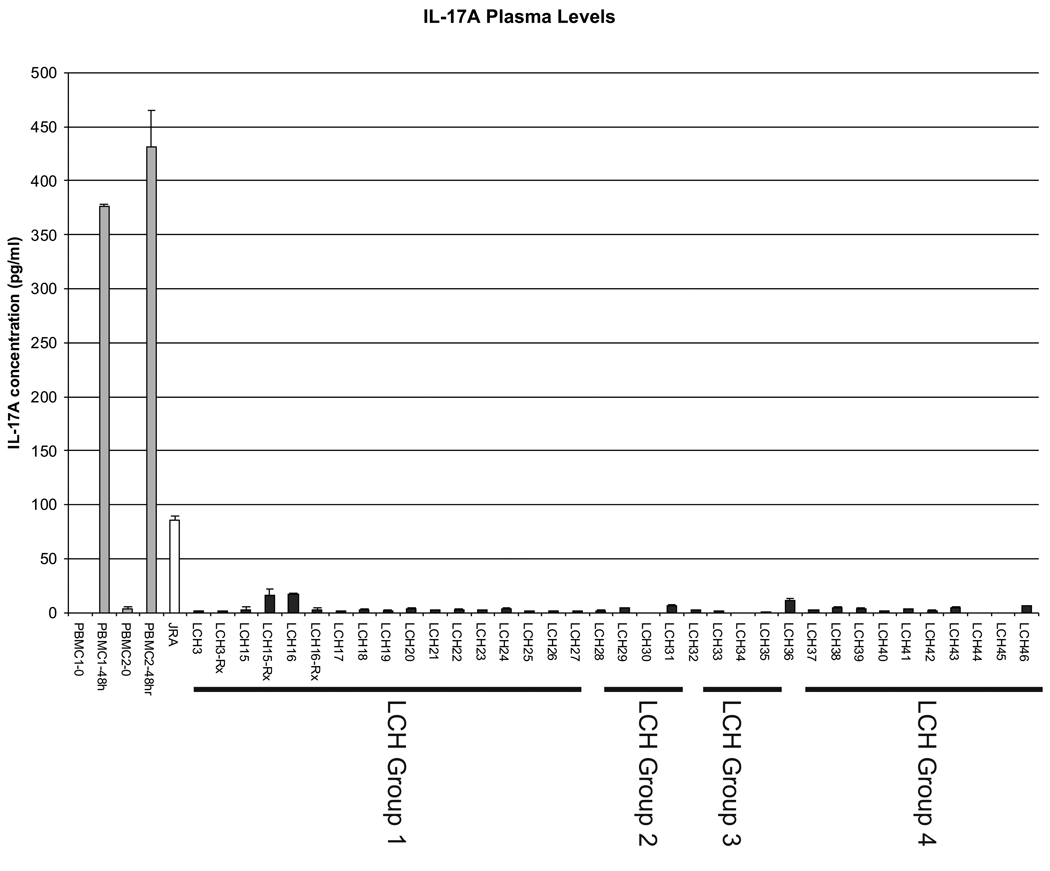
IL-17A plasma levels were determined using ELISA. As a control for the assay, media levels of IL-17A produced by peripheral blood mononuclear cells (PBMCs) from two healthy donors (PBMC1, PBMC2) after stimulation by PMA and ionomycin were used. A plasma sample from a patient with active JIA was also used as a positive control. The “LCH” number corresponds to the patient descriptions in Table 1. Group 1–3 correspond to clinical categories defined by the Histiocyte Society LCH-III protocol. Group 4 includes patients with single non-risk lesions.
Comment on
-
Langerhans cell histiocytosis reveals a new IL-17A-dependent pathway of dendritic cell fusion.Nat Med. 2008 Jan;14(1):81-7. doi: 10.1038/nm1694. Epub 2007 Dec 23. Nat Med. 2008. PMID: 18157139
References
-
- Arceci RJ. The histiocytoses: the fall of the Tower of Babel. Eur. J. Cancer. 1999;35:747–767. - PubMed
-
- McClain KL, Natkunam Y, Swerdlow SH. Atypical cellular disorders. Hematology. Am. Soc. Hematol. Educ. Program. 2004:283–296. - PubMed
-
- Coury F, et al. Langerhans cell histiocytosis reveals a new IL-17Adependent pathway of dendritic cell fusion. Nat. Med. 2008;14:81–87. - PubMed
-
- Dong C. TH17 cells in development: an updated view of their molecular identity and genetic programming. Nat. Rev. Immunol. 2008;8:337–348. - PubMed
Publication types
MeSH terms
Substances
Grants and funding
LinkOut - more resources
Full Text Sources
