. 2009 May;5(5):e1000423.
doi: 10.1371/journal.ppat.1000423. Epub 2009 May 8.

Implication of the mosquito midgut microbiota in the defense against malaria parasites

Affiliations

Implication of the mosquito midgut microbiota in the defense against malaria parasites

Yuemei Dong et al. PLoS Pathog. 2009 May.

Abstract

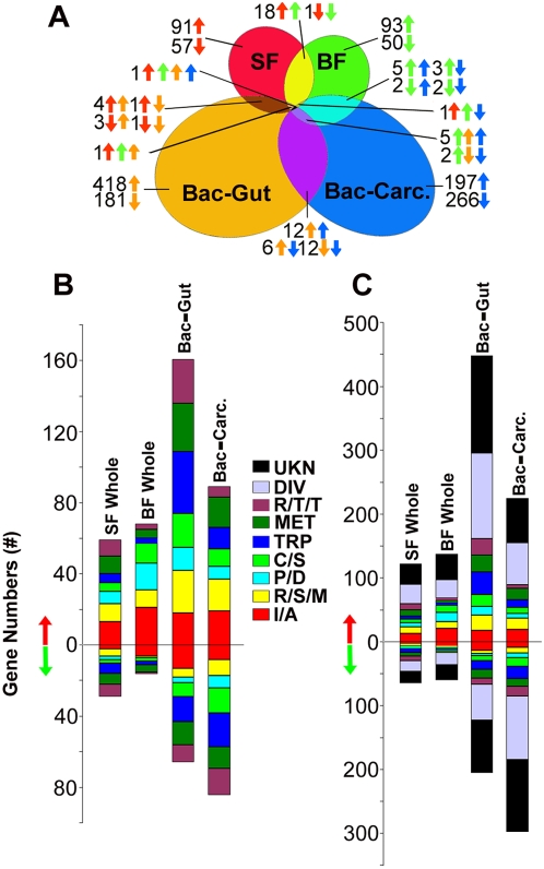
Malaria-transmitting mosquitoes are continuously exposed to microbes, including their midgut microbiota. This naturally acquired microbial flora can modulate the mosquito's vectorial capacity by inhibiting the development of Plasmodium and other human pathogens through an unknown mechanism. We have undertaken a comprehensive functional genomic approach to elucidate the molecular interplay between the bacterial co-infection and the development of the human malaria parasite Plasmodium falciparum in its natural vector Anopheles gambiae. Global transcription profiling of septic and aseptic mosquitoes identified a significant subset of immune genes that were mostly up-regulated by the mosquito's microbial flora, including several anti-Plasmodium factors. Microbe-free aseptic mosquitoes displayed an increased susceptibility to Plasmodium infection while co-feeding mosquitoes with bacteria and P. falciparum gametocytes resulted in lower than normal infection levels. Infection analyses suggest the bacteria-mediated anti-Plasmodium effect is mediated by the mosquitoes' antimicrobial immune responses, plausibly through activation of basal immunity. We show that the microbiota can modulate the anti-Plasmodium effects of some immune genes. In sum, the microbiota plays an essential role in modulating the mosquito's capacity to sustain Plasmodium infection.


Conflict of interest statement

The authors have declared that no competing interests exist.

Figures

Figure 1
Figure 1. Distribution of bacterial loads and major species composition of midguts microbiota in 5 individual laboratory-reared 5-d-old female A. gambiae mosquitoes from 5 consecutive generations.
The bacteria species were determined to be closely related to Enterobacter asburiae, Microbacterium sp., Sphingomonas sp., Serratia sp., and Chryseobacterium meningosepticum. G1 to G5 denotes generation 1st to 5th.
Figure 2
Figure 2. Antibiotic treatment eliminated the natural microbial flora from the mosquitoes' midguts.
(A) The bacterial staining of the midguts of septic mosquitoes (Untreated) and aseptic mosquitoes (Antibiotic-treated), arrows indicating the bacteria (upper panel). Lower panel shows the bacterial loads from the homogenates of midguts (Midgut) or whole mosquitoes (Whole) from septic (untreated) or aseptic (antibiotic-treated) mosquitoes that had fed on either sugar or uninfected blood. (B) Aseptic mosquitoes (antibiotic-treated) became more susceptible to P. falciparum infection compared to the control septic mosquitoes. The upper panel shows IFA (immuno-fluorescence assay) slides of oocysts in the midgut epithelium which were stained with anti-Pfs25 8 days post infection. The lower panel shows the ookinetes numbers in the midgut epithelium (28 hrs) and oocysts counts (10 days) in uninfected septic, aseptic (Antibiotic), and antibiotic-treated mosquitoes that had been re-challenged with natural floral bacteria (Rechallenge). Points indicate the absolute value of parasites counts in individual mosquitoes, and horizontal black bars in each column represent the median value of parasites from three replicates. A Kruskal-Wallis test was used to determine the significance of oocysts numbers (p<0.05 or p<0.01).
Figure 3
Figure 3. P. falciparum oocyst intensity in mosquitoes which had been co-fed with a mixture of live bacteria of E. coli and S. aureus (Ec/Sa) or heat-inactivated bacteria (HIA) in the blood meal, or mosquitoes that had been injected with live bacteria or heat-inactivated bacteria one day before the blood meal.
Mosquitoes that had co-fed or been pre-injected with PBS served as controls. Points indicate the absolute value of oocysts counts in individual mosquitoes, and horizontal black bars in each column represent the median value of oocysts from three replicates where the narrow black bars above or below the median values indicate the standard errors. p-value was calculated through a Kruskal-Wallis test. (A) Oocysts counts from P. falciparum infected midguts which had been co-fed with bacteria. (B) Oocysts counts from P. falciparum infected midguts which had been pre-injected with bacteria one day before the infected blood meal.
Figure 4
Figure 4. Ookinetes counts in the lumen or midgut epithelium of untreated septic, aseptic (Antibiotic), antibiotic treated mosquitoes that had been re-challenged with natural flora bacteria (Rechallenge), and mosquitoes that had been co-fed with live bacteria in the blood meal (Cofeeding) (upper panel).
Points indicate the absolute value of ookinetes counts in individual midguts, and bars represent the mean value of ookinetes from two replicates where the standard errors are included. An asterisk denotes p<0.01, and p-value was calculated by a Kruskal-Wallis test. Lower panel: immuno-fluoresence staining of ookinetes in the midgut lumen 24 hrs post infection where midgut homogenates were stained with anti-Pfs25 antibody followed by Alexa Fluor 488-conjugated (green) goat anti-mouse antibody staining. Scale bars: 5 µm.
Figure 5
Figure 5. Global gene regulation at the different conditions of infection.
(A) Comparison of transcript abundance between whole septic and aseptic mosquitoes after feeding on sugar (SF) or uninfected blood (BF), and in the midguts (Bac-Gut) or carcass tissues (Bac-Carc.) of mosquitoes 12 hrs post ingestion of uninfected blood supplemented with E. coli and S. aureus (substitution of bacteria with PBS as control). Colored arrows indicate genes that are up- or down- regulated in the corresponding treatment group. (B) Proportions and numbers of genes belonging to distinct functional groups which were up- or down- regulated in the corresponding treatment group. SF Whole: sugar-fed whole septic mosquitoes compared to aseptic ones; BF Whole: uninfected blood-fed whole septic mosquitoes compared to aseptic ones; Bac-Gut: mosquitoes midgut tissues 12 hrs post ingestion of experimental bacteria; Bac-Carc.: mosquitoes carcass tissues 12 hrs post ingestion of experimental bacteria (E. coli and S. aureus); I/A: putative immunity and apoptosis; R/S/M: oxidoreductive, stress-related and mitochondrial; C/S: cytoskeletal, structural; MET: metabolism; R/T/T: replication, transcription, translation; P/D: proteolysis, digestion; TRP: transport; DIV: diverse; UKN: unknown functions; gene functions were predicted based on Gene Ontology data and manual sequence homology searches. (C) Same as in (B), but also including genes of diverse functions (DIV) and unknown functions (UKN).
Figure 6
Figure 6. Immune gene-silencing influenced the bacterial loads of the mosquito midguts.
Bars represent the mean values of total CFUs (log10 transformed) from 10 midguts, and standard error bars are included. *, p<0.05; **, p<0.01.
Figure 7
Figure 7. The depletion of PGRP-LB, Cec3, and ClipA9 through RNAi gene silencing resulted in the changes of P. falciparum oocyst intensity in the septic (untreated) and aseptic (antibiotic-treated) mosquitoes.
Points indicate the absolute value of oocysts counts in individual mosquitoes, and horizontal black bars in each column represent the median value of oocysts from three replicates where the narrow black bars above or below the median values indicate the standard errors. p-values were calculated through a Kruskal-Wallis test. (A) P. falciparum oocyst intensity increased in aseptic mosquitoes (Antibiotic) when Cec3 or PGRP-LB was silenced. dsGFP injected mosquitoes (GFP) were used as controls. (B) P. falciparum oocyst intensity decreased in septic mosquitoes (Untreated) when ClipA9 was silenced.

References

    1. Sinden RE, Billingsley PF. Plasmodium invasion of mosquito cells: Hawk or dove? Trends Parasitol. 2001;17:209–212. - PubMed
    1. Ghosh A, Edwards MJ, Jacobs-Lorena M. The journey of the malaria parasite in the mosquito: hopes for the new century. Parasitol Today. 2000;16:196–201. - PubMed
    1. Michel K, Kafatos FC. Mosquito immunity against Plasmodium. Insect Biochem Mol Biol. 2005;35:677–689. - PubMed
    1. Vlachou D, Schlegelmilch T, Christophides GK, Kafatos FC. Functional genomic analysis of midgut epithelial responses in Anopheles during Plasmodium invasion. Curr Biol. 2005;15:1185–1195. - PubMed
    1. Dillon RJ, Dillon VM. The gut bacteria of insects: nonpathogenic interactions. Annu Rev Entomol. 2004;49:71–92. - PubMed

Publication types

MeSH terms

LinkOut - more resources