. 2009;4(5):e5494.
doi: 10.1371/journal.pone.0005494. Epub 2009 May 8.

Hippocampal lesions impair rapid learning of a continuous spatial alternation task

Affiliations

Hippocampal lesions impair rapid learning of a continuous spatial alternation task

Steve M Kim et al. PLoS One. 2009.

Abstract

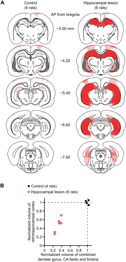
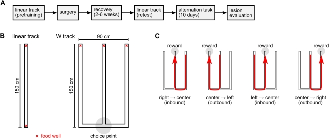
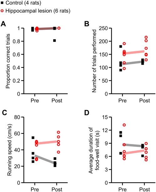
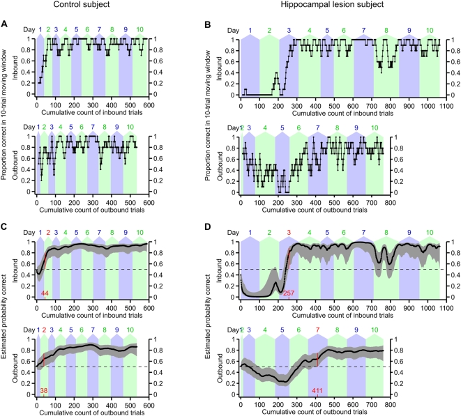
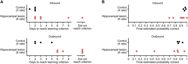
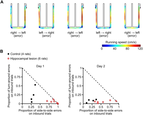
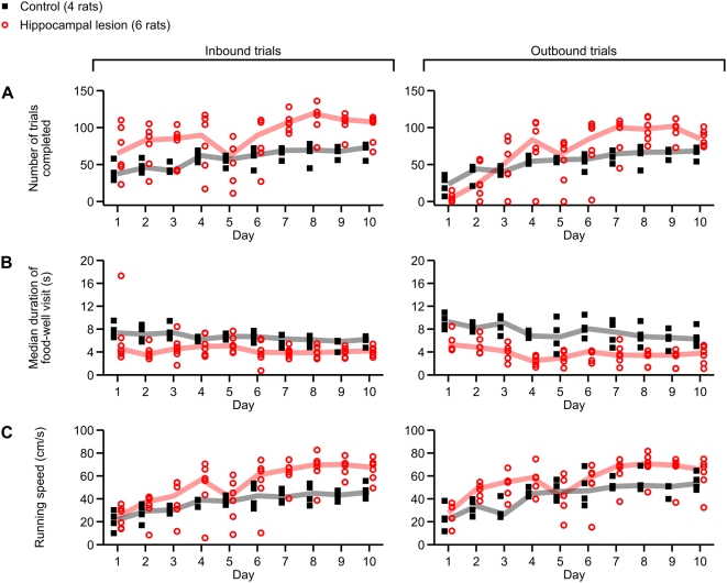
The hippocampus is essential for the formation of memories for events, but the specific features of hippocampal neural activity that support memory formation are not yet understood. The ideal experiment to explore this issue would be to monitor changes in hippocampal neural coding throughout the entire learning process, as subjects acquire and use new episodic memories to guide behavior. Unfortunately, it is not clear whether established hippocampally-dependent learning paradigms are suitable for this kind of experiment. The goal of this study was to determine whether learning of the W-track continuous alternation task depends on the hippocampal formation. We tested six rats with NMDA lesions of the hippocampal formation and four sham-operated controls. Compared to controls, rats with hippocampal lesions made a significantly higher proportion of errors and took significantly longer to reach learning criterion. The effect of hippocampal lesion was not due to a deficit in locomotion or motivation, because rats with hippocampal lesions ran well on a linear track for food reward. Rats with hippocampal lesions also exhibited a pattern of perseverative errors during early task experience suggestive of an inability to suppress behaviors learned during pretraining on a linear track. Our findings establish the W-track continuous alternation task as a hippocampally-dependent learning paradigm which may be useful for identifying changes in the neural representation of spatial sequences and reward contingencies as rats learn and apply new task rules.


Conflict of interest statement

Competing Interests: The authors have declared that no competing interests exist.

Figures

Figure 1
Figure 1. Histological reconstruction of hippocampal lesions.
(A) Drawings of coronal sections at different anteroposterior levels illustrate the extent and location of brain damage, for subjects in the control group (left) and in the lesion group (right). Damaged areas within each subject are shaded in light pink; where there is overlap among subjects, the opacities of the overlapping regions sum to give darker shading. The darkest shade of red indicates areas that were consistently damaged in all subjects. The coronal section outlines are adapted from . (B) Quantification of lesion extent. The horizontal axis is the estimated volume (combined over both hemispheres) of the dentate gyrus, CA fields, and fimbria. The vertical axis is the estimated volume (combined over both hemispheres) of the retrohippocampal cortex, which we define as the subiculum, presubiculum, parasubiculum, and entorhinal cortex. These volume estimates underrepresent the true loss of neurons because they include spared hippocampal white matter and partially-damaged shrunken tissue.
Figure 2
Figure 2. Experimental design and behavioral tasks.
(A) Timeline of the experiment. (B) Diagrams of the running tracks used in this experiment. The red X marks indicate locations of food wells. The gray circle indicates the choice-point intersection on the W track. (C) Sequential illustration of correct performance of the W-track continuous spatial alternation task. Rats were rewarded for visiting the three food wells of the W track in the correct repeating sequence.
Figure 3
Figure 3. Summary of performance on linear track task.
Plotted in each panel is a measure of linear track behavior before surgery (Pre) and after recovery from surgery (Post). Filled black symbols indicate values for the control group (4 rats), and open red symbols indicate the same for the hippocampal lesion group (6 rats). The correspondingly color-coded heavy lines are group medians. (A) Proportion of correct trials. (B) Number of trials completed. (C) Mean running speed, excluding times spent at food wells. (D) Median dwell time at food wells at the end of trials. Only running speed significantly differed between the groups (main effect of group, p = 0.0092; group×day interaction, p = 0.012). Post hoc within-day comparisons revealed that the difference in running speed between the two groups was not significant on the last day of pre-surgery training (p = 0.76), but was significant for the post-surgery test (p = 0.0095). Thus, hippocampal lesions caused an increase in running speed but did not disrupt task performance on the linear track.
Figure 4
Figure 4. Examples of learning curves on the W-track continuous alternation task for two subjects.
(A) 10-trial moving average of proportion correct for a control subject. The top plot shows performance on inbound trials, while the bottom plot shows performance on outbound trials. Trials are counted cumulatively along the horizontal axis, starting with the first trial on day 1 and ending with the last trial on day 10. The alternating blue and green background shading indicates the number of trials completed on each day. (B) 10-trial moving average of proportion correct for a lesion subject. (C) Smooth learning curve estimated using the state-space model of learning, for the same subject as in (A). The top plot shows the estimated learning curve for the inbound component of the task, while the bottom plot shows the estimated learning curve for the outbound component of the task. Trials are counted cumulatively along the horizontal axis in the same manner as in (A). Black dots indicate maximum-likelihood estimates of the probability of correct performance, and gray errors bars indicate point-wise 95% confidence intervals. Dashed horizontal lines indicate the chance performance level (1/2) that would be expected if the subject randomly chose the next destination food well. We defined the learning criterion (highlighted in red) as the earliest trial at which the 95% confidence interval of the learning curve exceeded this chance level and remained above chance for two full consecutive days. (D) Similar to (C), but for the same hippocampal lesion subject as in (B). The initial low dip of the inbound learning curve, and the paucity of outbound trials, reflects the many perseverative inbound errors that this subject made during the first two days of testing. This subject's peformance on the inbound component of the task regressed transiently on day 8 for unknown reasons.
Figure 5
Figure 5. Effect of hippocampal lesions on learning of the W-track continuous spatial alternation task.
(A) Number of days to reach learning criterion on the inbound (top) and outbound (bottom) components of the task. Compared to the control group, the hippocampal lesion group exhibited significantly slower learning of the outbound task component (p = 0.0095). (B) Mean estimated probability of correct performance on day 10 of testing. Although there is an apparent trend for the performance of the hippocampal lesion subjects to be skewed lower, this trend did not reach significance.
Figure 6
Figure 6. Effect of hippocampal lesions on inbound errors during early task experience.
(A) Example of perseverative errors made by a hippocampal lesion subject during the first session of the W-track continuous alternation task. Path-maps are shown for five consecutive incorrect inbound trials. The paths are color-coded to indicate the rat's instantaneous running speed. Arrows indicate the direction of travel. (B) Scatterplots showing the pattern of inbound errors on on day 1 (left) and day 2 (right) of the W-track continuous alternation task. The plotted symbols show, for each individual subject, the proportions of errors on inbound trials, classified according to destination: the proportion of inbound trials in which the subject ran from one side food-well to the opposite side, skipping the center arm (horizontal axis); and the proportion of inbound trials in which the subject returned to the outside arm food-well from which it had just departed (vertical axis). The dashed diagonal line indicates the maximum possible values of these error proportions. A larger proportion of lesioned animals' inbound trials were associated with side-to-side trajectories on both day 1 and day 2 as compared to controls (day 1, p<0.01; day 2, p<0.04).
Figure 7
Figure 7. Effect of hippocampal lesions on behavior on the W track.
Plotted in each panel is a measure of behavior across all 10 days of testing on the W track. Filled black symbols indicate values for the control group (4 rats), and open red symbols indicate the same for the hippocampal lesion group (6 rats). The correspondingly color-coded heavy lines are group medians. (A) Number of inbound (left) and outbound (right) trials performed on each day of testing. Compared to control subjects, subjects with hippocampal lesions tended to perform more inbound trials (p<0.02). (B) Average dwell time per food-well visit at the end of inbound (left) and outbound (right) trials. Compared to lesioned subjects, control subjects tended to dwell at the food well for a longer time after each trial (inbound, p<0.005; outbound, p<0.002), in part because they completed a greater proportion of trials correctly and thus spent more time consuming food reward. (C) Running speeds on inbound and outbound trials. Compared to control subjects, lesioned subjects ran at higher speeds on both inbound (p<0.05) and outbound (p<0.04) trials.

References

    1. Amaral DG, Witter MP. Hippocampal Formation. In: Paxinos C, editor. The Rat Nervous System. Academic Press; 1995. pp. 443–493.
    1. Scoville WB, Milner B. Loss of recent memory after bilateral hippocampal lesions. J Neurol Neurosurg Psychiatry. 1957;20:11–21. - PMC - PubMed
    1. Rempel-Clower NL, Zola SM, Squire LR, Amaral DG. Three cases of enduring memory impairment after bilateral damage limited to the hippocampal formation. J Neurosci. 1996;16:5233–5255. - PMC - PubMed
    1. Zola SM, Squire LR, Teng E, Stefanacci L, Buffalo EA, et al. Impaired recognition memory in monkeys after damage limited to the hippocampal region. J Neurosci. 2000;20:451–463. - PMC - PubMed
    1. Spiers HJ, Maguire EA, Burgess N. Hippocampal amnesia. Neurocase. 2001;7:357–382. - PubMed

Publication types

Substances