

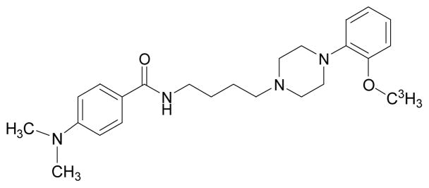
[(3)H]4-(Dimethylamino)-N-[4-(4-(2-methoxyphenyl)piperazin- 1-yl)butyl]benzamide, a selective radioligand for dopamine D(3) receptors. I. In vitro characterization
- PMID: 19425052
- PMCID: PMC2783604
- DOI: 10.1002/syn.20652
[(3)H]4-(Dimethylamino)-N-[4-(4-(2-methoxyphenyl)piperazin- 1-yl)butyl]benzamide, a selective radioligand for dopamine D(3) receptors. I. In vitro characterization
Abstract
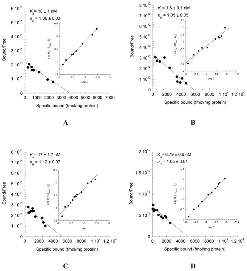
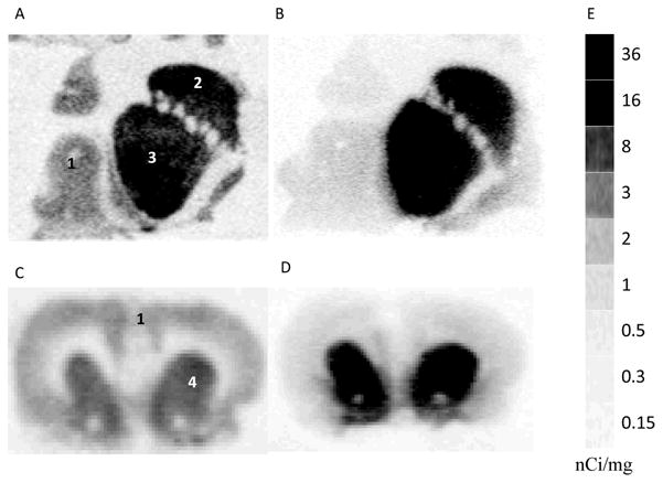
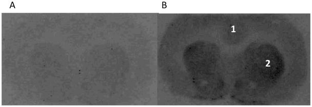
4-(Dimethylamino)-N-(4-(4-(2-methoxyphenyl)piperazin-1-yl)butyl)benzamide (WC-10), a N-phenyl piperazine analog, has been shown to have high affinity and selectivity for dopamine D(3) receptors versus dopamine D(2) receptors (Chu et al. [2005] Bioorg Med Chem 13:77-87). In this study, WC-10 was radiolabeled with tritium (specific activity = 80 Ci/mmol) and [(3)H]WC-10 binding to genetically cloned dopamine D(2L) and D(3) receptors was evaluated in vitro. [(3)H]WC-10 binds with a 66-fold higher affinity to human HEK D(3) than HEK D(2L) receptors, with a dissociation constant (K(d)) of 1.2 nM at HEK D(3) receptors. However, [(3)H]WC-10 binds to rat Sf9 rD(3) receptors with a K(d) of 3.9 nM, a value that is 3-fold lower than binding to human HEK D(3) receptors and 40-fold value higher than binding to rat Sf9 rD(2L) receptors. The K(d) values obtained from saturation binding experiments were consistent with the results determined from kinetic (k(on) and k(off)) studies. The pharmacologic profiles of a series of dopaminergic drugs for inhibiting the binding of [(3)H]WC-10 to D(3) receptors was in agreement with previously reported data. In vitro autoradiography studies of rat and monkey brains show that [(3)H]WC-10 labeled D(3) sites in the striatal region.
2009 Wiley-Liss, Inc.
Figures


References
-
- Ariano MA, Sibley DR. Dopamine receptor distribution in the rat CNS: elucidation using anti-peptide antisera directed against D1A and D3 subtypes. Brain Research. 1994;649:95–110. - PubMed
-
- Boundy VA, Luedtke RR, Gallitano AL, Smith JE, Filtz TM, Kallen RG, Molinoff PB. Expression and characterization of the rat D3 dopamine receptor: pharmacologic properties and development of antibodies. J Pharmacol Exp Ther. 1993;264:1002–1011. - PubMed
-
- Camacho-Ochoa M, Walker EL, Evans DL, Piercey MF. Rat brain binding sites for pramipexole, a clinically useful D3-preferring dopamine agonist. Neurosci Lett. 1995;196:97–100. - PubMed
Publication types
MeSH terms
Substances
Grants and funding
- NS048056/NS/NINDS NIH HHS/United States
- R01 DA012647/DA/NIDA NIH HHS/United States
- S10 RR021007/RR/NCRR NIH HHS/United States
- R01 NS041509/NS/NINDS NIH HHS/United States
- R21 DA016181/DA/NIDA NIH HHS/United States
- P30 NS048056/NS/NINDS NIH HHS/United States
- R01 DA029840/DA/NIDA NIH HHS/United States
- R01 DA013584/DA/NIDA NIH HHS/United States
- R03 NS090214/NS/NINDS NIH HHS/United States
- R01 NS050425/NS/NINDS NIH HHS/United States
- DA16181/DA/NIDA NIH HHS/United States
- R01 NS058714/NS/NINDS NIH HHS/United States
- DA12647/DA/NIDA NIH HHS/United States
LinkOut - more resources
Full Text Sources
Other Literature Sources
Miscellaneous