. 2009 Jun;84(6):354-61.
doi: 10.1002/ajh.21427.

Targeted deletion of the gamma-adducin gene (Add3) in mice reveals differences in alpha-adducin interactions in erythroid and nonerythroid cells

Affiliations

Targeted deletion of the gamma-adducin gene (Add3) in mice reveals differences in alpha-adducin interactions in erythroid and nonerythroid cells

Kenneth E Sahr et al. Am J Hematol. 2009 Jun.

Abstract

In red blood cells (RBCs) adducin heterotetramers localize to the spectrin-actin junction of the peripheral membrane skeleton. We previously reported that deletion of beta-adducin results in osmotically fragile, microcytic RBCs and a phenotype of hereditary spherocytosis (HS). Notably, alpha-adducin was significantly reduced, while gamma-adducin, normally present in limited amounts, was increased approximately 5-fold, suggesting that alpha-adducin requires a heterologous binding partner for stability and function, and that gamma-adducin can partially substitute for the absence of beta-adducin. To test these assumptions we generated gamma-adducin null mice. gamma-adducin null RBCs appear normal on Wright's stained peripheral blood smears and by scanning electron microscopy. All membrane skeleton proteins examined are present in normal amounts, and all hematological parameters measured are normal. Despite a loss of approximately 70% of alpha-adducin in gamma-adducin null platelets, no bleeding defect is observed and platelet structure appears normal. Moreover, systemic blood pressure and pulse are normal in gamma-adducin null mice. gamma- and beta-adducin null mice were intercrossed to generate double null mice. Loss of gamma-adducin does not exacerbate the beta-adducin null HS phenotype although the amount alpha-adducin is reduced to barely detectable levels. The stability of alpha-adducin in the absence of a heterologous binding partner varies considerably in various tissues. The amount of alpha-adducin is modestly reduced ( approximately 15%) in the kidney, while in the spleen and brain is reduced by approximately 50% with the loss of a heterologous beta- or gamma-adducin binding partner. These results suggest that the structural properties of adducin differ significantly between erythroid and various nonerythroid cell types.


Figures

Fig. 1
Fig. 1
Targeted disruption of the γ-adducin gene. (A) A targeting construct was made by inserting loxP sites (LP1 and LP2) upstream and downstream of the first protein coding exon (exon 2), and a Neor gene flanked by FRT sequences (Frt-neo) into the downstream intron. The following restriction enzyme sites are indicated: N, NdeI; Xh, XhoI; X, XbaI. Sizes of the predicted 5′ and 3′ restriction fragments and locations of the 5′ and 3′ probes (shaded boxes) are indicated. (B) A correctly targeted G418r ES cell clone (1A10) was identified by Southern analysis using 5′ and 3′ probes and used for blastocyst injection. Correctly targeted mice (γ-addCKO/+; littermates 4.2 and 4.3) were obtained by mating a high chimera male with a C57BL/6J female were identified by PCR of the neor gene and confirmed by Southern analysis. (C) PCR analysis of a litter generated by mating a γ-addCKO/+ male and wt female mouse is shown. The PCR primers used flank the upstream loxP site and generate a 239 bp and 275 bp product from wt and targeted DNA, respectively. (D) The neor gene was excised by mating a correctly targeted mouse to a homozygous Flp mouse. As indicated, PCR primers flanking the Neor gene insertion site generate 275 bp, 1500 bp, and 400 by products from wt, γ-addCKO, and γ-addΔneo DNA, respectively. (E) A null mouse lacking exon 1 was then generated by mating a γ-addΔneo/+ female with an EIIa-cre/EIIa-cre male mouse. Littermates with an excised exon 1 are identified by a 375 bp product. Representative PCR products of +/+ and heterozygous targeted DNA are shown in (D) and (E). (F) Total RNA isolated from the spleens of +/+, +/−, and −/− littermates was analyzed by northern hybridization using cDNA fragments corresponding to γ-adducin exon 2, γ-adducin exons 6-10, actin, and gapdh. 20 μg of total RNA was used for the γ-adducin analysis, while 2 μg of total RNA was used for actin and gapdh analysis. The absence of γ-adducin protein in null mice was verified by western analysis of spleen tissue homogenates (20 μg protein per lane) from littermates generated by a +/− intercross.
Fig. 2
Fig. 2
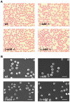
Red blood cell morphology. (A) Bright field light microscopy (original magnification 1000x) and (B) scanning electron microscopy of red blood cells isolated from wt, β-adducin null, γ-adducin null, and β/γ-adducin null mice. Scale bar is 10 μM.
Fig. 3
Fig. 3
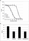
Analysis of red blood cells by osmotic fragility and ektacytometry. A. Osmotic fragility was measured using whole blood collected from wt, β-adducin null, γ-adducin null, and β/γ-adducin null mice. Three mice of each genotype were used, each measured in duplicate. B. Ektacytometry was performed using four mice per genotype. By either assay, RBCs isolated from wt and γ-adducin null mice have almost identical profiles, while RBCs isolated from β-adducin null and γ/β-adducin null mice display equally increased fragility.
Fig. 4
Fig. 4
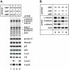
Western analysis of red blood cell ghost proteins. (A) Red blood cell ghost proteins were prepared from wt, β-adducin null, γ-adducin null, and γ/β-adducin null mice. The loss of γ-adducin alone (lane 3) has no significant impact on the relative amounts of several membrane skeleton proteins, including spectrin, ankyrin, band 3, proteins 4.1, 4.2, and 4.9 (dematin), p55, glycophorin C (GPC), tropomodulin (Tmod), α-tropomyosin (TM), E-capZα, and E-capZβ. While the loss of β-adducin affects the relative amounts of several of these proteins (lane 2), including actin, α-tropomyosin, E-capZα, and E-capZβ the additional loss of γ-adducin (lane 4) causes no further change in the relative amounts of these proteins. Tmod, TM, E-capZα, and E-capZβ protein amounts were measured using ghosts prepared in the presence of 2 mM MgCl2. As shown in (B), the loss of γ-adducin alone in RBCs has no significant effect on the relative amounts of α- or β-adducin (lane 7). As previously described, in the absence of β-adducin (lane 6) the relative amount of α-adducin is significantly decreased while the level of γ-adducin is increased. The loss of both β- and γ-adducin (lane 8) has a dramatic effect on the relative amount of α-adducin, reducing it to an almost undetectable level. (All lanes for each protein are from a single gel but rearranged). Representative results from two independent experiments are shown.
Fig 5
Fig 5
Analysis of blood platelets. A. The loss of γ-adducin in platelets (lanes 3 and 4) results in a significant decrease (~70%) in the amount of α-adducin. (β-adducin was not detected in platelets.) A representative result of two independent experiments is shown. B. Bleeding times (mean ± standard error) were compared using five wt and six γ-adducin KO age-matched mice. No significant difference was detected in the two groups. C. TEM of wt and γ-adducin KO platelets. Scale bar is 500 nm.
Fig. 6
Fig. 6
Western analysis of nonerythroid cells. Crude cytosol (c) and membrane (m) fractions were prepared from spleen, kidney, and brain tissues of wt, β-adducin null, γ-adducin null, and β/γ-adducin double null mice. The combined loss of β- and γ-adducin decreased α-adducin in the spleen (lanes 7, 8) and brain (lanes 23, 24) by ~50%, and by ~15% in the kidney (lanes 15 and 16). A representative result of two (kidney and spleen) or three (brain) independent experiments is shown.

References

    1. Suriyapperuma SP, Lozovatsky L, Ciciotte SL, Peters LL, Gilligan DM. The mouse adducin gene family: alternative splicing and chromosomal localization. Mamm Genome. 2000;11:16–23. - PubMed
    1. Gilligan DM, Lozovatsky L, Gwynn B, Brugnara C, Mohandas N, Peters LL. Targeted disruption of the beta adducin gene (Add2) causes red blood cell spherocytosis in mice. Proc Natl Acad Sci U S A. 1999;96:10717–10722. - PMC - PubMed
    1. Fowler VM. Identification and purification of a novel Mr 43,000 tropomyosin-binding protein from human erythrocyte membranes. J Biol Chem. 1987;262:12792–12800. - PubMed
    1. Dong L, Chapline C, Mousseau B, et al. 35H, a sequence isolated as a protein kinase C binding protein, is a novel member of the adducin family. J Biol Chem. 1995;270:25534–25540. - PubMed
    1. Bennett V, Baines AJ. Spectrin and ankyrin-based pathways: metazoan inventions for integrating cells into tissues. Physiol Rev. 2001;81:1353–1392. - PubMed

Publication types

MeSH terms