. 2009 Jul 20;515(3):269-94.
doi: 10.1002/cne.22040.

Subventricular zone neural progenitors from rapid brain autopsies of elderly subjects with and without neurodegenerative disease

Affiliations

Subventricular zone neural progenitors from rapid brain autopsies of elderly subjects with and without neurodegenerative disease

Brian W Leonard et al. J Comp Neurol. .

Abstract

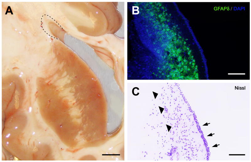
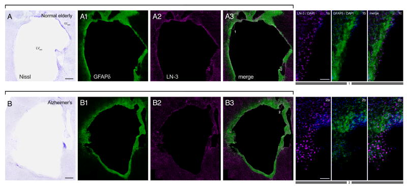
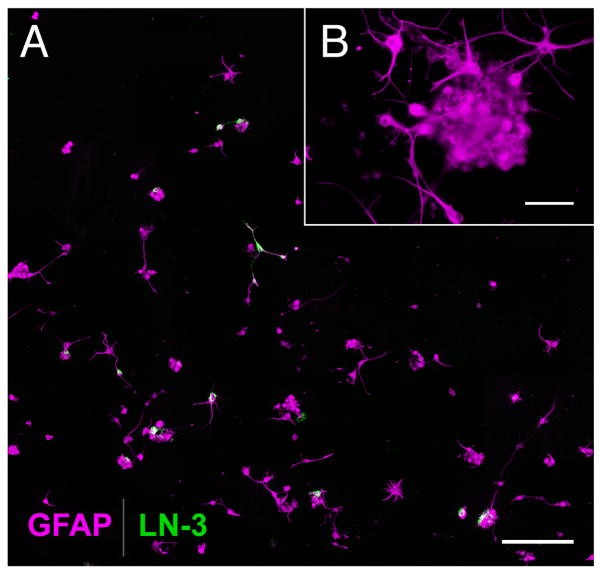
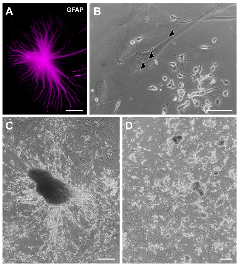
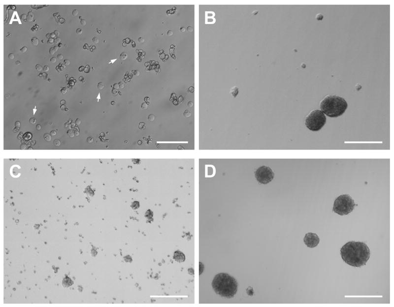
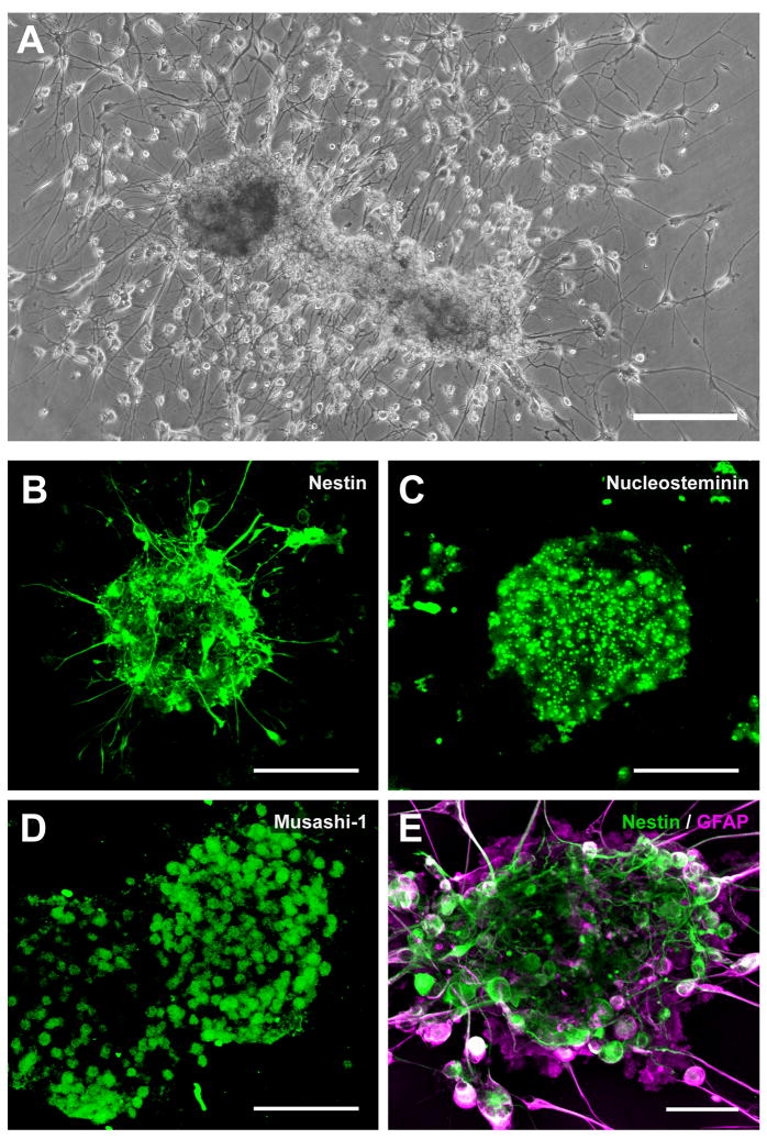
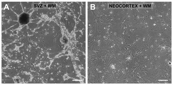
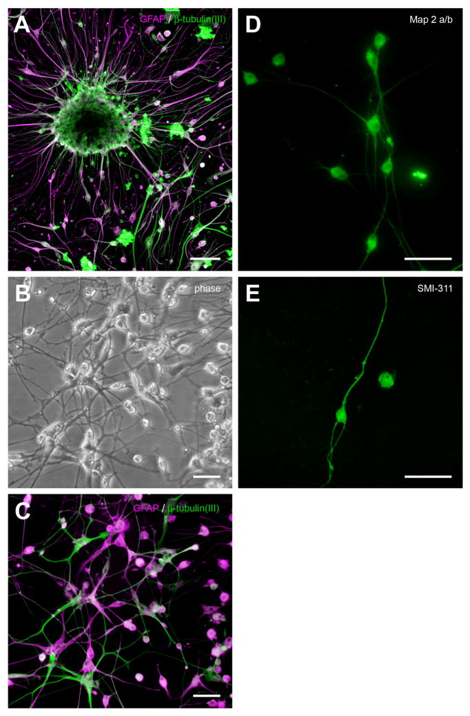
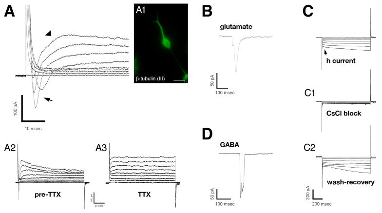
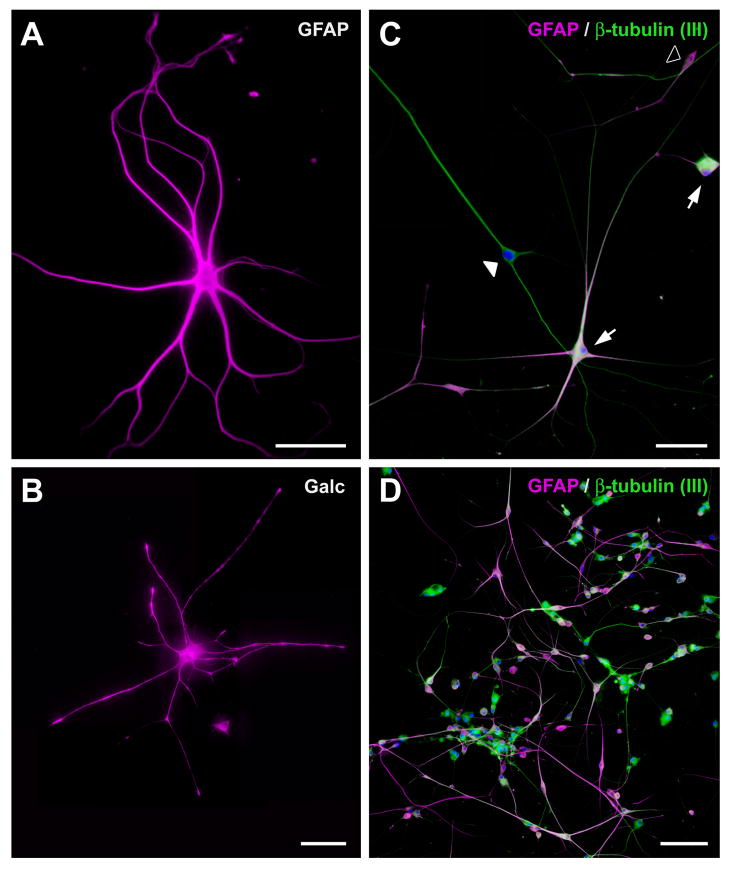
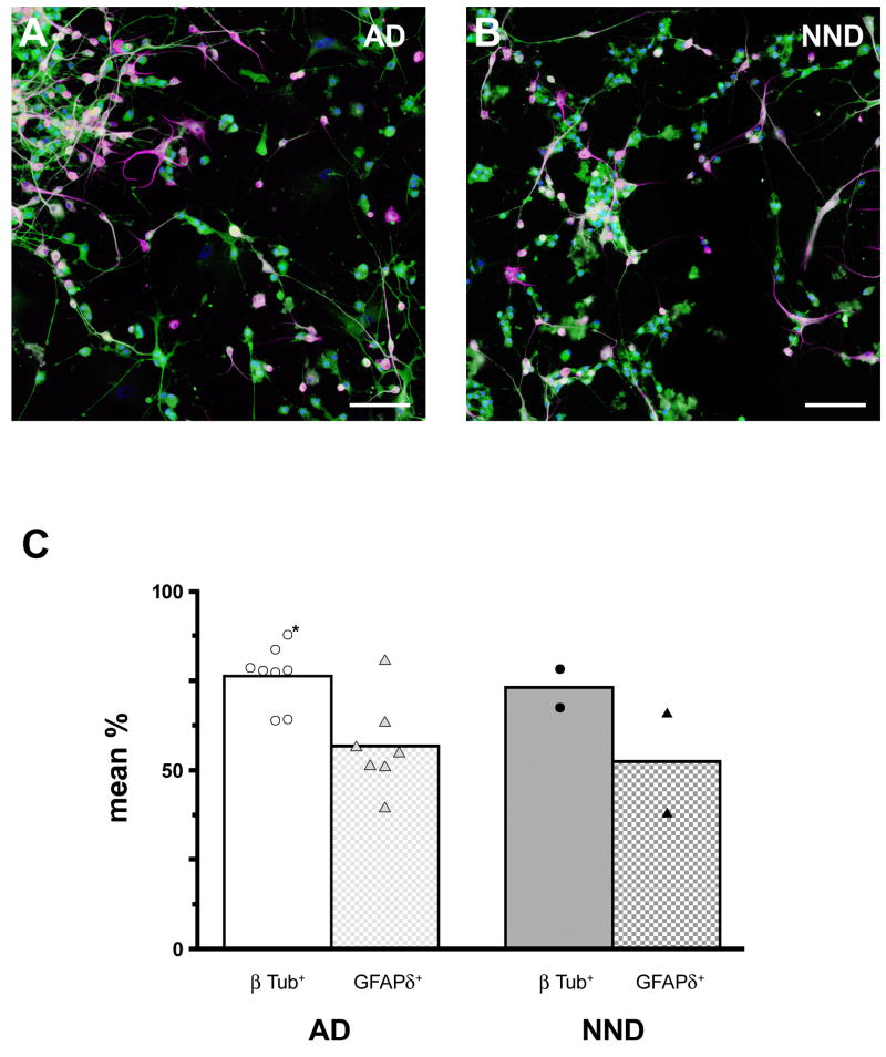
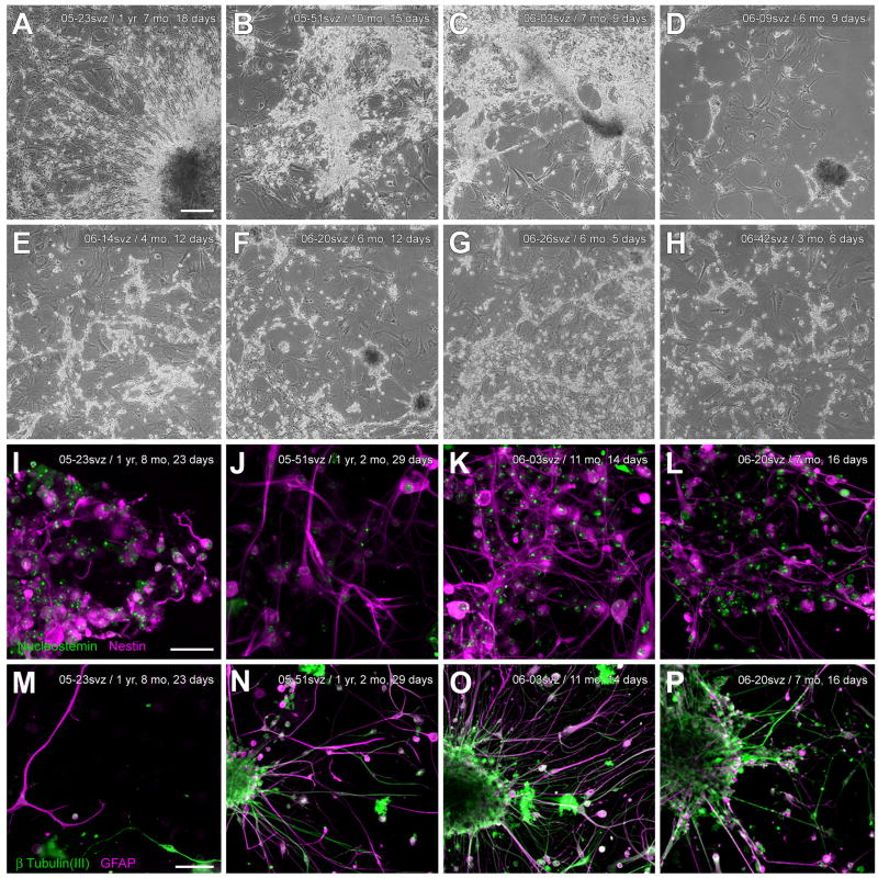
In mice and in young adult humans, the subventricular zone (SVZ) contains multipotent, dividing astrocytes, some of which, when cultured, produce neurospheres that differentiate into neurons and glia. It is unknown whether the SVZ of very old humans has this capacity. Here, we report that neural stem/progenitor cells can also be cultured from rapid autopsy samples of SVZ from elderly human subjects, including patients with age-related neurologic disorders. Histological sections of SVZ from these cases showed a glial fibrillary acidic protein (GFAP)-positive ribbon of astrocytes similar to the astrocyte ribbon in human periventricular white matter biopsies that is reported to be a rich source of neural progenitors. Cultures of the SVZ contained 1) neurospheres with a core of Musashi-1-, nestin-, and nucleostemin-immunopositive cells as well as more differentiated GFAP-positive astrocytes; 2) SMI-311-, MAP2a/b-, and beta-tubulin(III)-positive neurons; and 3) galactocerebroside-positive oligodendrocytes. Neurospheres continued to generate differentiated progeny for months after primary culturing, in some cases nearly 2 years postinitial plating. Patch clamp studies of differentiated SVZ cells expressing neuron-specific antigens revealed voltage-dependent, tetrodotoxin-sensitive, inward Na+ currents and voltage-dependent, delayed, slowly inactivating K+ currents, electrophysiologic characteristics of neurons. A subpopulation of these cells also exhibited responses consistent with the kinetics and pharmacology of the h-current. However, although these cells displayed some aspects of neuronal function, they remained immature, insofar as they did not fire action potentials. These studies suggest that human neural progenitor activity may remain viable throughout much of the life span, even in the face of severe neurodegenerative disease.


Figures

Fig. 1
Fig. 1
Dissection and anatomy of the human elderly subventricular zone. (A) Photograph of the right lateral ventricle and adjacent structures from an autopsied 96-year-old Alzheimer’s disease (AD) case with a postmortem interval (PMI) of 3.0 h. For cell culture, periventricular white matter samples (dashed line) that included the SVZ were dissected from the superior lateral wall of the lateral ventricle. Samples were collected from three to six levels of the frontally sectioned brain, typically from the anterior and mid-body of the lateral ventricle. The frontal section in panel A is just anterior to the level of the optic chiasm. Although samples for cell culture included white matter beyond the SVZ, neocortical control cultures from the same cases that included this white matter but excluded the SVZ did not develop neurospheres, as described elsewhere in the text (see Results). (B) GFAP-δ immunohistochemistry of the superior lateral wall of the human elderly lateral ventricle (frontal plane), showing a thin ribbon of GFAP-δ-immunoreactive astrocytes (green) lining the ventricular wall just deep to the ependyma. Cell nuclei counterstained with DAPI (blue). From an 85-year-old progressive supranuclear palsy case with a clinical history of dementia; PMI 4.45 h. (C) As can be seen in an adjacent cresyl violet-stained section of the same field, the characteristic ribbon of astrocytes (arrowheads) is actually separated from the ependymal layer (arrows) by a 50–100 μm hypocellular gap, similar to that observed in lateral ventricular biopsy samples from younger patients (e.g., Sanai et al., 2004; Quinones-Hinojosa et al., 2006). Scale bars: A, 2 mm; B, C, 100 μm.
Fig. 2
Fig. 2. Similar SVZ cellular architecture in Alzheimer’s disease and elderly control subjects
Low magnification (A, A1–A3; B, B1–B3) montages and high magnification (1a–1c; 2a–2c) photomicrographs of Nissl and immunostained frontal sections of the lateral ventricle anterior to the caudate nucleus obtained from a normal elderly control subject (A) and an Alzheimer’s disease subject (B). GFAPδ immunoreactivity shows distribution of astrocytes, and LN-3 immunoreactivity (anti-HLA-DR) shows distribution of microglia. Boxes in low-magnification merge panels show where high-magnifications images were taken. DAPI counterstain (blue) in high magnification panels shows cell nuclei. In this figure and in all subsequent ones, red-green fluorescence was converted to magenta-green (see Materials and Methods). Portions of the ependymal layer were often denuded in all elderly cases. Panels in A from a 92 year-old control case with microscopic changes of AD but insufficient for diagnosis of AD due to a lack of dementia in their clinical history; PMI 3 h. Panels in B from a 78 year-old AD case; PMI 4 h. Scale bars: 1 mm, low magnification; 100 μm high magnification. Abbreviations: LVant, anterior lateral ventricle; CCant, anterior corpus callosum.
Fig. 3
Fig. 3
(A) Early-stage SVZ cultures are depleted of microglia and enriched with astrocytes. Relatively pure astrocyte-progenitor cultures were produced by first allowing microglia in the initial SVZ cell suspension to become adherent, then replating the remaining, non-adherent cells into a new flask, as shown here. These secondary flasks were therefore relatively depleted of microglia, as shown by the near absence of immunoreactivity for HLA-DR (green), a microglial marker. By contrast, nearly all the cells at this early stage of culture were immunoreactive for the astrocyte marker, GFAP (magenta). B shows a small GFAP-immunoreactive cluster of cells from the same culture reminiscent of an early-stage neurosphere. From an 89-year-old AD case; PMI 3.5 h. Scale bars: 250 μm; inset, 50 μm.
Fig. 4
Fig. 4
Morphology of neurospheres derived from human elderly autopsies of the SVZ. Microglia-depleted SVZ supernatants developed spherical aggregates of free-floating cells, some of which bore the characteristic morphology of neurospheres. (A) Phase-contrast micrograph of neurospheres and cell aggregates in suspension, shown here seven days after initial culturing. From a 69-year-old AD case; PMI 2.5 h. (B) Neurospheres from the same case as in A on the third passage. (C) Within hours of plating on laminin or poly-L-lysine substrates, adherent, neurosphere-like clusters of cells were observed with many fine processes and cells radiating outward, shown here at 2 weeks post-plating. From an 84-year-old AD case; PMI 3.0 h. Scale bars: A, B, 100 μm; C, 250 μm.
Fig. 5
Fig. 5
Association of neurospheres co-cultured with type 1 astrocytes. (A, B) Neurospheres and clusters of small, phase-bright cells were often observed attached to the surface of beds of GFAP-immunoreactive astrocytes having type 1 morphology (Raff et al., 1983). Arrowheads in B indicate possible contacts between clusters of putative progenitors and a type 1 astrocyte. (A, 96-year-old AD case; PMI 1.66 h; B, 88-year-old control case with microscopic changes of AD but insufficient for diagnosis of AD due to a lack of dementia in their clinical history; PMI 3.75 h. (C, D) In confluent cultures, these cells typically surrounded or provided a bed for neurosphere-like cell aggregates, as well as for more loosely organized groups of bipolar and multipolar phase-bright cells (C, 69-year-old AD case; PMI 2.75 h; D, 84-year-old AD case; PMI 3.0 h). Scale bars: A, B, 100 μm; C, D; 250 μm.
Fig. 6
Fig. 6
Mitogen expansion of neurospheres derived from human elderly autopsies of the SVZ. (A) Microglia-depleted, SVZ single-cell suspensions treated with hEGF (20 ng/ml) and hFGF-b (10 ng/ml) in serum-free medium (see Materials and Methods) adhere initially to the uncoated flask surface, shown here (arrows) at 15 h (91-year-old AD case; PMI 2.5 h). (Some cellular debris is also present). (B) Within 1–3 weeks of continued mitogen exposure, fewer adherent cell clusters and more free-floating neurospheres were observed in suspension (15 days exposure, same case and flask as in A). (C) At early time points (5 days exposure) shown here in a different case (84-year-old AD case; PMI 3.0 h) many clusters of cells are still adherent, and diameters and morphology of neurospheres in suspension vary widely. (D) With continued exposure to hEGF and hFGF-b, neurospheres were mainly in suspension and had more spherical morphology (~9 weeks exposure; same case as in C). In parallel cultures derived from cortical tissue and adjoining periventricular white matter that intentionally discarded the SVZ, we never observed neurospheres develop with this mitogen expansion protocol, nor develop spontaneously in DMEM complete. Scale bars: A, 50 μm; B, C, D; 250 μm.
Fig. 7
Fig. 7
Neurospheres produced from human elderly SVZ are immunoreactive for neural stem cell proteins. (A) Within 2 weeks of plating on uncoated surfaces, adherent, neurosphere-like clusters of cells were observed with many fine processes and cells radiating outward. From an 84-year-old AD case; PMI 3.0 h. (B, C, D) Confocal micrographs of neurospheres immunoreactive for neural stem cell markers. Plating on PLL/laminin quickened the development and complexity of processes, as shown previously. SVZ neurospheres were expanded from a single-cell suspension for ~4.5 weeks in SFM with EGF/FGF, plated for one day on PLL/laminin to allow adherence, then fixed and immunostained for the neural stem cell proteins nestin, nucleostemin, and Musashi-1. From a 79-year-old Parkinson’s disease case who also had AD; PMI 2.5 h. Deletion of the primary antibodies resulted in no specific immunostaining. (E) Confocal micrograph of a neurosphere 2 weeks after plating, showing nestin-immunoreactive core (green) and differentiating GFAP-immunoreactive cells (red) at the periphery (same case and culture as in A). Scale bars: A, 250 μm; B, C, D, 100 μm; E, 50 μm.
Fig. 8
Fig. 8
Neurospheres develop only from human elderly autopsied periventricular white matter (WM) that includes the SVZ. Cultures were produced and maintained in parallel using postmortem tissue from the same case (69-year-old AD case; PMI 2.5 h) that contained either (A) periventricular WM and adjoining SVZ (cf. Fig 1A) or (B) frontal cortex and adjoining subcortical WM excluding the SVZ. Microglia were depleted as before (see Materials and Methods). Neurospheres growing on top of putative astrocytes were observed only in cultures derived from SVZ+WM; only astrocytes developed in cultures derived from neocortex+WM. Images taken 28 weeks after plating. Scale bars: A, B, 250 μm.
Fig. 9
Fig. 9
Development of neuron-like cells from elderly SVZ neurospheres. (A) When neurospheres were transferred to poly-L-lysine/laminin substrates and subcultured, they became adherent within 24 h, and cells with a more differentiated, bi- or multi-polar morphology began to appear at the neurosphere periphery, shown here about 2 weeks postplating. Confocal micrograph showing neurosphere immunostained for GFAP (red) and β-tubulin(III) (green) (88-year-old control case with microscopic changes of AD but insufficient for diagnosis of AD due to a lack of dementia in their clinical history; PMI 3.75 h). (B) Phase-contrast photomicrograph of differentiated cells about 200 μm away from a different neurosphere that was plated after the 18 weeks growing in culture, shown here at two weeks after subculturing (84-year-old AD case; PMI 3.0 h). (C) Epifluorescent photomicrograph of the same subculture and field, immunostained for β-tubulin(III) and GFAP. (D, E) Epifluorescent photomicrographs of putative neurons differentiated from elderly SVZ neurospheres that were immunoreactive for MAP2a/b and SMI-311, the latter a pan-neuronal neurofilament marker (D, same case as A, different subculture; E, 82-year-old progressive supranuclear palsy case; PMI 2.33 h). Scale bars: A, 100 μm; B–E, 50 μm; F, 25 μm.
Fig. 10
Fig. 10
Electrophysiology of putative neurons differentiated from elderly human SVZ neurospheres. Patch-clamp recordings made in putative neurons differentiated from neurospheres derived from elderly, postmortem SVZ. (A) Example voltage-clamp records from a β-tubulin (III)-positive cell (A1) showing transmembrane currents in response to increasing voltage steps. Fast, voltage-dependent inward current (arrow) and slowly activating/inactivating outward current (arrowhead) were observed. Lower traces (A2, A3) show that the inward current was abolished by 1 μM TTX, suggestive of neuronal Na+ channels (upper extreme of traces are truncated for display purposes); the outward current, presumably carried by K+ ions was left intact (holding potential = −50 mV) (88-year-old control case with microscopic changes of AD but insufficient for diagnosis of AD due to a lack of dementia in their clinical history; PMI 3.75 h). (B) Responses of other putative neurons to glutamate (1 mM) (same case and subculture as in A, different cell) and GABA (100 μM) (90-year-oldcase diagnosed as argyrophilic grain disease (with clinical dementia); PMI 2.5 h). (C) Other putative neurons displayed an inward-rectified current (arrow) that was activated at hyperpolarizing membrane potentials and was blocked by CsCl (1 mM), suggesting the h current. From a 69-year-old AD case; PMI 2.75 h. Scale bar: A1, immunofluorescent image, 25 μm.
Fig. 11
Fig. 11
Development of non-neuronal and hybrid cells in human SVZ cultures. (A) The most common type of cell that developed from SVZ neurospheres was the type 2 astrocyte, immunoreactive for GFAP (96-year-old AD; PMI 1.66 h). (B) Much less common were galactocerebroside-immunopositive (Galc) cells with oligodendrocyte morphology (same case as in A, different subculture). (C) As reported in rodent SVZ cultures (Laywell et al., 2005), a hybrid cell, the “asteron,” was also observed in human elderly SVZ cultures. These cells were immunoreactive for both the astrocyte marker GFAP (magenta) and the neuron-specific markerβ-tubulin (III) (green; DAPI counterstain). Note that with the GFAP and β-tubulin (III) double label, some cells, putative neurons, are labeled only with β-tubulin (III) (filled arrowhead); some cells, putative astrocytes, are labeled only with GFAP (open arrowhead); and some cells, asterons, distinctly show both markers (filled arrows). Such a finding in the same culture suggests that the failure of GFAP to stain putative neurons or, conversely, the failure of neuronal markers to stain putative astrocytes in samples double-labeled with both antibodies was not due, here or in the previous figures, to inadequate immunocytochemical methods with one or the other set of antibodies. From a 69-year-old Parkinson’s disease case; PMI 4.16 h. (D) Asterons often appeared to surround clusters of neurons (same case and subculture as in C). Scale bars: A, B, C, 50 μm; D, 100 μm.
Fig. 12
Fig. 12
Quantitation of neuronal differentiation of cells from elderly SVZ neurospheres. (A, B) Representative epifluorescent micrographs of β-tubulin(III) (green) and GFAP-δ (magenta) immunostaining of differentiated cells from SVZ neurospheres of an AD case (85-year-old; PMI 3.5 h) and NND case (91-year-old; PMI 2.0 h). DAPI counterstain shows cell nuclei. Scale bar: A, B 100 μm. (C) Mean percentage of β-tubulin(III)-labeled cells (open bars) and GFAP-δ-labeled cells (gray hatched bars) in AD and NND cases were not statistically different (p = 0.669 and p = 0.712, respectively). Individual case values are also plotted. Asterisk indicates value of a PD case (69-year-old; PMI 4.16 h) having dementia, plotted for comparison.
Fig. 13
Fig. 13
Long-term viability of elderly human SVZ cultures. (A–H) Low-power, phase-contrast photomicrographs of representative flasks of viable SVZ astrocytes, progenitors, and neurospheres that had been maintained for many months to over a year (N=11). Adherent cells and neurospheres could be gently dislodged and passaged to produce viable secondary, tertiary, and quaternary flasks of astrocytes, progenitors, and neurospheres. For six of the flasks, case ID and age of culture is indicated (A, 96-year-old AD case; PMI 1.66 h; B, 69-year-old AD case; PMI 2.75 h; C, 88-year-old control case with microscopic changes of AD but insufficient for diagnosis of AD due to a lack of dementia in their clinical history; PMI 3.75 h; D, 91-year-old AD case; PMI 3.50 h; E, 91-year-old AD case; PMI 2.5 h; F, 69-year-old AD case; PMI 2.5 h; G, 84-year-old AD case; PMI 3.00 h; H, 89-year-old AD case; PMI 3.50 h.) Magnification in panels A–H is identical. (I–J) For a subset of the flasks shown in panels A–H, cells and neurospheres were dislodged, plated onto PLL/laminin at the ages indicated, allowed to develop for several weeks, then fixed and immunostained for neural stem cell markers (see I–L) or GFAP and β-tubulin (III) (see M–P). Long-term cultures shared virtually all the characteristics described earlier in this report for cultures maintained and sacrificed within a month or two of plating, including nestin- and nucleostemin-positive cores within neurospheres, the elaboration of dense networks of processes, and the presence of differentiated cells with neuronal- and astrocyte-immunoreactivity. Magnification in panels I–L is identical; magnification in panels M–P is identical. Scale bars: A–H, 250 μm; I–L 50 μm; M–P, 100 μm.

References

    1. Alafuzoff I, Pikkarainen M, Arzberger T, Thal DR, Al-Sarraj S, Bell J, Bodi I, Budka H, Capetillo-Zarate E, Ferrer I, Gelpi E, Gentleman S, Giaccone G, Kavantzas N, King A, Korkolopoulou P, Kovacs GG, Meyronet D, Monoranu C, Parchi P, Patsouris E, Roggendorf W, Stadelmann C, Streichenberger N, Tagliavini F, Kretzschmar H. Inter-laboratory comparison of neuropathological assessments of beta-amyloid protein: a study of the BrainNet Europe consortium. Acta neuropathologica. 2008;115(5):533–546. - PubMed
    1. Alvarez-Buylla A, Garcia-Verdugo JM. Neurogenesis in adult subventricular zone. J Neurosci. 2002;22(3):629–634. - PMC - PubMed
    1. Alvarez-Buylla A, Herrera DG, Wichterle H. The subventricular zone: source of neuronal precursors for brain repair. Prog Brain Res. 2000;127:1–11. - PubMed
    1. Banerjee A, Roach MC, Trcka P, Luduena RF. Increased microtubule assembly in bovine brain tubulin lacking the type III isotype of beta-tubulin. J Biol Chem. 1990;265(3):1794–1799. - PubMed
    1. Benjamins JA, Callahan RE, Montgomery IN, Studzinski DM, Dyer CA. Production and characterization of high titer antibodies to galactocerebroside. J Neuroimmunol. 1987;14(3):325–338. - PubMed

Publication types

MeSH terms