Sensory innervation of the calvarial bones of the mouse
- PMID: 19425099
- PMCID: PMC2710390
- DOI: 10.1002/cne.22049
Sensory innervation of the calvarial bones of the mouse
Abstract
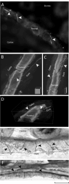
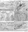
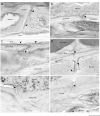
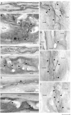
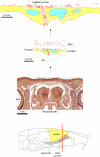
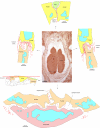
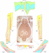
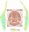
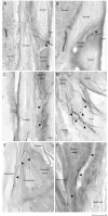
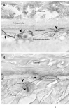
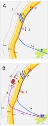
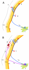
Migraine sufferers frequently testify that their headache feels as if the calvarial bones are deformed, crushed, or broken (Jakubowski et al. [2006] Pain 125:286-295). This has lead us to postulate that the calvarial bones are supplied by sensory fibers. We studied sensory innervation of the calvaria in coronal and horizontal sections of whole-head preparations of postnatal and adult mice, via immunostaining of peripherin (a marker of thinly myelinated and unmyelinated fibers) or calcitonin gene-related peptide (CGRP; a marker more typical of unmyelinated nerve fibers). In pups, we observed nerve bundles coursing between the galea aponeurotica and the periosteum, between the periosteum and the bone, and between the bone and the meninges; as well as fibers that run inside the diploë in different orientations. Some dural fibers issued collateral branches to the pia at the frontal part of the brain. In the adult calvaria, the highest concentration of peripherin- and CGRP-labeled fibers was found in sutures, where they appeared to emerge from the dura. Labeled fibers were also observed in emissary canals, bone marrow, and periosteum. In contrast to the case in pups, no labeled fibers were found in the diploë of the adult calvaria. Meningeal nerves that infiltrate the periosteum through the calvarial sutures may be positioned to mediate migraine headache triggered by pathophysiology of extracranial tissues, such as muscle tenderness and mild trauma to the skull. In view of the concentration of sensory fibers in the sutures, it may be useful to avoid drilling the sutures in patients undergoing craniotomies for a variety of neurosurgical procedures.
Copyright 2009 Wiley-Liss, Inc.
Figures







References
-
- Alberius P, Skagerberg G. Adrenergic innervation of the calvarium of the neonatal rat. Its relationship to the sagittal suture and developing parietal bones. Anat Embryol (Berl) 1990;182(5):493–498. - PubMed
-
- Alvarez FJ, Morris HR, Priestley JV. Sub-populations of smaller diameter trigeminal primary afferent neurons defined by expression of calcitonin gene-related peptide and the cell surface oligosaccharide recognized by monoclonal antibody LA4. J Neurocytol. 1991;20(9):716–731. - PubMed
-
- Bolay H, Reuter U, Dunn AK, Huang Z, Boas DA, Moskowitz MA. Intrinsic brain activity triggers trigeminal meningeal afferents in a migraine model. Nat Med. 2002;8(2):136–142. - PubMed
-
- Caterina MJ, Schumacher MA, Tominaga M, Rosen TA, Levine JD, Julius D. The capsaicin receptor: a heat-activated ion channel in the pain pathway. Nature. 1997;389(6653):816–824. - PubMed
-
- de Gray LC, Matta BF. Acute and chronic pain following craniotomy: a review. Anaesthesia. 2005;60(7):693–704. - PubMed
Publication types
MeSH terms
Substances
Grants and funding
LinkOut - more resources
Full Text Sources
Other Literature Sources
Research Materials

