. 2009 Sep;13(9B):3720-9.
doi: 10.1111/j.1582-4934.2009.00785.x. Epub 2009 May 6.

VEGFR2+PDGFRbeta+ circulating precursor cells participate in capillary restoration after hyperoxia acute lung injury (HALI)

Affiliations

VEGFR2+PDGFRbeta+ circulating precursor cells participate in capillary restoration after hyperoxia acute lung injury (HALI)

Rosemary Jones et al. J Cell Mol Med. 2009 Sep.

Abstract

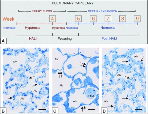
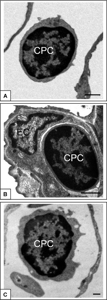
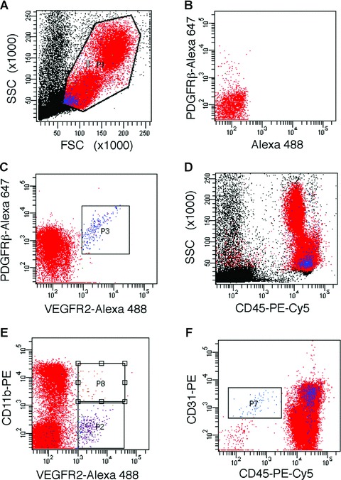
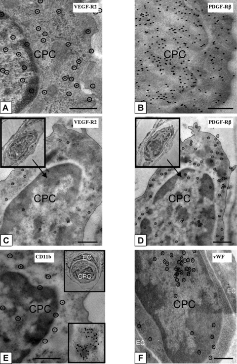
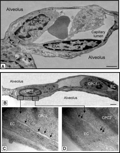
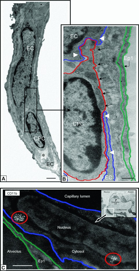
The in vivo morphology and phenotype of circulating cells that spontaneously contribute to new vessel formation in adults remain unclear. Here, we use high-resolution imaging and flow cytometry to characterize the morphology and phenotype of a distinct population of circulating mononuclear cells contributing to spontaneous new vessel formation after hyperoxia acute lung injury (HALI). We identify a subpopulation of myeloid (CD11b/Mac1(+)) haematopoietic cells co-expressing vascular endothelial growth factor receptor 2 (VEGFR2) and platelet derived growth factor receptor beta (PDGFRbeta). Moreover, we show that these CD11b(+)VEGFR2(+)PDGFRbeta(+) circulating precursor cells (CPCs) contribute structurally to the luminal surface of capillaries re-forming 2 weeks post-HALI. This indicates that these myeloid CPCs may function, at least transiently, as putative vascular precursors, and has important implications for capillary growth and repair in injury and in pathologies of the lung and other organs.


Figures

Figure 1
Figure 1
In vivo model of capillary remodelling in HALI and post-HALI. Schema of in vivo model (a) and representative brightfield images illustrating patent capillaries in normal lung (b, arrows), their loss in HALI (c, arrows, week 4) and the restoration of patent capillaries early in the post-HALI phase following spontaneous repair (d, arrow, week 6). In the model, rodents breathe 87% oxygen for 4 weeks (HALI) or 87% oxygen for 4 weeks, followed by weaning to air for 1 week (week 5) and air breathing for up to 4 weeks (post-HALI, weeks 6–9). Initially in HALI (week 1), ECs of small vessels and capillaries in the alveolar-capillary membrane are severely injured. As these cells (and epithelial cells) adapt to the high oxygen tension, the membrane remodels; however there is extensive capillary loss (HALI, weeks 2–4). Because of the greatly reduced capillary bed present at this time, the oxygen tension is lowered (∼less 10%) daily during the transition to breathing air (week 5) to prevent asphyxia, dyspnoea and cyanosis—the lower oxygen tension of air (i.e. relative hypoxia) triggering a burst of spontaneous vascular repair post-HALI (weeks 6 and 7). Spontaneous capillary repair and expansion continue to restore the membrane post-HALI until, as patent capillary networks approach their normal distribution, the response wanes (week 9) [8]. Two-μm-thick resin sections stained with toluidine blue. Bars = 25 μm (b–d).
Figure 2
Figure 2
CPC in capillaries of normal lung, in HALI and post-HALI. CPCs were identified by their similar morphology as freely circulating cells in patent capillaries of normal lung (a), in the narrowed capillary segments in HALI (b), and again as freely circulating cells in patent capillary segments post-HALI (c, week 9). Characterized by a high ratio of nucleus to cytoplasm and dense arrangement of heterochromatin CPCs were clearly identified from adjacent capillary ECs (see b). Eighty-nm-thick epon resin sections stained with uranyl acetate and lead citrate. Bars = 1 μm (a–c).
Figure 3
Figure 3
Phenotypic characterization of CPCs. LSR-II flow cytometric analyses of (gated, a) mononuclear cells in peripheral blood, identified a distinct population of CPCs that co-express VEGFR2 and PDGFRβ (c); single-colour controls were used for compensation (e.g. see staining for PDGFRβ-Alexa Fluor 647 in b). These CPCs (in blue) are positive for CD45 (d) and show scattering properties typical of small cells with a high nucleus to cytoplasm ratio (a, d). A subset of CPCs is positive for CD11b (e), whereas all VEGFR2+PDGFRβ+ CPCs are positive for CD31 (f); CPCs are distinct from cells with a phenotype consistent with ‘circulating ECs’, i.e. CD31+CD45 (see gated population in f).
Figure 4
Figure 4
Phenotypic characterization of CPCs in lungs post-HALI. Antigenic sites, visualized with 10-nm protein A-gold by high-resolution microscopy, demonstrated that CPCs were VEGFR2+ (a) and PDGFRβ+ (b) post-HALI (weeks 6 and 7). Representative images of the same CPC profile in adjacent sections of lung tissue (c and d, and insets) demonstrated co-expression of VEGFR2 and PDGFRβ antigenic sites post-HALI (week 7). Other representative images of additional antigenic sites expressed by CPCs post-HALI (week 7): CD11b (e and inset) and for vWF (f). Typically, the sites for CD11b were uniformly distributed over CPCs but also appeared as clusters (e, see bottom inset, illustrating an example of a cluster of ∼40 CD11b+ labelled sites). Ten-nm gold-labelled antigenic sites (a, c–f) are circled for clarity to distinguish these from 7 to 8 nm ribosomes on cell profiles. Ninety-nm-thick Unicryl resin sections stained with uranyl acetate and lead citrate. Bars = 0.5 μm (a–f).
Figure 5
Figure 5
CPCs contact and adhere to endothelium post HALI. (a) Representative image of CPC aligning and adhering to a capillary EC (see arrows) post-HALI (week 7). At this time, approximately 35% of CPCs form contacts with lung capillary ECs. (b) Representative image of two CPCs in a lung capillary post-HALI (week 6): CPC1 appears free in the lumen and CPC2 is in the process of adhering to the adjacent EC (see boxed areas): higher magnification shows regional separation of two distinct cell membranes (lower left image, arrows), and regional loss of membrane delineation between the two cells (lower right image, arrows). Eighty-nm-thick epon resin sections stained with uranyl acetate and lead citrate. Bars = 1 μm (a) and 2 μm (b).
Figure 6
Figure 6
CD11b+ CPCs and cells integrate into capillary surfaces post HALI. Illustrations of CD11b+ CPC (a) and capillary EC in a residual capillary structure post-HALI (week 6). An asterisk marks the restricted capillary lumen at this time. A region of the CPC cytosol is inserted between the processes of two capillary ECs (b, black arrows). Note the location of the CPC within the capillary lumen in relation to the processes of adjacent ECs (white arrowheads), to (an un-delineated) perivascular cell process, and to the underlying epithelial (Ep1) cell process. The blue line delineates the plasmalemmal membrane of the capillary EC, the red line outlines the CPC, and the green line demarcates the adjacent epithelial type 1 (Ep1). (c) Representative image of lung capillary (see boxed area of inset) and higher magnification of the luminal surface illustrating cell clusters of antigenic sites for CD11b (circled in red) typical of CPCs expressing this protein post-HALI (week 7). Image is inverted to highlight the location of this cell with antigenic sites. The other EC indicated in the same capillary (see inset, small arrow) does not express CD11b. Eighty-nm-thick epon resin section (a, b), and 90-nm-thick Unicryl resin section (c), stained with uranyl acetate and lead citrate. Bars = 1 μm (a), 0.5 μm (b, c).

References

    1. Dos Santos CC. Hyperoxic acute lung injury and ventilator-induced/associated lung injury: new insights into intracellular signaling pathways. Crit Care. 2007;11:126. - PMC - PubMed
    1. Li LF, Liao SK, Ko YS, et al. Hyperoxia increases ventilator-induced lung injury via mitogen-activated protein kinases: a prospective, controlled animal experiment. Crit Care. 2007;11:R25. - PMC - PubMed
    1. Rafii S, Lyden D. Therapeutic stem and progenitor cell transplantation for organ vascularization and regeneration. Nat Med. 2003;9:702–12. - PubMed
    1. Du R, Lu KV, Petritsch C, et al. HIF1alpha induces the recruitment of bone marrow-derived vascular modulatory cells to regulate tumor angiogenesis and invasion. Cancer Cell. 2008;13:206–20. - PMC - PubMed
    1. Rafii S, Lyden D, Benezra R, et al. Vascular and haematopoietic stem cells: novel targets for anti-angiogenesis therapy. Nat Rev Cancer. 2002;2:826–35. - PubMed

Publication types

Substances