. 2009 Sep;13(9B):4014-22.
doi: 10.1111/j.1582-4934.2009.00763.x. Epub 2009 May 1.

High kallikrein-related peptidase 6 in non-small cell lung cancer cells: an indicator of tumour proliferation and poor prognosis

Affiliations

High kallikrein-related peptidase 6 in non-small cell lung cancer cells: an indicator of tumour proliferation and poor prognosis

Heuzé-Vourc'h Nathalie et al. J Cell Mol Med. 2009 Sep.

Abstract

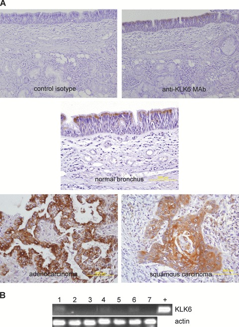
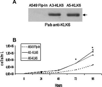
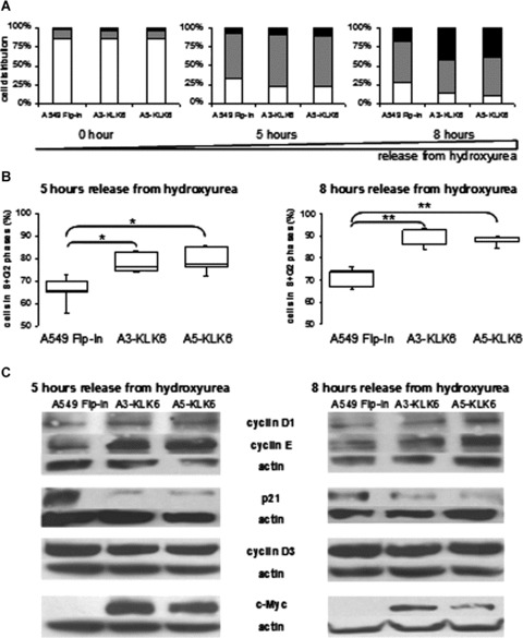
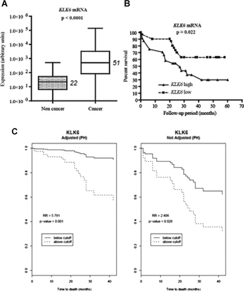
The human kallikrein-related peptidases (KLK) are serine proteases whose concentrations are often abnormal in common human malignancies and contribute to neoplastic progression through multifaceted roles. However, little attention has been paid to their synthesis and involvement in the development and dissemination of lung cancer, the leading cause of cancer mortality worldwide. We have analysed the production of KLK6 in normal lung and tumour tissues from patients with non-small cell lung cancer (NSCLC). KLK6 immunoreactivity was restricted to epithelial cells of the normal bronchi, but most of the cancer samples were moderately or highly immunoreactive, regardless of the histological subtype. In contrast, little or no KLK6 was detected in NSCLC cells. We have developed NSCLC lines expressing wild-type KLK6 in order to investigate the role of KLK6 in lung cancer biology, and analysed its impact on proliferation. Ectopic KLK6 dramatically enhanced NSCLC cell growth and KLK6-producing NSCLC cells had accelerated cell cycles, between the G1 and S phases. This was accompanied by a marked increase in cyclin E and decrease in p21. KLK6 production was also associated with enhanced synthesis of c-Myc, which is known to promote cell-cycle progression. Finally, examination of specimens from patients with NSCLC revealed that KLK6 mRNA is overexpressed in tumour tissue, and high KLK6 concentrations were associated with lower survival rates. We conclude that a high concentration of KLK6 is an indicator of tumour proliferation and an independent predictive factor in NSCLC.


Figures

Figure 1
Figure 1
KLK6 in tissues from patients with NSCLC but not in NSCLC-derived cell lines. (A) Immunohistochemistry of KLK6 in normal and malignant lung from 46 patients with primary NSCLC using a monoclonal antibody (original magnification: ×200; haematoxylin counterstain). KLK6 immunostaining (dark) in the apical region of normal bronchial epithelial cells (thin arrow) and in the cytoplasm of most cancer cells (large arrow). (B) KLK6 production in a panel of NSCLC lines by RT-PCR. Total RNA from NSCLC cell lines was extracted and reverse transcribed. KLK6 and β-actin (internal control) mRNAs were amplified from cDNA by PCR and the amplification products were separated on a 1% agarose gel containing ethidium bromide. (1) A549; (2) Calu-1; (3) H23; (4) H460; (5) H520; (6) H522; (7) H1838; +: plasmid containing KLK6 cDNA.
Figure 2
Figure 2
Ectopic KLK6 in NSCLC promotes proliferation. (A) Secretion of KLK6 by A549 Flp-In and stable isogenic clones (A3- and A5-KLK6) assessed by Western blotting using a polyclonal anti-KLK6 antibody. Equal volumes of cell culture supernatant were loaded onto 12% SDS-PAGE gels. (B) Control A549 Flp-In (dashed line) and A3-KLK6 (black full line) and A5-KLK6 (grey full line) (2.5 × 10³ cells of each) were seeded and cells were counted at the indicated times. Points, median value of triplicate determinations from one representative experiment (of three). Bars, lower and upper quartiles. P-values were determined using a Kruskal and Wallis test. *P < 0.05.
Figure 3
Figure 3
Effect of KLK6 on NSCLC cell growth and regulation of cell cycle progression. (A) Distribution of control A549 Flp-In and A3-KLK6 and A5-KLK6 cells within the G1 (white), S (grey) and G2 (black) phases of the cell cycle 0, 5 or 8 hrs after release from hydroxyurea. Results are expressed as the median values of five independent experiments. (B) Box plot of control A549 Flp-In and A3-KLK6 and A5-KLK6 cells passed from G1 into G2+S phases 5 and 8 hrs after release from hydroxyurea. Results are representative of five independent experiments. P-values were determined using a Kruskal and Wallis test. *P < 0.05, **P < 0.025. (C) Assay of cell-cycle proteins and c-Myc by Western blotting of whole-cell extracts (50 μg) from the control A549 Flp-In, A3-KLK6 and A5-KLK6 lines 5 and 8 hrs after release from hydroxyurea. Actin protein was used to check for equal loading. Results are from one of three experiments.
Figure 4
Figure 4
KLK6 overexpression in tumour tissues from patients with NSCLC, and patient survival. (A) Box plot of KLK6 transcript concentration by quantitative real-time RT-PCR in 57 tumours and matched non tumour samples from patients with primary NSCLC undergoing an initial surgical resection of the lung tumour (aged 44–83 years, median: 65). Gene expression was normalized to the amount of 18S rRNA and is reported in arbitrary units. Median values are indicated in italics. The difference between the concentration of KLK6 transcripts in non-tumour and tumour tissues was determined by a Wilcoxon test for matched pairs. (B) Kaplan–Meier curves for overall survival of patients with high and low concentrations of KLK6 mRNA in lung tumour tissues. The optimal cut-off value for survival analysis was identified by the chi-squared test, based on the ability of KLK6 to predict the overall survival of the population studied. Using this cut-off, KLK6 mRNA concentration was categorized as high or low. A cut-off of 600 (in arbitrary units) for KLK6 mRNA was equal to the 56th percentile. (C) Survival curves obtained from Cox regression models after adjusting for gender, age, histological type, tumour size, nodal tumour status, stage and smoking status (packs/ year)

References

    1. Borgoño CA, Diamandis EP. The emerging roles of human tissue kallikreins in cancer. Nat Rev Cancer. 2004;4:876–90. - PubMed
    1. Hutchinson S, Luo LY, Yousef GM, et al. Purification of human kallikrein 6 from biological fluids and identification of its complex with alpha(1)-antichymotrypsin. Clin Chem. 2003;49:746–51. - PubMed
    1. Petraki CD, Karavana VN, Skoufogiannis PT, et al. The spectrum of human kallikrein 6 (zyme/protease M/neurosin) expression in human tissues as assessed by immunohistochemistry. J Histochem Cytochem. 2001;49:1431–41. - PubMed
    1. Petraki CD, Papanastasiou PA, Karavana VN, et al. Cellular distribution of human tissue kallikreins: immunohistochemical localization. Biol Chem. 2006;387:653–63. - PubMed
    1. Shaw JL, Diamandis EP. Distribution of 15 human kallikreins in tissues and biological fluids. Clin Chem. 2007;53:1423–32. - PubMed

Publication types

MeSH terms