Review
. 2009 May;229(1):12-26.
doi: 10.1111/j.1600-065X.2009.00770.x.

CD28 and CTLA-4 coreceptor expression and signal transduction

Affiliations
Review

CD28 and CTLA-4 coreceptor expression and signal transduction

Christopher E Rudd et al. Immunol Rev. 2009 May.

Abstract

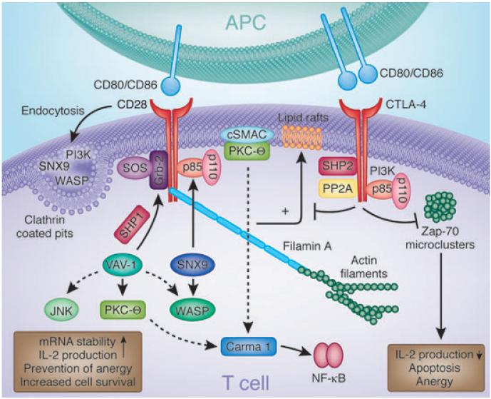
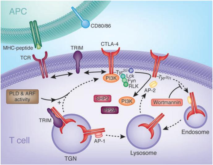
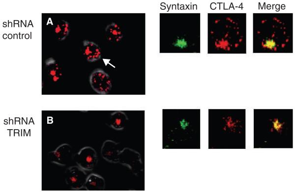
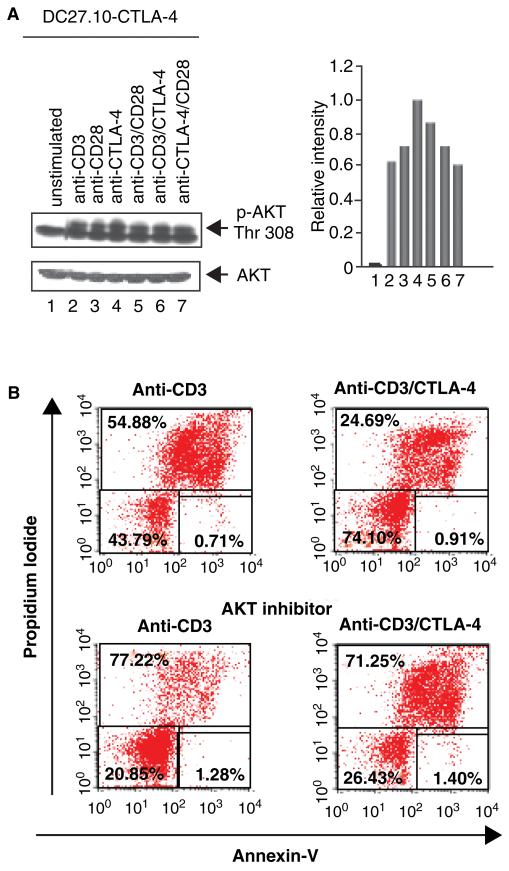
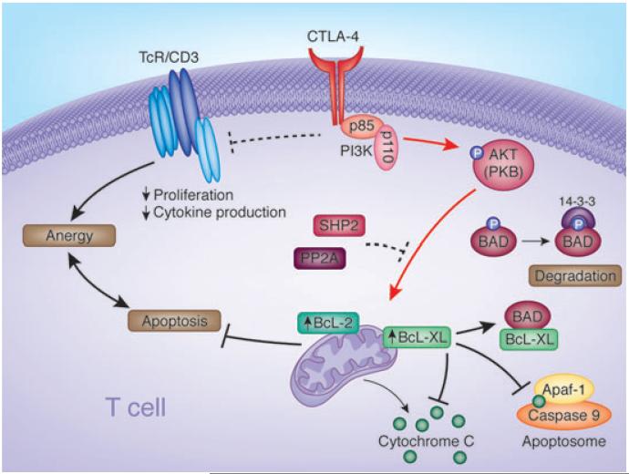
T-cell activation is mediated by antigen-specific signals from the TCRzeta/CD3 and CD4-CD8-p56lck complexes in combination with additional co-signals provided by coreceptors such as CD28, inducible costimulator (ICOS), cytotoxic T-lymphocyte antigen-4 (CTLA-4), programmed death (PD-1), and others. CD28 and ICOS provide positive signals that promote and sustain T-cell responses, while CTLA-4 and PD-1 limit responses. The balance between stimulatory and inhibitory co-signals determines the ultimate nature of T-cell responses where response to foreign pathogen is achieved without excess inflammation and autoimmunity. In this review, we outline the current knowledge of the CD28 and CTLA-4 signaling mechanisms [involving phosphatidylinositol 3 kinase (PI3K), growth factor receptor-bound protein 2 (Grb2), Filamin A, protein kinase C theta (PKCtheta), and phosphatases] that control T-cell immunity. We also present recent findings on T-cell receptor-interacting molecule (TRIM) regulation of CTLA-4 surface expression, and a signaling pathway involving CTLA-4 activation of PI3K and protein kinase B (PKB)/AKT by which cell survival is ensured under conditions of anergy induction.


Figures

Fig. 1
Fig. 1. Signaling molecules involved in CD28 and CTLA-4 function
CD28 and CTLA-4 associate with various intracellular signaling proteins. CD28 associates with PI3K and Grb2 via SH2 domain binding to the Tyr–Val–Asn–Met (YMNM) motif. PI3K generates PI3,4P2 and PI3,4,5P3 lipids, while Grb2 binds to the exchange factor Sos1, an activator of the GTPase p21ras. CD28–Grb2 binding is also needed for the phosphorylation and activation of Vav1. In turn, Vav1 can activate Rac1 that activates the serine/threonine kinase JNK, may activate Cdc42 in the activation of WASP, and is needed for the membrane association of PKCθ. Protein tyrosine phosphatase 1 (SHP-1) can also associate with Vav1, Grb2, and Sos1, dampening signals from CD28. The CD28–PYAPP motif (located further to the C-terminus) has been reported to bind to FLNA and colocalizes with PKCθ and CD28. Mutations in the PYAPP motif block PKCθ formation in a cSMAC. CD28 endocytosis is regulated by the combination of mediators, PI3K, WASP, and SNX9. Mutations that disrupt PI3K binding impair internalization via a clathrin-dependent mechanism. SNX9 binds WASP via its SH3 domain and uses its PX domain to interact with the p85 subunit of PI3K and its product PIP3. WASP, SNX9, PI3K, and CD28 colocalize within clathrin-containing endocytic vesicles after TCR/CD28 costimulation. On the other hand, CTLA-4 binds to PI3K using its YVKM motif as well as phosphatases SHP-2 and PP2A. Phosphatases have been postulated to generate negative signals. CTLA-4 can also block the expression of lipid rafts (GEMs) and the induction by the TCR of ZAP-70 microcluster formation. CTLA-4, cytotoxic T-lymphocyte antigen-4; PI3K, phosphatidylinositol 3-kinase; SH2, src homology 2; WASP, Wiskott–Aldrich syndrome protein; PKCθ, protein kinase C h; cSMAC, central supramolecular activation cluster; SNX, sorting nexin; TCR, T-cell receptor; YVKM, Val–Tyr–Val–Lys–Met; SHP-1, SH2-domain-containing protein tyrosine phosphatase 1; GEM, glycolipid-enriched microdomain.
Fig. 2
Fig. 2. Regulation of CTLA-4 surface expression and internalization
Newly synthesized CTLA-4 binds to the transmembrane adapter TRIM in the TGN promoting the formation of CTLA-4-containing vesicles and their transport to the cell surface. On the cell surface, CTLA-4 and TRIM no longer associate allowing TRIM to interact with other receptors, possibly the TCR complex. CTLA-4 externalization is also dependent on general factors such as PLD and GTPase ARF-1. Shuttling to the lysosomal compartment from the TGN occurs due to adapter AP-1 binding to GVY201VKM motif in CTLA-4. On the surface, CTLA-4 becomes phosphorylated on the Y201VKM by kinases Lck, Fyn, and Rlk leading to the association of PI3K and possibly other proteins. Phosphorylation retards internationalization. Dephosphorylation allows binding to the clathrin adapter AP-2 to the GVY201VKM motif and rapid internalization to endosomes and lysosomes. Upon T-cell activation, CTLA-4 enriched lysosomes (and endosomes) are recycled to the cell surface. This process involves Wortmannin-sensitive lipid kinases but not PI3K (174). CTLA-4, cytotoxic T-lymphocyte antigen-4; TRIM, T-cell receptor-interacting molecule; TGN, trans-Golgi network; TCR, T-cell receptor; PLD, phospholipase D; ARF, adenosine diphosphate ribosylation factor; AP, adapter protein; PI3K, phosphatidylinositol 3-kinase.
Fig. 3
Fig. 3. TRIM controls the generation of TGN proximal vesicles with CTLA-4
(A) Pattern of intracellular CTLA-4 staining in wildtype T cells (shRNA control) (left panel). Arrows point to CTLA-4-labeled vesicles. Right panels: cells labeled with anti-Syntaxin (TGN marker), anti-CTLA-4, and merged images. Anti-CTLA-4 labels both TGN and intracellular vesicles (TGN proximal and endosomal/lysosomal). (B) Pattern of intracellular CTLA-4 staining in TRIM knockdown cells (left panel). There is a general loss of CTLA-4 labeled vesicles. Instead, CTLA-4 is restricted to the Golgi area. Right panels: This loss of CTLA-4 vesicles was confirmed by analysis of a magnified single-cell using anti-Syntaxin and anti-CTLA-4 followed by merging of the images. TRIM, T-cell receptor-interacting molecule; TGN, trans-Golgi network; CTLA-4, cytotoxic T-lymphocyte antigen-4; shRNA, short-hairpin RNA.
Fig. 4
Fig. 4. CTLA-4 coligation can rescue T cells from AICD under conditions of anergy induction
(A) CTLA-4 coligation induces AKT–Thr–308 phosphorylation. DC27.10–CTLA-4 cells were left untreated (lane 1) or stimulated for 30 min with anti-CD3 (lane 2), anti-CD28 (lane 3), anti-CTLA-4 (lane 4), anti-CD3/CD28 (lane 5), anti-CD3/CTLA-4 (lane 6), and anti-CD28/CTLA-4 (lane 7) antibodies. The DC27.10 cell is a mouse hybridoma that has been stably transfected with CTLA-4 (i.e. DC27.10–CTLA-4). Cell lysates were subjected to immunoblotting with anti-phospho-PKB/AKT (Thr-308) antibody (lanes 1–7). Lower panel: equal amounts of cell lysates were detected by anti-PKB/AKT blotting (lanes 1–7). Histogram depiction of phosphorylated PKB/AKT as detected by densitometric reading. Similar observations were made in peripheral T cells (86). (B) CTLA-4 coligation can rescue T cells from AICD in an AKT/PKB-dependent manner. Pre-activated PBLs were stimulated with anti-CD3 (left panel) and anti-CD3/CTLA-4 (right panel) in the absence (upper panel) or presence of AKT inhibitor (AKT inhibitor II) (lower panel). CTLA-4 coligation reversed cell death induced by anti-CD3 (i.e. 54% to 24% cell death), an event reversed by the inhibition of AKT/PKB (i.e. 77% and 71% cell death) and PI3K (data not shown). Cell viability was assessed by staining with Annexin V-Cy5 and propidium iodide and analyzed using FACS. CTLA-4, cytotoxic T-lymphocyte antigen-4; AICD, antigen-induced cell death; PKB, protein kinase B; PBL, peripheral blood lymphocyte; PI3K, phosphatidylinositol 3-kinase.
Fig. 5
Fig. 5. Diagram outlining the mechanism of CTLA-4 induced prosurvival signaling pathways
CTLA-4 coligation can reverse AICD induced by ligation of the TCR complex. In the signaling pathway, CTLA-4–PI3K activates PKB/Akt by phosphorylation at Thr-308. CTLA-4 also inactivates proapoptotic BAD by phosphorylation at Ser-136. BAD phosphorylation at Ser-136 leads to binding to 14–3–3 proteins and frees Bcl-XL and Bcl-2 to mediate mitochondrial-dependent cell survival. BAD otherwise promotes cell death by sequestering Bcl-XL/Bcl-2 leading to the release of cytochrome c and apoptosis. Inactivated BAD induced by CTLA-4 ligation is also accompanied by increased Bcl-XL/Bcl-2 expression. CTLA-4–PI3K-PKB/Akt maintenance of cell survival under conditions of anergy induction provides a potential mechanism to ensure long-term tolerance in immunity. CTLA, cytotoxic T-lymphocyte antigen; AICD, antigen-induced cell death; TCR, T-cell receptor; PI3K, phosphatidylinositol 3-kinase; PKB, protein kinase B; BAD, Bcl-2 antagonist of cell death.

References

    1. Naluai A, et al. The CTLA4/CD28 gene region on chromosome 2q33 confers susceptibility to celiac disease in a way possibly distinct from that of type 1 diabetes and other chronic inflammatory disorders. Tissue Antigens. 2001;56:350–355. - PubMed
    1. Howard T, Rochelle J, Seldin M. Cd28 and Ctla-4, two related members of the Ig supergene family, are tightly linked on proximal mouse chromosome 1. Immunogenetics. 1991;33:74–76. - PubMed
    1. Balzano C, Buonavista N, Rouvier E, Golstein P. CTLA-4 and CD28: similar proteins, neighbouring genes. Int J Cancer Suppl. 1992;7:28–32. - PubMed
    1. van der Merwe P, Bodian D, Daenke S, Linsley PS, Davis SJ. CD80 (B7-1) binds both CD28 and CTLA-4 with a low affinity and very fast kinetics. J Exp Med. 1997;185:393–403. - PMC - PubMed
    1. Ikemizu S, et al. Structure and dimerization of a soluble form of B7-1. Immunity. 2000;12:51–60. - PubMed

MeSH terms