Interplay between the heterotrimeric G-protein subunits Galphaq and Galphai2 sets the threshold for chemotaxis and TCR activation
- PMID: 19426503
- PMCID: PMC2694176
- DOI: 10.1186/1471-2172-10-27
Interplay between the heterotrimeric G-protein subunits Galphaq and Galphai2 sets the threshold for chemotaxis and TCR activation
Abstract
Background: TCR and CXCR4-mediated signaling appears to be reciprocally regulated pathways. TCR activation dampens the chemotactic response towards the CXCR4 ligand CXCL12, while T cells exposed to CXCL12 are less prone to subsequent TCR-activation. The heterotrimeric G proteins Galphaq and Galphai2 have been implicated in CXCR4-signaling and we have recently also reported the possible involvement of Galphaq in TCR-dependent activation of Lck (Ngai et al., Eur. J. Immunol., 2008, 38: 32083218). Here we examined the role of Galphaq in migration and TCR activation.
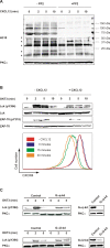
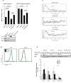
Results: Pre-treatment of T cells with CXCL12 led to significantly reduced Lck Y394 phosphorylation upon TCR triggering indicating heterologous desensitization. We show that knockdown of Galphaq significantly enhanced basal migration in T cells and reduced CXCL12-induced SHP-1 phosphorylation whereas Galphai2 knockdown inhibited CXCL12-induced migration.
Conclusion: Our data suggest that Galphai2 confers migration signals in the presence of CXCL12 whereas Galphaq exerts a tonic inhibition on both basal and stimulated migrational responses. This is compatible with the notion that the level of Galphaq activation contributes to determining the commitment of the T cell either to migration or activation through the TCR.
Figures


Similar articles
-
The heterotrimeric G-protein alpha-subunit Galphaq regulates TCR-mediated immune responses through an Lck-dependent pathway.Eur J Immunol. 2008 Nov;38(11):3208-18. doi: 10.1002/eji.200838195. Eur J Immunol. 2008. PMID: 18991294
-
Differential regulation of CXCR4-mediated T-cell chemotaxis and mitogen-activated protein kinase activation by the membrane tyrosine phosphatase, CD45.J Biol Chem. 2003 Mar 14;278(11):9536-43. doi: 10.1074/jbc.M211803200. Epub 2003 Jan 8. J Biol Chem. 2003. PMID: 12519755
-
p52Shc is required for CXCR4-dependent signaling and chemotaxis in T cells.Blood. 2007 Sep 15;110(6):1730-8. doi: 10.1182/blood-2007-01-068411. Epub 2007 May 30. Blood. 2007. PMID: 17537990
-
Intracellular mediators of CXCR4-dependent signaling in T cells.Immunol Lett. 2008 Jan 29;115(2):75-82. doi: 10.1016/j.imlet.2007.10.012. Epub 2007 Nov 20. Immunol Lett. 2008. PMID: 18054087 Review.
-
TAOK3, a Regulator of LCK-SHP-1 Crosstalk during TCR Signaling.Crit Rev Immunol. 2019;39(1):59-81. doi: 10.1615/CritRevImmunol.2019030480. Crit Rev Immunol. 2019. PMID: 31679194 Review.
Cited by
-
A single amino acid substitution in CXCL12 confers functional selectivity at the beta-arrestin level.Oncotarget. 2018 Jun 22;9(48):28830-28841. doi: 10.18632/oncotarget.25533. eCollection 2018 Jun 22. Oncotarget. 2018. PMID: 29989007 Free PMC article.
-
Phosphoinositide 3-kinase-dependent antagonism in mammalian olfactory receptor neurons.J Neurosci. 2011 Jan 5;31(1):273-80. doi: 10.1523/JNEUROSCI.3698-10.2011. J Neurosci. 2011. PMID: 21209212 Free PMC article.
-
MAPK signaling drives inflammation in LPS-stimulated cardiomyocytes: the route of crosstalk to G-protein-coupled receptors.PLoS One. 2012;7(11):e50071. doi: 10.1371/journal.pone.0050071. Epub 2012 Nov 30. PLoS One. 2012. PMID: 23226236 Free PMC article.
-
Co-receptor signaling in the pathogenesis of neuroHIV.Retrovirology. 2021 Aug 24;18(1):24. doi: 10.1186/s12977-021-00569-x. Retrovirology. 2021. PMID: 34429135 Free PMC article. Review.
-
Gq-Coupled Receptors in Autoimmunity.J Immunol Res. 2016;2016:3969023. doi: 10.1155/2016/3969023. Epub 2016 Jan 17. J Immunol Res. 2016. PMID: 26885533 Free PMC article. Review.
References
-
- Ma Q, Jones D, Borghesani PR, Segal RA, Nagasawa T, Kishimoto T. Impaired B-lymphopoiesis, myelopoiesis, and derailed cerebellar neuron migration in CXCR4- and SDF-1-deficient mice. Proceedings of the National Academy of Sciences of the United States of America. 1998;95:9448–9453. doi: 10.1073/pnas.95.16.9448. - DOI - PMC - PubMed
Publication types
MeSH terms
Substances
LinkOut - more resources
Full Text Sources
Molecular Biology Databases
Miscellaneous

