Review
. 2009:68:133-216.
doi: 10.1016/S0065-2164(09)01204-0.

The capsule of the fungal pathogen Cryptococcus neoformans

Affiliations
Review

The capsule of the fungal pathogen Cryptococcus neoformans

Oscar Zaragoza et al. Adv Appl Microbiol. 2009.

Abstract

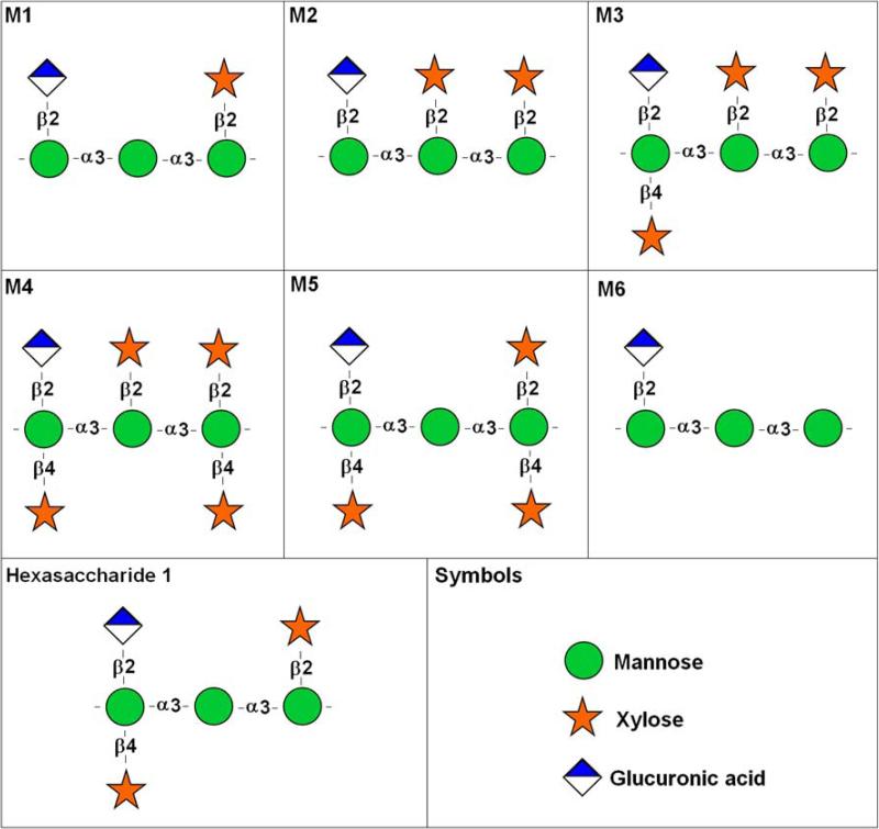
The capsule of the fungal pathogen Cryptococcus neoformans has been studied extensively in recent decades and a large body of information is now available to the scientific community. Well-known aspects of the capsule include its structure, antigenic properties and its function as a virulence factor. The capsule is composed primarily of two polysaccharides, glucuronoxylomannan (GXM) and galactoxylomannan (GalXM), in addition to a smaller proportion of mannoproteins (MPs). Most of the studies on the composition of the capsule have focused on GXM, which comprises more than 90% of the capsule's polysaccharide mass. It is GalXM, however, that is of particular scientific interest because of its immunological properties. The molecular structure of these polysaccharides is very complex and has not yet been fully elucidated. Both GXM and GalXM are high molecular mass polymers with the mass of GXM equaling roughly 10 times that of GalXM. Recent findings suggest, however, that the actual molecular weight might be different to what it has traditionally been thought to be. In addition to their structural roles in the polysaccharide capsule, these molecules have been associated with many deleterious effects on the immune response. Capsular components are therefore considered key virulence determinants in C. neoformans, which has motivated their use in vaccines and made them targets for monoclonal antibody treatments. In this review, we will provide an update on the current knowledge of the C. neoformans capsule, covering aspects related to its structure, synthesis and particularly, its role as a virulence factor.


Figures

Figure 1
Figure 1
Different micrographs and compositions showing the polysaccharide capsule of C. neoformans. A) Suspension of the cells in India Ink; B) Scanning electron microscopy; C-H) Immunofluorescence using specific mAbs to the capsule (green and red fluorescence) showing also the cell wall localization (blue flurorescence). D) 3D image composition of a C. neoformans cell labeled with two different mAbs to the capsule. In blue, the cell wall. E) Side view of a section of cell shown in D. F-H) Sections showing the 3 dimension of the capsule, visualized after staining with mAbs (green and red). Pictures by Oscar Zaragoza, and from (Maxson et al, 2007b).
Figure 2
Figure 2. Repeating polysaccharide motifs of GXM
Motifs M1 to M6, which were described by Cherniak and colleagues (1998) as the major structure reporter groups (SRG) of GXM, are shown. Hexasaccharide 1, an additional GXM substituted triad, is also presented. Symbol nomenclature for glycans followed the format available in http://grtc.ucsd.edu/symbol.html.
Figure 3
Figure 3. Basic GalXM structure
A GalXM motif has a α (1→6) galactan backbone. The branches are β- 3-O-linked to the backbone and consist of an α (1→3)-Man, α (1→4)-Man, β -galactosidase trisaccharide with variable amounts of β(1→2)- or β (1→3)-xylose side groups. Symbol nomenclature for glycans followed the format available in http://grtc.ucsd.edu/symbol.html.
Figure 4
Figure 4
Model of capsule growth and capsule rearrangements during budding in C. neoformans (From Zaragoza et al, 2006b). A. Model of capsule growth. After capsule enlarges a newly synthesized capsule (light green) accumulates at the edge of the capsule, with the old capsule (light blue) remaining close to the cell wall. B. Model of adaptation to non-capsule growth inducing conditions. Cells with large capsules, when transferred to a medium that does not induce capsule growth, cannot degrade the capsule, but the new emerging buds have a small capsule. C. Rearrangements of the capsule during budding. Panels 1–6 illustrate schematically different stages of budding. When bud arises, a dimple and a tunnel are formed (2, 3), which allows the separation of the bud. Bud growth is accompanied by capsule growth in the bud (3, 4). Capsule of the mother cell closes as the bud separates from the mother cell, at the same time that the bud completes the capsule without taking any polysaccharide of the mother cell (5), which will allow for the complete separation of the bud (6).
Figure 5
Figure 5. Proposed model for GXM traffic in C. neoformans
GXM is synthesized in the Golgi apparatus and released in post-Golgi vesicles. These vesicles would be integrated into endocytic compartments / multivesicular bodies that are further addressed to the plasma membrane. After fusion of endosome-derived structures with the plasma membrane, small vesicles are released into the periplasmic space to be translocated to the extracellular space. Extracellular vesicles would then release their content, which includes capsular polysaccharides. Abbreviations: N, nucleus; G, Golgi apparatus; PM, plasma membrane; E, endosome; MVB, multivesicular body; CW, cell wall; C, capsule.
Figure 6
Figure 6. Cell wall-capsule connections in C. neoformans
Staining of capsular structures with a monoclonal antibody to GXM (green), cell wall chitin with calcofluor white (blue), and chitin-derived oligomers with WGA (red) followed by confocal analysis reveals that projected structures recognized by WGA connect the cell wall and the capsule. For experimental details, see (Rodrigues et al., 2008a).
Figure 7
Figure 7
Scheme summarizing all the levels at which capsular polysaccharides induce apoptosis on antigen presenting cells and T-cells. GXM and GalXM induce apoptosis on antigen presenting cells, such as macrophages, and T-cells. In macrophages, GXM and GalXM induce accumulation of molecules involved in apoptosis induction, such as FasL and Fas. These molecules induce apoptosis in both macrophages and T-cells. In addition, in T-cells, GalXM can directly induce apoptosis through Fas molecules. Apoptosis occurs through caspase pathway.
Figure 8
Figure 8
Comparison of model prediction with experimental results obtained for C. neoformans cells using immunofluorescence. Green shows the concentration of complex. The boundaries of the cell wall are shown in blue, detected using calcoflour. Shown are data for two cell sizes (adapted from Rakesh et.al, 2008).

References

    1. Aksenov SI, Babyeva IP, Golubev VI. On the mechanism of adaptation of micro-organisms to conditions of extreme low humidity. Life Sci Space Res. 1973;11:55–61. - PubMed
    1. Alspaugh JA, Perfect JR, Heitman J. Cryptococcus neoformans mating and virulence are regulated by the G-protein alpha subunit GPA1 and cAMP. Genes Dev. 1997;11(23):3206–17. - PMC - PubMed
    1. Alspaugh JA, Pukkila-Worley R, Harashima T, Cavallo LM, Funnell D, Cox GM, Perfect JR, Kronstad JW, Heitman J. Adenylyl cyclase functions downstream of the Galpha protein Gpa1 and controls mating and pathogenicity of Cryptococcus neoformans. Eukaryot Cell. 2002;1(1):75–84. - PMC - PubMed
    1. Alvarez M, Casadevall A. Phagosome extrusion and host-cell survival after Cryptococcus neoformans phagocytosis by macrophages. Curr Biol. 2006;16(21):2161–5. - PubMed
    1. Alvarez M, Casadevall A. Cell-to-cell spread and massive vacuole formation after Cryptococcus neoformans infection of murine macrophages. BMC Immunol. 2007;8:16. - PMC - PubMed

Publication types

MeSH terms

LinkOut - more resources