. 2009:457:63-80.
doi: 10.1016/S0076-6879(09)05004-6.

32P labeling of protein phosphorylation and metabolite association in the mitochondria matrix

Affiliations

32P labeling of protein phosphorylation and metabolite association in the mitochondria matrix

Angel M Aponte et al. Methods Enzymol. 2009.

Abstract

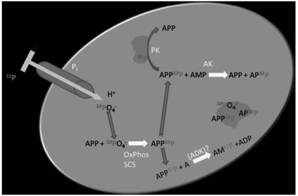
Protein phosphorylations, as well as phosphate metabolite binding, are well characterized post-translational mechanisms that regulate enzyme activity in the cytosol, but remain poorly defined in mitochondria. Recently extensive matrix protein phosphorylation sites have been discovered but their functional significance is unclear. Herein we describe methods of using (32)P labeling of intact mitochondria to determine the dynamic pools of protein phosphorylation as well as phosphate metabolite association. This screening approach may be useful in not only characterizing the dynamics of these pools, but also provide insight into which phosphorylation sites have a functional significance. Using the mitochondrial ATP synthetic capacity under appropriate conditions, inorganic (32)P was added to energized mitochondria to generate high specific activity gamma-P(32)-ATP in the matrix. In general, SDS denaturing and gel electrophoresis was used to primarily follow protein phosphorylation, whereas native gel techniques were used to observe weaker metabolite associations since the structure of mitochondrial complexes was minimally affected. The protein phosphorylation and metabolite association within the matrix was found to be extensive using these approaches. (32)P labeling in 2D gels was detected in over 40 proteins, including most of the complexes of the cytochrome chain and proteins associated with intermediary metabolism, biosynthetic pathways, membrane transport, and reactive oxygen species metabolism. (32)P pulse-chase experiments further revealed the overall dynamics of these processes that included phosphorylation site turnover as well as phosphate-protein pool size alterations. The high sensitivity of (32)P resulted in many proteins being intensely labeled, but not identified due to the sensitivity limitations of mass spectrometry. These low concentration proteins may represent signaling proteins within the matrix. These results demonstrate that the mitochondrial matrix phosphoproteome is both extensive and dynamic. The use of this, in situ, labeling approach is extremely valuable in confirming protein phosphorylation sites as well as examining the dynamics of these processes under near physiological conditions.


Figures

Figure 1
Figure 1
Schematic diagram of mitochondrial matrix generating γ-32P-ATP from extramitochondrial 32P as well as other phosphate labeled metabolites. Pt: phosphate transport protein. ADK: adenosine kinase, it is not clear that this enzyme is present and active in the matrix. AK: adenylate kinase. PK: protein kinase. OxPhos: Oxidative phosphorylation. SCS: Succinate CoA Synthetase. Grey masses represent proteins that have metabolites bound or are phosphorylated.
Figure 2
Figure 2
32P Dose Response in Heart Mitochondria. Porcine heart mitochondria were incubated in standard conditions for 20 min where A) 1μCi B) 25μCi, C) 50μCi and D) 250μCi of 32P was added. Proteins were separated in the horizontal direction by isoelectric focusing point (pI), from pH ~4 to 10, and vertically by molecular weight, from ~150 to 10 kDa. The relative amplitude for each image was arbitrarily set.
Figure 3
Figure 3
32P labeling assignments in isolated heart mitochondria. Mitochondria were incubated for 20 minutes with 250 uCi of 32P. Chromatography is the same at in Figure 2. Protein assignments: 1 ATP synthase, mitochondrial F1 complex, β subunit. 2) 60 kDa heat shock protein, mitochondrial precursor. 3) NADH dehydrogenase (ubiquinone) Fe-S protein 1 (75kDa). 4) 70kDa heat shock protein, mitochondrial precursor. 5) Pyruvate dehydrogenase complex, E2 subunit. 6) ATP synthase d-chain, mitochondrial precursor. 7) Pyruvate dehydrogenase complex, E1 subunit. 8) Elongation factor Tu, mitochondrial precursor. 9) Aconitase hydratase, mitochondrial precursor. 10) Isocitrate dehydrogenase (NAD) subunit alpha, mitochondrial precursor 11) Succinyl-CoA ligase (GDPforming) alpha-chain, mitochondrial precursor. 12) Mn superoxide dismutase. 13) Creatine kinase, sarcomeric mitochondrial precursor. 14) Cytochrome c oxidase polypeptide Va, mitochondrial precursor. 15) Voltage-dependent anion channel 1. 16) ATP synthase, mitochondrial F1 complex, α subunit.
Figure 4
Figure 4
32P Pulse-Chase Experiments. Control, 32P was added to porcine heart mitochondria and incubated for 20 min 32P was added to heart mitochondria and incubated for 10 min then cold inorganic phosphate was added to the incubation media for an additional 10 min. Heart mitochondria incubated with cold inorganic phosphate for 10 min then 32P was added to the incubation media for an additional 10 min. Proteins were separated in the horizontal direction by isoelectric focusing point (pI), from pH ~4 to 10, and vertically by molecular weight, from ~150 to 10 kDa. All exposures were identical along with window level values in the display.
Figure 5
Figure 5
The Effects of Dichloroacetate (DCA) and Pyruvate on 32P Labeled Heart Mitochondria. Control: Heart mitochondria incubated in standard conditions for 20 min with 32P. DCA/Pyruvate: Heart mitochondria treated with 0.1 mM DCA and 5 mM pyruvate for 1 h on ice then 32P was added during the standard 20 min incubation at 37°C. Box indicates region of the phosphorylated pyruvate dehydrogenase E1 protein.
Figure. 6
Figure. 6
Purified Labeled Complex V from Porcine Heart Mitochondria. Subunit identifications were obtained by mass spectrometry. Purified proteins were separated by two-dimensional gel electrophoresis, first in the horizontal direction by isoelectric focusing point (pI), from pH ~4 to 10, and then vertically by molecular weight, from ~150 to 10 kDa.
Figure 7
Figure 7
Native PAGE of 32P labeled porcine heart mitochondrial complexes. A) cold water destain, no acid for 2 min B) BN-PAGE gel was fixed in an hot fixative followed by hot acid destain for additional 15 min.

References

    1. Aprille JR. Mechanism and regulation of the mitochondrial ATP-Mg/P(i) carrier. J. Bioenerg. Biomembr. 1993;25:473–481. - PubMed
    1. Ballif BA, Villen J, Beausoleil SA, Schwartz D, Gygi SP. Phosphoproteomic analysis of the developing mouse brain. Mol. Cell Proteomics. 2004;3:1093–1101. - PubMed
    1. Bender E, Kadenbach B. The allosteric ATP-inhibition of cytochrome c oxidase activity is reversibly switched on by cAMP-dependent phosphorylation. FEBS Lett. 2000;466:130–134. - PubMed
    1. Berger F, Lau C, Dahlmann M, Ziegler M. Subcellular compartmentation and differential catalytic properties of the three human nicotinamide mononucleotide adenylyltransferase isoforms. J. Biol. Chem. 2005;280:36334–36341. - PubMed
    1. Biondi RM, Nebreda AR. Signalling specificity of Ser/Thr protein kinases through docking-site-mediated interactions. Biochem. J. 2003;372:1–13. - PMC - PubMed

MeSH terms

Substances

LinkOut - more resources