Review
. 2009 Jun;30(6):278-86.
doi: 10.1016/j.tips.2009.04.001. Epub 2009 May 6.

Shuttling and translocation of heterotrimeric G proteins and Ras

Affiliations
Review

Shuttling and translocation of heterotrimeric G proteins and Ras

Deepak K Saini et al. Trends Pharmacol Sci. 2009 Jun.

Abstract

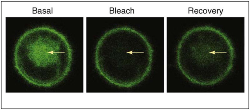
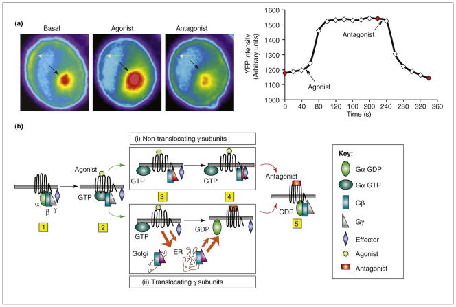
Heterotrimeric G proteins (alphabetagamma) and Ras proteins are activated by cell-surface receptors that sense extracellular signals. Both sets of proteins were traditionally thought to be constrained to the plasma membrane and some intracellular membranes. Live-cell-imaging experiments have now shown that these proteins are mobile inside a cell, shuttling continually between the plasma membrane and intracellular membranes in the basal state, maintaining these proteins in dynamic equilibrium in different membrane compartments. Furthermore, on receptor activation, a family of G protein betagamma subunits translocates rapidly and reversibly to the Golgi and endoplasmic reticulum enabling direct communication between the extracellular signal and intracellular membranes. A member of the Ras family has similarly been shown to translocate on activation. Although the impact of this unexpected intracellular movement of signaling proteins on cell physiology is likely to be distinct, there are striking similarities in the properties of these two families of signal-transducing proteins.


Figures

Figure 1
Figure 1
Shuttling of the G protein γ2 subunit from the plasma membrane to the Golgi. Chinese hamster ovary (CHO) cells transiently expressing yellow fluorescent protein (YFP)-γ2 were used. Arrows indicate the Golgi region, which was photobleached in the basal cell by laser and was monitored for fluorescence recovery (FRAP). The recovery shown in the right panel reflects the retrograde movement of YFP-γ2 from the plasma membrane to the Golgi, corresponding to the shuttling movement of this protein. Note that the γ2 subunit types are not capable of translocation on receptor activation, indicating that the ability to shuttle between plasma membrane and intracellular membranes in the basal state does not necessarily confer the ability to translocate on receptor activation.
Figure 2
Figure 2
Palmitoylation-dependent shuttling of signaling proteins. Shuttling of G protein heterotrimers occurs with equal rapidity in both retrograde and anterograde directions through simple diffusion (solid arrows). Shuttling of the Ras proteins involves retrograde movement from the plasma membrane to the Golgi by simple diffusion, whereas the anterograde movement is through vesicle-mediated trafficking and is distinctly slower. Palmitoyl (red) attached to the α subunit and Ras is removed resulting in reduced affinity for the plasma membrane even though the γ subunit and Ras contain a prenyl moiety (black). Palmitoyl transferase in the Golgi acylates the depalmitoylated α subunit and is thought to acylate Ras. Thus, a common mechanism – deacylation at the plasma membrane and palmitoylation at the Golgi – is thought to mediate the shuttling of these two signaling proteins.
Figure 3
Figure 3
Translocation of G protein βγ9 to the Golgi from the plasma membrane in response to M2 cetylcholine receptor activation. CHO cells stably expressing the M2 acetylcholine receptor and transiently transfected with G protein αo, β1 and YFP-γ9 subunits were used. Left hand side: images of the YFP-γ9 subunit from the cells were captured before agonist treatment and followed by agonist and antagonist treatment at 20 s intervals. Cells were exposed to 100 μM carbachol (an agonist) followed by 100 μM atropine (an antagonist) at defined time points. (a) Images showing translocation of YFP-γ9 to the Golgi. Arrows represent the regions in the cell where intensity changes in response to receptor activation and deactivation are observed. Yellow arrows indicate the plasma membrane and black arrows indicate the Golgi region. The decrease in fluorescence intensity over time is due to photobleaching. Right hand side: plot representing YFP intensity changes over time in the Golgi. Arrows indicate the time of addition of agonist and antagonist. Points shown in red represent the images shown in left panel. (b) Model for the mechanistic basis of translocation. After receptor activation by an agonist (steps 1 and 2), the G protein follows pathways (i) or (ii) depending on its γ subunit type. (i) Non-translocating γ subunits enable the βγ complex to stay on the activated receptor after the release of the α subunit (step 3). Competing affinities for an effector (blue diamond) and the receptor determine dissociation of βγ from the receptor (step 4). (ii) Translocation-capable γ subunits have low affinity for the activated receptor compared with a target in the Golgi or ER and translocate as a βγ complex to these membranes (step 3). Orange arrows indicate that translocation rates vary among βγ types depending on their relative affinities for the activated receptor or effectors. Receptor inactivation leads to an increase in affinity for the βγ complex and higher α-GDP concentration (step 4). Both pathways finally lead to increased α-GDP βγ binding to the inactivated receptor (step 5).
Figure 4
Figure 4
The evolutionary relationship between the primary structures of G protein γ subunits and Ras proteins. Protein sequences of γ subunits and Ras proteins of human origin were used to generate a phylogenetic tree based on sequence alignment at ClustalW website (www.ebi.ac.uk/ClustalW). Color codes are given to proteins based on their intracellular movement observed in living cells: red, Ras proteins; blue, rapidly translocating Gγ subunits; green, slowly translocating Gγ subunits; black, non-translocating Gγ subunits.

References

    1. Denker SP, et al. Differential distribution of α subunits and βγ subunits of heterotrimeric G proteins on Golgi membranes of the exocrine pancreas. J Cell Biol. 1996;133:1027–1040. - PMC - PubMed
    1. Pimplikar SW, Simons K. Regulation of apical transport in epithelial cells by a Gs class of heterotrimeric G protein. Nature. 1993;362:456–458. - PubMed
    1. Stow JL, et al. A heterotrimeric G protein, Gαi–3, on Golgi membranes regulates the secretion of a heparan sulfate proteoglycan in LLC-PK1 epithelial cells. J Cell Biol. 1991;114:1113–1124. - PMC - PubMed
    1. Slessareva JE, et al. Activation of the phosphatidylinositol 3- kinase Vps34 by a G protein α subunit at the endosome. Cell. 2006;126:191–203. - PubMed
    1. Garcia-Regalado A, et al. G protein-coupled receptor-promoted trafficking of Gβ1γ2 leads to AKT activation at endosomes via a mechanism mediated by Gβ1γ2–Rab11a interaction. Mol Biol Cell. 2008;19:4188–4200. - PMC - PubMed

Publication types