. 2009 May 8;4(5):453-63.
doi: 10.1016/j.stem.2009.03.017.

Cell of origin and microenvironment contribution for NF1-associated dermal neurofibromas

Affiliations

Cell of origin and microenvironment contribution for NF1-associated dermal neurofibromas

Lu Q Le et al. Cell Stem Cell. .

Abstract

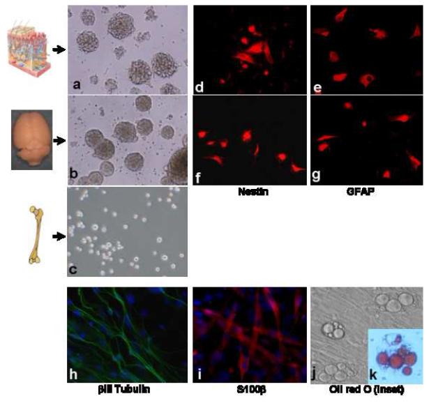
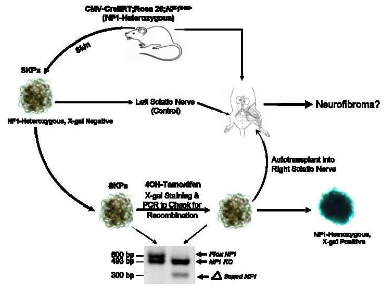
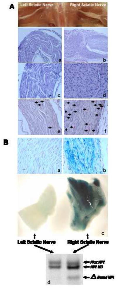
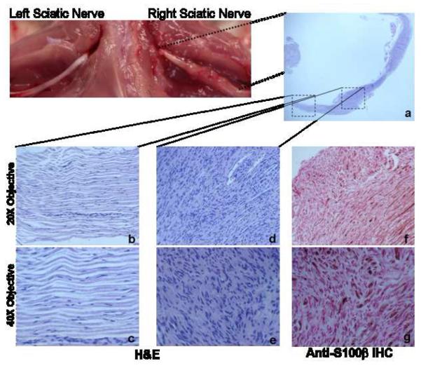
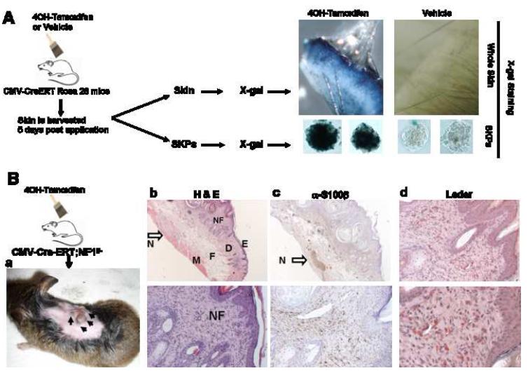
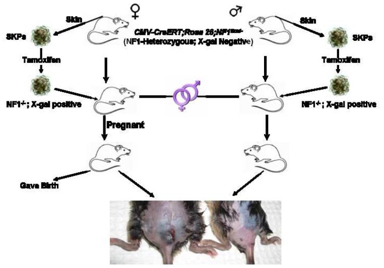
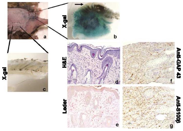
The tumor predisposition disorder neurofibromatosis type I (NF1) is one of the most common genetic disorders of the nervous system. It is caused by mutations in the Nf1 tumor-suppressor gene, which encodes a GTPase-activating protein (GAP) that negatively regulates p21-RAS. Development of malignant nerve tumors and neurofibromas occurs frequently in NF1. However, little is known about the molecular mechanisms mediating the initiation and progression of these complex tumors, or the identity of the specific cell type that gives rise to dermal or cutaneous neurofibromas. In this study, we identify a population of stem/progenitor cells residing in the dermis termed skin-derived precursors (SKPs) that, through loss of Nf1, form neurofibromas. We propose that SKPs, or their derivatives, are the cell of origin of dermal neurofibroma. We also provide evidence that additional signals from nonneoplastic cells in the tumor microenvironment play essential roles in neurofibromagenesis.


Figures

Figure 1
Figure 1. SKPs are Multipotent Progenitor Cells in the Dermis
Isolation of neural stem/progenitor cells from the skin and brain. Phase photomicrograph of neurospheres derived from mouse skin (a) and subventricular zone (SVZ) (b). Under the same culturing conditions, bone marrow cells did not form spheres (c). Both neurosphere cells purified from skin (d&f) and subventricular zone (e&g) express the neural stem cell markers nestin and GFAP, respectively. Under differentiating conditions, SKP-derived spheres differentiate into neurons that express βIII tubulin (h), glial cells that express S100β(l) and adipocytes containing characteristic intracellular lipid droplets (j) as demonstrated by Oil Red O staining (k).
Figure 2
Figure 2. Schematic Representation of the Experimental Design to Generate a Novel Dermal Neurofibroma Model
SKPs are purified from skin on neck, ears, or back for deletion of Nf1 ex vivo with 4OH-tamoxifen. The SKPs are then analyzed by X-gal staining and PCR genotyping to ensure successful recombination at the ROSA26 and Nf1 loci, respectively. The Nf1-/- SKPs are then autologously transplanted back into the same animal either intradermally/subcutaneously in the right flank or in proximity to the right sciatic nerve. As controls, Nf1+/- or Nf1+/+ SKPs are transplanted on the left side in the same manner.
Figure 3
Figure 3. Neurofibroma Formation Requires Proximity to the Nerves and Develops from Implanted SKPs
3A. H&E staining of enlarged right sciatic nerve where Nf1-/-;LacZ+ SKPs had been implanted (b: original magnification 20X; d: original magnification 40X) showing features consistent with plexiform neurofibroma, including disordered, convoluted bundles of cells exhibiting spindle-cell morphology, with ovoid and spindle-shaped nuclei and adjacent fine fibrillar stroma. There is also massive mast cell infiltration (black arrows) within the tumor as demonstrated by Leder staining (f), which stains mast cells red. As shown by H&E staining, these features are absent in the left sciatic nerve where control Nf1+/-;LacZ- SKPs were implanted (a: original magnification 20X; c: original magnification 40X) and Leder staining (e). 3B. SKPs were purified from skin taken from neck, ears, or back of CMV-CreERT2;Nf1flox/-;ROSA26flox-stop-flox mice and exposed to 4OH-tamoxifen to induce recombination at the ROSA26 and Nf1 loci. Two to three months after SKP implantation, mice underwent total body perfusion with 4% paraformaldehyde and left and right sciatic nerves were harvested for gross X-gal staining of whole nerves. The nerves were then post-fixed in formalin for paraffin sectioning and subsequently counter-stained with nuclear fast red. Representative X-gal stainings of whole left and right sciatic nerves show that the enlarged right sciatic nerve is X-gal positive (c, right panel) but not the control left sciatic nerve (c, left panel). The corresponding paraffin sections also show histological evidence of neurofibroma with X-gal-positive, disordered spindle-cell morphology, with ovoid and spindle-shaped nuclei for the right sciatic nerve (b) but not the control left sciatic nerve (a). The left and right sciatic nerves were also subjected to semiquantitative PCR genotyping (d). The recombined loxP sites (Δfloxed Nf1) at the Nf1 locus were detected in the tumor on the right sciatic nerve (d), further indicating that the neurofibromas are derived from the implanted SKPs. (Flox Nf1 = floxed Nf1 allele; Nf1 KO = mutant Nf1 allele).
Figure 4
Figure 4. Neurofibroma Formation at the Site of NF1-/- SKP Implantation
H&E staining of a longitudinal section of right sciatic nerve (a, original magnification 4X) showing neurofibroma formation with features consistent with plexiform neurofibroma, only at the site of SKP implantation (d: original magnification 20X; e: original magnification 40X) and not at the site further away from where the Nf1-/- SKPs were implanted (b: original magnification 20X; c: original magnification 40X). These neurofibromas are positive for the Schwann cell marker S100β (f: original magnification 20X; g: original magnification 40X).
Figure 5
Figure 5. Topical Application of Tamoxifen to the Skin for In Vivo Deletion of Nf1 in SKPs Induced Dermal Neurofibromas
5A. The CMV-CreERT2;ROSA26flox-stop-flox mice were painted with tamoxifen topically onto the back skin twice a day for 5 consecutive days. Five days after the last application, the skin was harvested for both X-gal staining and purification of SKPs for subsequent X-gal staining. Only skin and isolated SKPs derived from tamoxifen painted sites were positive for X-gal staining, indicating that the tamoxifen was able to penetrate the skin and recombine the flox sites within the SKP nuclei in vivo. 5B. a. Tamoxifen was painted topically onto the upper back skin of neonatal CMV-CreERT2;Nf1flox/- mice twice a day for 5 consecutive days. Six to seven months post tamoxifen application, these mice developed cutaneous nodules similar to human dermal neurofibroma at the tamoxifen application sites. b. Representative H&E staining of these cutaneous nodules showing circumscribed, non-encapsulated, intradermal, spindle cell proliferation within the dermis. (E=epidermis; D=dermis; F=fat; M=muscle). The dermal neurofibroma (NF=neurofibroma) stroma has fine wavy strands of collagen with associated spindle cells containing elongated wavy nuclei. There is an enlarged cutaneous nerve just beneath the tumor, between the subcutaneous fat and muscle layer (open arrow, N=Nerve). These are classic features of dermal neurofibroma (upper panel: original magnification 10X; lower panel: original magnification 40X). c. Immunohistochemical stains show spindle cells within neurofibromas are positive for S-100β protein. The enlarged cutaneous nerve just beneath the tumor (arrow) served as the positive control and the epidermis served as the negative control (upper panel: original magnification 10X; lower panel: original magnification 40X). d. Leder staining showed heavy infiltration of mast cells (Red) within the dermal neurofibroma (upper panel: original magnification 20X; lower panel: original magnification 40X).
Figure 6
Figure 6. Role of Steroid Hormones in Dermal Neurofibroma Development
SKPs were purified from skin on occipital scalp, ears, and superior back of both male and female mice for deletion of NF1 ex vivo with 4OH-tamoxifen. The SKPs were then X-gal stained and PCR genotyped to ensure successful recombination at the ROSA26 and Nf1 loci, respectively. The Nf1-/- SKPs were then autologously transplanted back into the same animal either intradermally/subcutaneously at dorsal, distal back (sacral area). Both male and female mice are then housed in the same cage to facilitate breeding. All female mice eventually became pregnant. Subsequently, within three months post SKP implantation, the female mice but not the male mice, developed cutaneous nodules at the graft site.
Figure 7
Figure 7. Dermal Neurofibromas Arise from Implanted Nf1-/- SKPs
a. Cutaneous nodule formation at the site of SKP implantation in female mice. b. X-gal staining showing the tumor/nodule within the dermis and subcutaneous tissue is LacZ-positive. However, the epidermis of the same tissue (arrow), which is genetically unmodified host tissue, is LacZ-negative, indicating that the tumor is derived from the implanted SKPs. c. X-gal staining of skin, where there is no SKP implantation, showing this control skin is LacZ-negative. d. Representative H&E staining of these cutaneous nodules showing classic features of dermal neurofibroma (original magnification 20X). e. Leder staining shows heavy infiltration of mast cells (Red) within the dermal neurofibroma (original magnification 20X). f. anti-GAP 43 and g. anti—S100β immunohistochemistry shows that spindle cells in the neurofibroma are positive for the Schwann cell markers GAP43 and S100β (original magnification 20X).

Comment in

References

    1. Al-Hajj M, Clarke MF. Self-renewal and solid tumor stem cells. Oncogene. 2004;23:7274–7282. - PubMed
    1. Biernaskie J, Sparling JS, Liu J, Shannon CP, Plemel JR, Xie Y, Miller FD, Tetzlaff W. Skin-derived precursors generate myelinating Schwann cells that promote remyelination and functional recovery after contusion spinal cord injury. J Neurosci. 2007;27:9545–9559. - PMC - PubMed
    1. Biernaskie JA, McKenzie IA, Toma JG, Miller FD. Isolation of skin-derived precursors (SKPs) and differentiation and enrichment of their Schwann cell progeny. Nat Protoc. 2006;1:2803–2812. - PubMed
    1. Cichowski K, Jacks T. NF1 tumor suppressor gene function: narrowing the GAP. Cell. 2001;104:593–604. - PubMed
    1. Cichowski K, Shih TS, Schmitt E, Santiago S, Reilly K, McLaughlin ME, Bronson RT, Jacks T. Mouse models of tumor development in neurofibromatosis type 1. Science. 1999;286:2172–2176. - PubMed

Publication types

Substances