

Review
. 2009 May;93(3):583-604, Table of Contents.
doi: 10.1016/j.mcna.2009.02.008.
Arterial aging and subclinical arterial disease are fundamentally intertwined at macroscopic and molecular levels
Affiliations
- PMID: 19427493
- PMCID: PMC2943242
- DOI: 10.1016/j.mcna.2009.02.008
Item in Clipboard
Review
Arterial aging and subclinical arterial disease are fundamentally intertwined at macroscopic and molecular levels
Med Clin North Am.
2009 May.
Abstract
The structure and function of arteries change throughout a lifetime. Age is the dominant risk factor for hypertension, coronary heart disease, congestive heart failure, and stroke. The cellular/molecular proinflammatory alterations that underlie arterial aging are novel putative candidates to be targeted by interventions aimed at attenuating arterial aging as a major risk factor for cardiovascular diseases. This review provides a landscape of central arterial aging and age-disease interactions, integrating perspectives that range from humans to molecules, with the goal that future therapies for cardiovascular diseases, such as hypertension, also will target the prevention or amelioration of unsuccessful arterial aging.
Figures

Age-associated macroscopic changes in central arterial structure in humans. Central arterial lumen (A) and intimal-medial wall thickness (B) increase with advancing age in men (red) and women (blue). Best-fitting age regression curves are shown for men (solid lines) and women (dashed lines).

Age-associated aortic structural remodeling. (A) Arterial immunostaining for α-SMA (brown) from monkeys. Arrowhead indicates internal elastin lamina. (B) Movat staining (upper panels) and immunohistochemical staining (brown color) for α-SMA (middle panels) and for CD68 (brown color) (lower panels) in aorta from humans. (C) Toludine blue staining for mast cells in old human aorta (left panel) and observed under an oil lens (right panel). I, intima; L, lumen; M, media. Up-down arrows indicate thickened intima. (From Wang M, Takagi G, Asai K, et al. Aging increases aortic MMP-2 activity and angiotensin II in nonhuman primates. Hypertension 2003;41(6):1308–16 and Wang M, Zhang J, Jiang LQ, et al. Proinflammatory profile within the grossly normal aged human aortic wall. Hypertension 2007;50(1):219–7; with permission.)

VSMC during aging. (A) Chemotatic response to a PDGF-BB gradient is increased in early passage VSMC from the aortic media of old rats compared with those from younger rats. VSMC within the older aorta are primed to respond to the growth factor. (From Pauly RR, Passaniti A, Crow M, et al. Experimental models that mimic the differentiation and dedifferentiation of vascular cells. Circulation 1992;86(Suppl):III68–73; with permission.) (B) Growth curves of VSMC cultured from young and old rat aortae. The number of VSMC obtained from old rats was significantly higher at days 3, 7, and 14. (From Li Z, Cheng H, Lederer WJ, et al. Enhanced proliferation and migration and altered cytoskeletal proteins in early passage smooth muscle cells from young and old rat aortic explants. Exp Mol Pathol 1997;64(1):1–11; with permission.)

Age-associated changes in arterial function in humans. (A) Flow-mediated induced dilation in the brachial artery of apparently healthy women. (Adapted from Celermajer DS, Sorensen KE, Spiegelhalter DJ, et al. Aging is associated with endothelial dysfunction in healthy men years before the age-related decline in women. J Am Coll Cardiol 1994;24(2):471–6; with permission.) Age-associated increase in carotid-femoral PWV (B), an index of central arterial stiffness, and in augmentation index (C) in healthy men (red) and women (blue). Best-fitting age regression curves are shown for men (solid lines) and women (dashed lines).

Ang II and its converting enzymes increase in the aged aortic wall from rats and nonhuman primates. (A) Immunolabeled Ang II (red) in the en face medial aortic sections from young (left panel) and old rats (right panels). (From Jiang L, Wang M, Zhang J, et al. Increased aortic calpain-1 activity mediates age-associated angiotensin II signaling of vascular smooth muscle cells. PLoS ONE 2008;3:e2231.) (B) The immunofluorescent staining for Ang II (red color) on frozen sections of aortae from monkeys. (C) The immunostaining for ACE (red color) on frozen sections of aortae from monkeys. (D) The immunostaining for chymase in an old monkey aorta. Inset, rectangular region under high power. A, adventitia; L, lumen; M, media. (From Wang M, Takagi G, Asai K, et al. Aging increases aortic MMP-2 activity and angiotensin II in nonhuman primates. Hypertension 2003;41(6): 1308–16; with permission.)

Components of the RAS in the human aortic wall. Immunofluorescence staining for (A) Ang II (red color); (B) AT1 (green color) (upper panels); and (C) ACE (green color) (upper panel). L, lumen; M, media. (From Wang M, Zhang J, Jiang LQ, et al. Proinflammatory profile within the grossly normal aged human aortic wall. Hypertension 2007;50(1):219–7; with permission.)

Simplified Ang II signaling cascades. (From Wang M, Lakatta EG. Central arterial aging: humans to molecules. In: Safar M, editor. Handbook of hypertension: arterial stiffness in hypertension. Amsterdam: Elsevier; 2006. p. 137–60; with permission.)
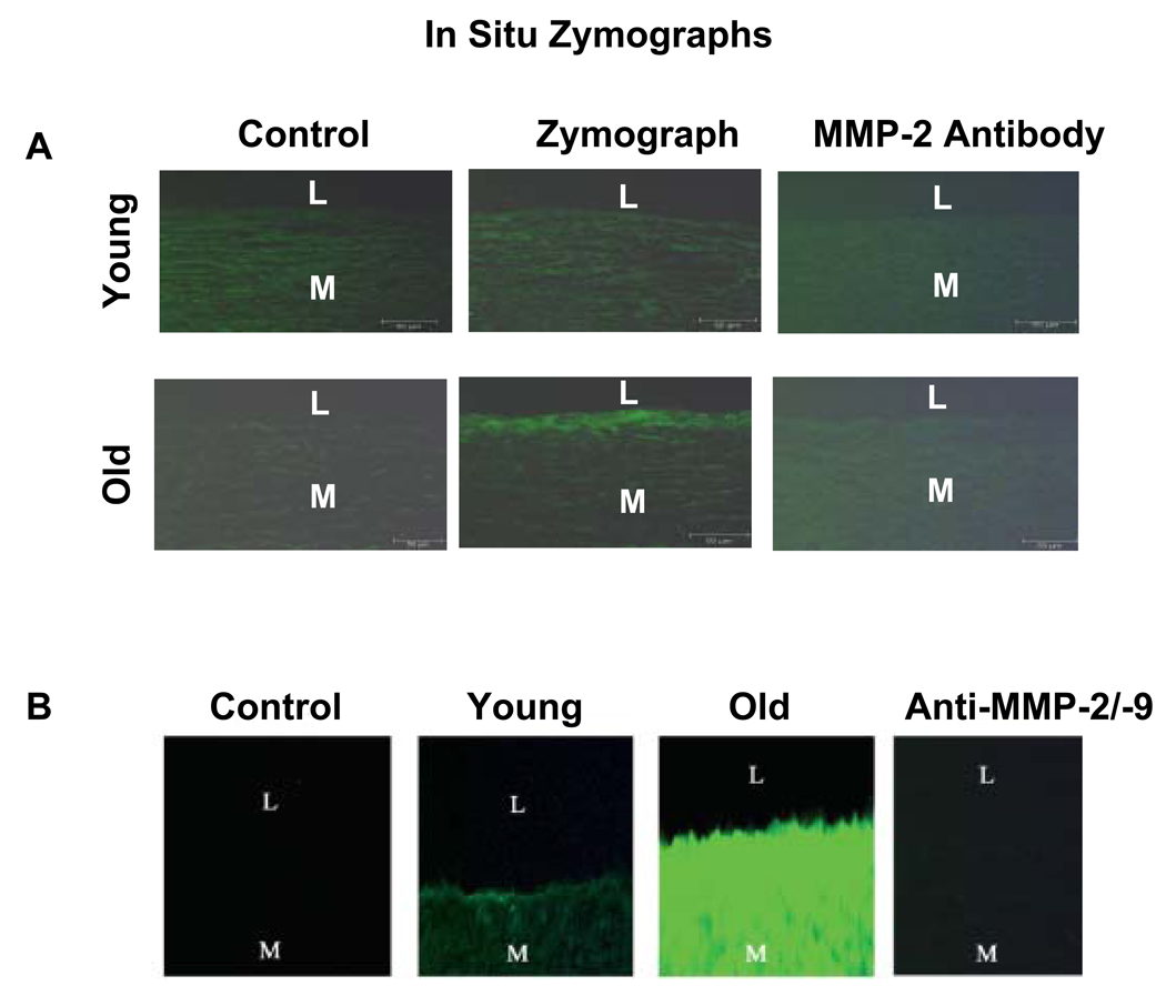
In situ gelatin zymograms. (A) In situ gelatin zymographs of monkey aorta. Protease activity (green color) is localized mainly in the older aortic intima (middle panel). (From Wang M, Takagi G, Asai K, et al. Aging increases aortic MMP-2 activity and angiotensin II in nonhuman primates. Hypertension 2003;41(6):1308–16.) (B) In situ gelatin zymographs (green color) of humans. I, intimae; L, lumen; M, media. *P<.05, young versus old. (From Wang M, Zhang J, Jiang LQ, et al. Proinflammatory profile within the grossly normal aged human aortic wall. Hypertension 2007;50(1):219–7; with permission.)
References
-
- Najjar SS, Scuteri A, Lakatta EG. Arterial aging: is it an immutable cardiovascular risk factor? Hypertension. 2005;46(3):454–462. - PubMed
-
- Lakatta EG, Levy D. Arterial and cardiac aging: major shareholders in cardiovascular disease enterprises: part I: aging arteries: a “set-up” for vascular disease. Circulation. 2003;107(1):139–146. - PubMed
-
- Najjar SS, Lakatta EG. Vascular aging: from molecular to clinical cardiology. In: Patterson WC, Runge M, editors. Principles of molecular cardiology. Totowa (NJ): Humana Press; 2005. pp. 517–547.
-
- Wang M, Lakatta EG. Central arterial aging: humans to molecules. In: Safar M, editor. Handbook of hypertension: arterial stiffness in hypertension. Amsterdam: Elsevier; 2006. pp. 137–160.
-
- Lakatta EG. Central arterial aging and the epidemic of systolic hypertension and atherosclerosis. J Am Soc Hypertens. 2007;1(5):302–340. - PubMed
Publication types
MeSH terms
Substances
Grants and funding
LinkOut - more resources
Full Text Sources
Medical
